. 2002 May 1;540(Pt 3):843-50.
doi: 10.1113/jphysiol.2001.013192.

Modulation of K(+) currents in Xenopus spinal neurons by p2y receptors: a role for ATP and ADP in motor pattern generation

Affiliations

Modulation of K(+) currents in Xenopus spinal neurons by p2y receptors: a role for ATP and ADP in motor pattern generation

Paul Brown et al. J Physiol. .

Abstract

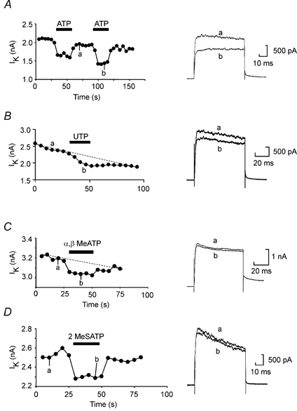
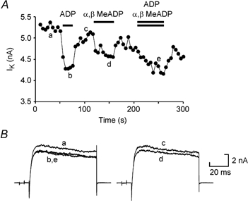
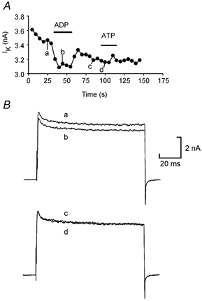
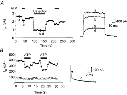
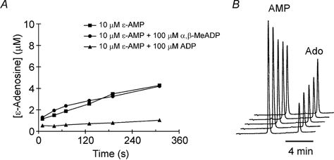
We have investigated the pharmacological properties and targets of p2y purinoceptors in Xenopus embryo spinal neurons. ATP reversibly inhibited the voltage-gated K(+) currents by 10 +/- 3 %. UTP and the analogues alpha,beta-methylene-ATP and 2-methylthio-ATP also inhibited K(+) currents. This agonist profile is similar to that reported for a p2y receptor cloned from Xenopus embryos. Voltage-gated K(+) currents could be inhibited by ADP (9 +/- 0.8 %) suggesting that a further p2y1-like receptor is also present in the embryo spinal cord. Unexpectedly we found that alpha,beta-methylene-ADP, often used to block the ecto-5'-nucleotidase, also inhibited voltage-gated K(+) currents (7 +/- 2.3 %). This inhibition was occluded by ADP, suggesting that alpha,beta-methylene-ADP is an agonist at p2y1 receptors. We have directly studied the properties of the ecto-5'-nucleotidase in Xenopus embryo spinal cord. Although ADP inhibited this enzyme, alpha,beta-methylene-ADP had no action. Caution therefore needs to be used when interpreting the actions of alpha,beta-methylene-ADP as it has previously unreported agonist activity at P2 receptors. Xenopus spinal neurons possess fast and slow voltage-gated K(+) currents. By using catechol to selectively block the fast current, we completely occluded the actions of ATP and ADP. Furthermore, the purines appeared to block only the fast relaxation component of the tail currents. We therefore conclude that the p2y receptors target only the fast component of the delayed rectifier. As ATP breakdown to ADP is rapid and ADP may accumulate at higher levels than ATP, the contribution of ADP acting through p2y1-like receptors may be an important additional mechanism for the control of spinal motor pattern generation.


Figures

Figure 1
Figure 1. Voltage-gated K+ currents are modulated by the triphosphate nucleotides ATP, UTP and artificial analogues of ATP
A, repeated voltage steps from −50 to 20 mV applied every 5 s evoked stable outward currents carried by the delayed rectifier in these neurons. ATP (100 μm) reversibly inhibited the current. B, in a different neuron UTP at 100 μm also inhibited the K+ currents. C and D, the non-hydrolysable ATP analogues α,β-methylene-ATP and 2-methylthio-ATP (at 100 μm) inhibited the K+ currents reversibly too.
Figure 2
Figure 2. Voltage-gated K+ currents are modulated by the diphosphate nucleotides ADP and α,β-methylene-ADP acting at the same receptor
A, graph showing the amplitude of the K+ currents evoked by repeated voltage steps from −50 to 20 mV applied every 5 s. ADP (100 μm) and α,β-methylene-ADP (100 μm) were applied separately and reversibly inhibited the currents. When the two agonists were applied simultaneously, the amount of block was no greater than that evoked by ADP alone. B, representative traces from the same experiment.
Figure 3
Figure 3. Some neurons are sensitive to ADP but not ATP suggesting that different receptors mediate the two responses
A, graph showing the amplitude of the K+ currents evoked by repeated voltage steps from −50 to 20 mV applied every 5 s. ADP at 200 μm inhibited the currents, but ATP at 200 μm did not. B, representative current traces from the same experiment.
Figure 4
Figure 4. Modulation of the voltage-gated K+ currents is confined to the fast activating component and is occluded by catechol
A, graph showing the amplitude of the K+ currents evoked by repeated voltage steps from −50 to 20 mV applied every 5 s. ATP (100 μm) inhibits the current (trace b). Catechol at 500 μm selectively blocks the fast activating component of the delayed rectifier. ATP applied during the catechol block has no effect on the remaining K+ currents (trace c, ATP washout trace d). Note that catechol (traces c and d) blocks only the fast component of the tail current. B, analysis of the tail currents shows that ATP only modulates the fast relaxation component (measured at 1 ms, •), but not the slow relaxation component (measured at 10 ms, ○). Representative tail current records illustrating modulation of the fast component by ATP are shown on the right.
Figure 5
Figure 5. Direct measurement of ecto-5′-nucleotidase activity in the spinal cord
HPLC techniques were used to monitor the conversion of etheno-AMP (ε-AMP) to ε-adenosine. A, the enzyme was inhibited by ADP but not α,β-methylene-ADP, the classical antagonist of this enzyme. B, HPLC chromatograms showing the elution of ε-AMP and ε-adenosine (Ado). These chromatograms were obtained in the presence of α,β-methylene-ADP and represent the conversion of ε-AMP to ε-adenosine after 40, 70, 130, 190 and 310 s. The vertical axis is in units of absorbance. The ε-AMP clearly falls in size as the ε-adenosine peak rises.

References

    1. Bogdanov YD, Dale L, King BF, Whittock N, Burnstock G. Early expression of a novel nucleotide receptor in the neural plate of Xenopus embryos. Journal of Biological Chemistry. 1997;272:12583–12590. - PubMed
    1. Brown DA, Filippov AK, Barnard EA. Inhibition of potassium and calcium currents in neurones by molecularly-defined P2Y receptors. Journal of the Autonomic Nervous System. 2000;81:31–36. - PubMed
    1. Brown P, Dale N. Adenosine A1 receptors modulate high voltage-activated Ca2+ currents and motor pattern generation in the Xenopus embryo. Journal of Physiology. 2000;525:655–667. - PMC - PubMed
    1. Dale N. The isolation and identification of spinal neurons that control movement in the Xenopus embryo. European Journal of Neuroscience. 1991;3:1025–1035. - PubMed
    1. Dale N. A large, sustained Na+-dependent and voltage-dependent K+ current in spinal neurons of the frog embryo. Journal of Physiology. 1993;462:349–372. - PMC - PubMed

Publication types

MeSH terms

LinkOut - more resources