. 2002 May 1;540(Pt 3):851-60.
doi: 10.1113/jphysiol.2001.013193.

Spike-independent release of ATP from Xenopus spinal neurons evoked by activation of glutamate receptors

Affiliations

Spike-independent release of ATP from Xenopus spinal neurons evoked by activation of glutamate receptors

Paul Brown et al. J Physiol. .

Abstract

As the release of ATP from neurons has only been directly studied in a few cases, we have used patch sniffing to examine ATP release from Xenopus spinal neurons. ATP release was detected following intracellular current injection to evoke spikes. However, spiking was not essential as both glutamate and NMDA could evoke release of ATP in the presence of TTX. Neither acetylcholine nor high K(+) was effective at inducing ATP release in the presence of TTX. Although Cd(2+) blocked glutamate-evoked release of ATP suggesting a dependence on Ca(2+) entry, neither omega-conotoxin-GVIA nor nifedipine prevented ATP release. N-type and L-type channels are thus not essential for glutamate-evoked ATP release. That glutamate receptors can elicit release in the absence of spiking suggests a close physical relationship between these receptors, the Ca(2+) channels and release sites. As the dependence of ATP release on the influx of Ca(2+) through Ca(2+) channel subtypes differs from that of synaptic transmitter release, ATP may be released from sites that are distinct from those of the principal transmitter. In addition to its role as a fast transmitter, ATP may thus be released as a consequence of the activation of excitatory glutamatergic synapses and act to signal information about activity patterns in the nervous system.


Figures

Figure 1
Figure 1. Patch sniffing can detect transient ATP release from isolated Xenopus neurons
A, whole cell recording from a HEK cell expressing P2X2 receptors showing that application of 500 μm ATP resulted in a large inward current. Other transmitters and agonists (all at 200 μm) did not evoke currents. B1, schematic diagram to show patch-sniffing arrangement for spike-induced ATP release. B2, simultaneous recording from Xenopus neuron (upper trace) and sniffer patch showing that a train of action potentials in the neuron evokes a current in the sniffer patch (lower trace). The artifacts in the sniffer patch are field potentials evoked in the recording from the spikes in the neuron. B3, similar records from a different cell and sniffer patch. C1, schematic diagram to show patch-sniffing arrangement for glutamate-induced ATP release. C2 and C3, currents evoked in a sniffer patch (C2) and a sniffer cell (C3) by application of 1 mm glutamate (bars) to two different Xenopus neurons. C4, 200 μm NMDA could also induce ATP release (different neuron, sniffer cell).
Figure 2
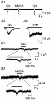
Figure 2. Glutamate or NMDA can stimulate ATP release from isolated spinal cords
A1, currents in a sniffer cell evoked by ATP release resulting from application of 200 μm NMDA (bar). A2, magnified portion of part of the same trace to show inward current has increased noise characteristic of channel activity. A3, currents from the same sniffer cell and spinal cord, showing that a smaller amount of ATP release could be detected after a recovery period of 4 min. B1, similar recording from a different experiment with a different sniffer cell, showing that 1 mm glutamate also evoked ATP release from isolated spinal cord. B2, part of the same record on a different time scale illustrating that the period of inward current shows increased noise characteristic of channel activity. C, sniffer cell current record from a separate experiment illustrating ATP detection on the first application of NMDA but no further release after re-application of glutamate or NMDA.
Figure 3
Figure 3. PPADS blocks the sniffer currents
A, in the presence of PPADS, 3 applications of glutamate to an isolated spinal cord failed to evoke a response in the nearby sniffer cell. B, after washout of PPADS, sniffer responses were recorded in response to 2 of 3 applications of glutamate. Lower trace shows an expanded region from the trace above indicated by the bar to illustrate ATP release evoked by glutamate. Note the large amplitude fluctuations indicative of channel gating and the increase of baseline noise in the absence of PPADS suggesting some spontaneous release of ATP from the spinal cord.
Figure 4
Figure 4. Spikes are not necessary for glutamate receptor-evoked ATP release
A1, the sniffer patch detects ATP release on application of 1 mm glutamate and 200 μm NMDA to a cord in the presence of TTX (500 nm). A2, part of the same record on a different time scale illustrating that the period of inward current shows increased noise characteristic of channel activity. A3, exogenous ATP (500 μm) evoked very similar currents in the same sniffer patch to those recorded during stimulation of endogenous release by glutamate (A2). B1, ATP release from an isolated neuron in the presence of TTX could still be detected following application of 1 mm glutamate (recorded by a sniffer patch). Inset shows an expanded portion of the trace to illustrate the sniffer currents following the application of glutamate. B2, NMDA application was also able to evoke release of ATP from neurons in the presence of TTX.
Figure 5
Figure 5. Glutamate but not high K+ or acetylcholine causes release of ATP from isolated spinal cord in the presence of TTX
A, a high K+ saline failed to evoke sniffer currents from the isolated spinal cord. B, application of 100 μm acetylcholine (ACh) to the same piece of spinal cord did not evoke a response in the sniffer cell either. C, two subsequent applications of glutamate to the same spinal cord evoked sniffer currents. The same sniffer cell was used for A, B and C.
Figure 6
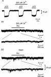
Figure 6. ATP release requires a Ca2+ influx
A, repeated responses to 500 μm ATP in a sniffer cell were unaffected by 200 μm Cd2+, demonstrating that Cd2+ does not affect the sensitivity of ATP detection. B1, current record (sniffer cell) showing that glutamate cannot evoke release of ATP in the presence of 200 μm Cd2+. Lower trace is an expanded portion of the above record during glutamate application. B2, after washout of Cd2+, a response in the sniffer cell was seen in response to glutamate application indicating that ATP release had been restored. Lower trace is an expanded portion of the above record during glutamate application.
Figure 7
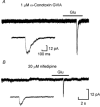
Figure 7. ATP release can occur when N-type or L-type channels are blocked
A, in the presence of ω-conotoxin GVIA, glutamate can evoke ATP release from an isolated cord. Inset shows expanded portion of the sniffer current record. B, nifedipine is also unable to block glutamate-induced ATP release from an isolated spinal cord (different experiment to that in A). Inset shows expanded portion of the response (at the same scale as the inset in A). Sniffer cells used in A and B.

References

    1. Anderson CM, Baldwin SA, Young JD, Cass CE, Parkinson FE. Distribution of mRNA encoding a nitrobenzylthioinosine-insensitive nucleoside transporter (ENT2) in rat brain. Brain Research. Molecular Brain Research. 1999a;70:293–297. - PubMed
    1. Anderson CM, Xiong W, Geiger JD, Young JD, Cass CE, Baldwin SA, Parkinson FE. Distribution of equilibrative, nitrobenzylthioinosine-sensitive nucleoside transporters (ENT1) in brain. Journal of Neurochemistry. 1999b;73:867–873. - PubMed
    1. Bardoni R, Goldstein PA, Lee CJ, Gu JG, MacDermott AB. ATP P2X receptors mediate fast synaptic transmission in the dorsal horn of the rat spinal cord. Journal of Neuroscience. 1997;17:5297–5304. - PMC - PubMed
    1. Brown PE, Dale N. Adenosine A1 receptors modulate high voltage-activated Ca2+ currents and motor pattern generation in the Xenopus embryo. Journal of Physiology. 2000;525:655–667. - PMC - PubMed
    1. Dale N. Reciprocal inhibitory interneurones in the Xenopus embryo spinal cord. Journal of Physiology. 1985;363:61–70. - PMC - PubMed

Publication types

MeSH terms

LinkOut - more resources