. 2002 May 1;540(Pt 3):883-93.
doi: 10.1113/jphysiol.2001.016188.

Phosphorylation and functional regulation of ClC-2 chloride channels expressed in Xenopus oocytes by M cyclin-dependent protein kinase

Affiliations

Phosphorylation and functional regulation of ClC-2 chloride channels expressed in Xenopus oocytes by M cyclin-dependent protein kinase

Tetsushi Furukawa et al. J Physiol. .

Abstract

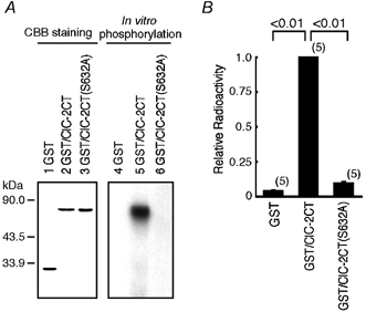
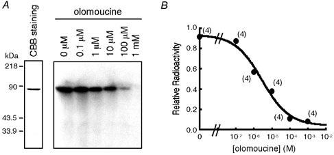
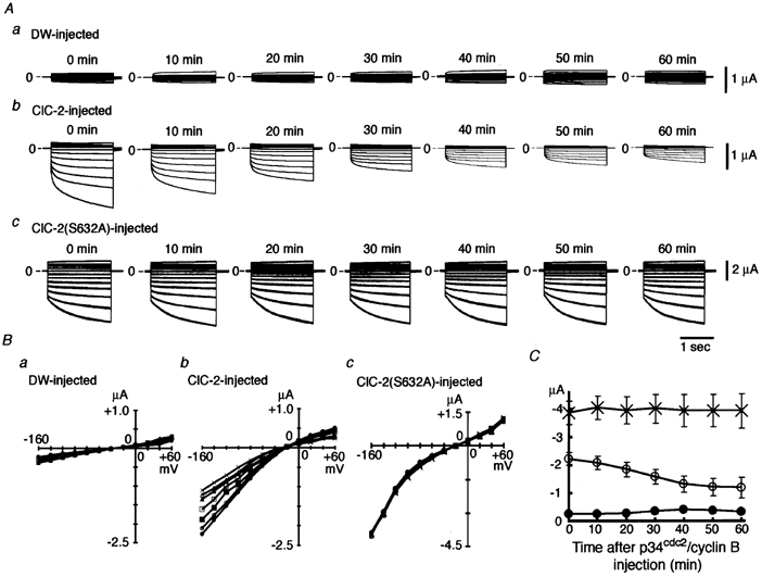
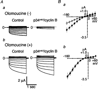
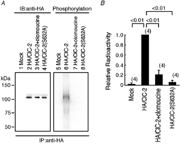
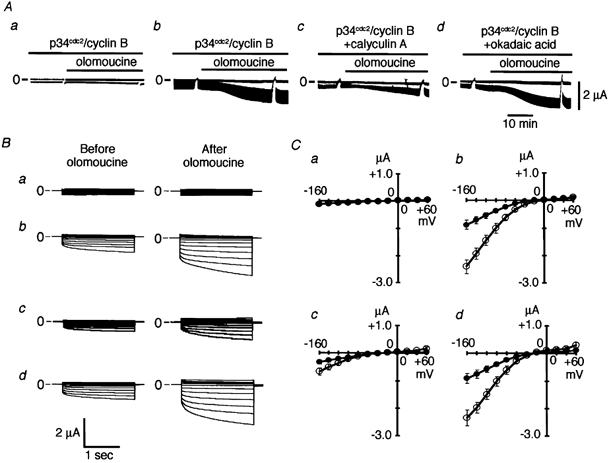
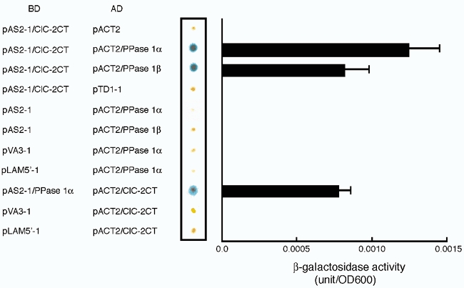
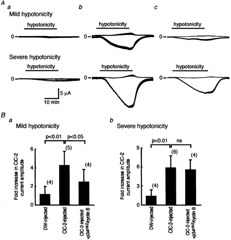
Many dramatic alterations in various cellular processes during the cell cycle are known to involve ion channels. In ascidian embryos and Caenorhabditis elegans oocytes, for example, the activity of inwardly rectifying Cl(-) channels is enhanced during the M phase of the cell cycle, but the mechanism underlying this change remains to be established. We show here that the volume-sensitive Cl(-) channel, ClC-2 is regulated by the M-phase-specific cyclin-dependent kinase, p34(cdc2)/cyclin B. ClC-2 channels were phosphorylated by p34(cdc2)/cyclin B in both in vitro and cell-free phosphorylation assays. ClC-2 phosphorylation was inhibited by olomoucine and abolished by a (632)Ser-to-Ala (S632A) mutation in the C-terminus, indicating that (632)Ser is a target of phosphorylation by p34(cdc2)/cyclin B. Injection of activated p34(cdc2)/cyclin B attenuated the ClC-2 currents but not the S632A mutant channel currents expressed in Xenopus oocytes. ClC-2 currents attenuated by p34(cdc2)/cyclin B were increased by application of the cyclin-dependent kinase inhibitor, olomoucine (100 microM), an effect that was inhibited by calyculin A (5 nM) but not by okadaic acid (5 nM). A yeast two-hybrid system revealed a direct interaction between the ClC-2 C-terminus and protein phosphatase 1. These data suggest that the ClC-2 channel is also counter-regulated by protein phosphatase 1. In addition, p34(cdc2)/cyclin B decreased the magnitude of ClC-2 channel activation caused by cell swelling. As the activities of both p34(cdc2)/cyclin B and protein phosphatase 1 vary during the cell cycle, as does cell volume, the ClC-2 channel could be regulated physiologically by these factors.


Figures

Figure 1
Figure 1. Phosphorylation of ClC-2 C-terminus in vitro
A, lanes 1–3 show results of Coomassie brilliant blue (CBB) staining: loaded with 0.2 μg of purified glutathione-S-transferase (GST) protein (lane 1); GST/ClC-2CT (lane 2); and GST/ClC-2CT (S632A) (lane 3). Lanes 4–6 show representative results of in vitro phosphorylation assay: loaded with reaction mixture containing GST (lane 4); GST/ClC-2CT (lane 5); and GST/ClC-2CT(S632A) (lane 6). B, quantification of in vitro phosphorylation by filter phosphorylation assay. Thirty microlitres of terminated kinase assay mixtures were spotted onto P-81 phosphocellulose paper, and radioactivity was counted with a liquid scintillation counter normalised to the value for GST/ClC-2CT. The mean radioactive value for GST/ClC-2CT was 14855 ± 1441 cpm (n = 5). The bar indicates standard deviation, and the number in parentheses attached to the bar indicates the number of experiments in this and in the following figures.
Figure 2
Figure 2. Effects of olomoucine on phosphorylation of the ClC-2 C-terminus in vitro
A, the left panel shows the results of CBB staining; 0.2 μg of purified GST/ClC-2CT was loaded. The right panel shows representative results of experiments testing the effects of olomoucine on in vitro phosphorylation of GST/ClC-2CT by activated p34cdc2/cyclin B. The phosphorylation reaction was carried out in the absence (lane 1) or the presence of olomoucine at a concentration of 0.1 μm (lane 2), 1 μm (lane 3), 10 μm (lane 4), 100 μm (lane 5), and 1 mm (lane 6). B, dose-response curve for inhibition of in vitro phosphorylation by olomoucine. Incorporated radioactivity was measured by a liquid scintillation counter in a filter phosphorylation assay in four independent experiments. Values were normalised to those obtained in the absence of olomoucine.
Figure 3
Figure 3. Effects of p34cdc2/cyclin B injection on ClC-2 currents
A, representative experiments showing the effects of activated p34cdc2/cyclin B injected into oocytes on ClC-2 currents. Panel a, an oocyte injected with distilled water (DW); panel b, an oocyte injected with ClC-2 cRNA; and panel c, an oocyte injected with ClC-2(S632A) cRNA. Step pulses to various potentials between −160 mV and +60 mV in 20 mV increments were applied every 10 min after injection of p34cdc2/cyclin B (0.04 u) for 60 min, and the superimposed currents at each time point are shown. B, current-voltage curves from five oocytes injected with DW (panel a), from seven oocytes injected with ClC-2 cRNA (panel b) and from four oocytes injected with ClC-2(S632A) cRNA (panel c) before (•) and 10 (○), 20 (▪), 30 (□), 40 (▴), 50 (▵), and 60 min (×) after injection of p34cdc2/cyclin B. Bars showing standard deviation have been omitted for clarity. C, time course of changes in ClC-2 channel currents at −160 mV after injection of p34cdc2/cyclin B (0.04 u). •, oocytes injected with distilled water; ○, oocytes injected with ClC-2 mRNA; ×, oocytes injected with ClC-2(S632A) mRNA.
Figure 4
Figure 4. Effects of olomouocine on p34cdc2/cyclin B-inhibition of ClC-2 currents
A, representative membrane currents before (left panel) and 60 min after (right panel) injection of activated p34cdc2/cyclin B. Panel a, an oocyte injected with ClC-2 cRNA; panel b, an oocyte injected with ClC-2 cRNA and pre-incubated with 100 μm olomoucine. Olomoucine was applied 30 min before injection of p34cdc2/cyclin B. Step pulses to various potentials between −160 mV and +60 mV in 20 mV increments were applied, and the superimposed currents are shown. B, current-voltage curves from seven oocytes injected with ClC-2 cRNA (panel a), from five oocytes injected with ClC-2 cRNA and pre-treated with olomoucine (panel b) before (•) and 60 min after (○) injection of activated p34cdc2/cyclin B.
Figure 5
Figure 5. Phosphorylation of full-length ClC-2 by p34cdc2/cyclin B in oocyte microsomes
A, the left panel shows representative data demonstrating biosynthetic labelling with [35S]-methionine of oocytes injected with DW (lane 1), oocytes injected with haemagglutinin (HA)/ClC-2 cRNA (lane 2), oocytes injected with HA/ClC-2 cRNA and incubated with olomoucine for 1.5 h (lane 3), and oocytes injected with HA/ClC-2(S632A) cRNA (lane 4). The right panel shows representative data demonstrating results of phosphorylation of full-length ClC-2 in oocyte microsomes. Radioactive gels were dried and subjected to autoradiography. IB, immunoblotting; IP, immunoprecipitation. B, quantification of phosphorylation of oocyte microsomes by a BAS1000 image analyser. Radioactivity was normalised to the value for HA/ClC-2.
Figure 6
Figure 6. Effects of protein phosphatase (PPase) inhibitors
A, representative continuous recordings of membrane currents showing the effects of extracellularly applied 100 μm olomoucine. Activated p34cdc2/cyclin B (0.04 u) was injected 30 min before olomoucine application in a DW-injected oocyte (panel a), in a ClC-2- injected oocyte (panel b), in a ClC-2-injected oocyte pre-treated with calyculin A (5 nm; panel c) and in a ClC-2-injected oocyte pre-treated with okadaic acid (5 nm; panel d); 2 s step pulses to −160 mV from a holding potential of −30 mV were applied every 5 s. Before and 30 min after application of olomoucine, step pulses at various potentials between −160 mV and +60 mV in 20 mV increments were applied. The upper lines show the current level at a holding potential of −30 mV and lower lines at a step pulse to −160 mV. Upward represents the outward current and downward represents the inward current. B, representative superimposed current traces before (left panel) and 30 min after olomoucine application (right panel) are shown. Panel a shows data from a DW-injected oocyte, panel b from a ClC-2-injected oocyte, panel c from a ClC-2-injected oocyte pre-treated with calyculin A (5 nm) and panel d from a ClC-2-injected oocyte pre-treated with okadaic acid (5 nm). C, the current-voltage curves from five oocytes injected with DW (panel a), from five oocytes injected with ClC-2 cRNA (panel b), from five oocytes injected with ClC-2 cRNA and pre-treated with calyculin A (5 nm; panel c) and from five oocytes injected with ClC-2 cRNA and pre-treated with okadaic acid (5 nm; panel d) before (•) and 30 min after (○) olomoucine application.
Figure 7
Figure 7. Interaction between ClC-2CT and PPase 1 shown by yeast two-hybrid assay
The combinations of transformed plasmids for the GAL4-binding domain (BD)-fusion protein and the GAL4-activation domain (AD)-fusion protein are listed on the left. Results of representative qualitative β-galactosidase filter lift assay (middle panel, out of three experiments) and quantitative β-galactosidase assay (right panel, n = 4) for each combination of plasmid transformation are shown. pVA3-1, murine p53 in the pAS2-1 vector; pLAM5′-1, human Lamin C in the pAS2-1 vector; pTD1-1, SV40 large T antigen in the pACT2 vector.
Figure 8
Figure 8. Relationship between regulation of ClC-2 currents by volume and p34cdc2/cyclin B phosphorylation
A, representative continuous recordings of ClC-2 currents in a DW-injected oocyte (panel a), in a ClC-2-injected oocyte (panel b), and in a ClC-2-injected oocyte also injected with p34cdc2/cyclin B (0.04 u). The extracellular solution was changed from an isotonic solution to a mild hypotonic solution (upper panel) or to a severe hypotonic solution (lower panel) for 30 min. Step pulses from a holding potential of −30 mV to −160 mV were applied every 5 s. The upper lines show the current level at a holding potential of −30 mV and the lower lines at step pulses to −160 mV. Upward represents the outward current and downward represents the inward current. B, the fold increase in ClC-2 current amplitude at −160 mV, 30 min after superfusion with hypotonic solution relative to that in the isotonic solution with mild hypotonic solution (panel a) and with a severe hypotonic solution (panel b).

References

    1. Block ML, Moody WJ. A voltage-dependent chloride current linked to the cell cycle in ascidian embryos. Science. 1990;247:1090–1092. - PubMed
    1. Bosl MR, Stein V, Hubner C, Zdebik AA, Jordt S -E, Mukhopadhyay AK, Davidoff MS, Holstein A -F, Jentsch TJ. Male germ cells and photoreceptors, both dependent on close cell-cell interactions, degenerate upon ClC-2 Cl− channel disruption. EMBO Journal. 2001;20:1289–1299. - PMC - PubMed
    1. Bruggemann A, Stuhmer W, Pardo LA. Mitosis-promoting factor-mediated suppression of a cloned delayed rectifier potassium channel expressed in Xenopus oocytes. Proceedings of the National Academy of Sciences of the USA. 1997;94:537–542. - PMC - PubMed
    1. Bubien JK, Kork KL, Rado TA, Frizzell RA. Cell cycle dependence of chloride permeability in normal and cystic fibrosis lymphocytes. Science. 1990;248:1416–1419. - PubMed
    1. Campbell KS, Buder A, Deuschle U. Interactions between the amino-terminal domain of p56lck and cytoplasmic domains of CD4 and CD8 alpha in yeast. European Journal of Immunology. 1995;25:2408–2412. - PubMed

Publication types

LinkOut - more resources