. 2002 May 1;540(Pt 3):989-1002.
doi: 10.1113/jphysiol.2001.013086.

Calcitonin gene-related peptide immunoreactivity and afferent receptive properties of dorsal root ganglion neurones in guinea-pigs

Affiliations

Calcitonin gene-related peptide immunoreactivity and afferent receptive properties of dorsal root ganglion neurones in guinea-pigs

S N Lawson et al. J Physiol. .

Abstract

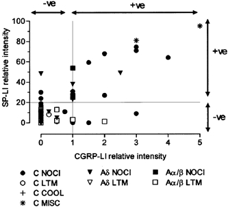
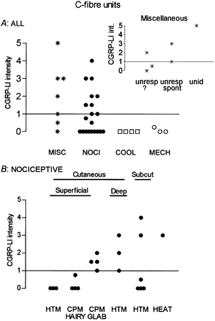
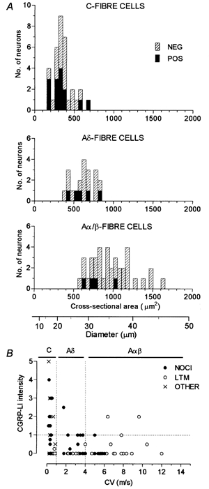
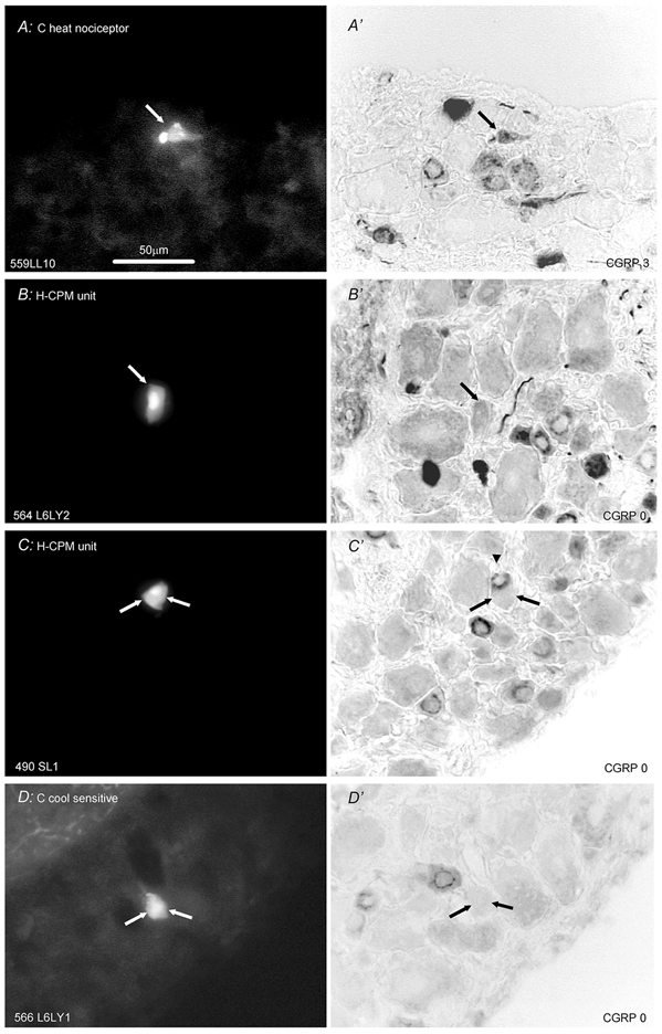
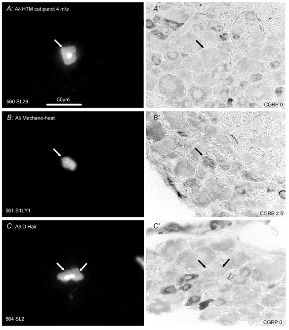
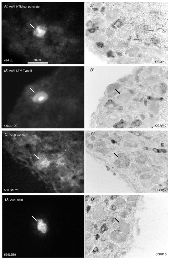
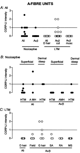
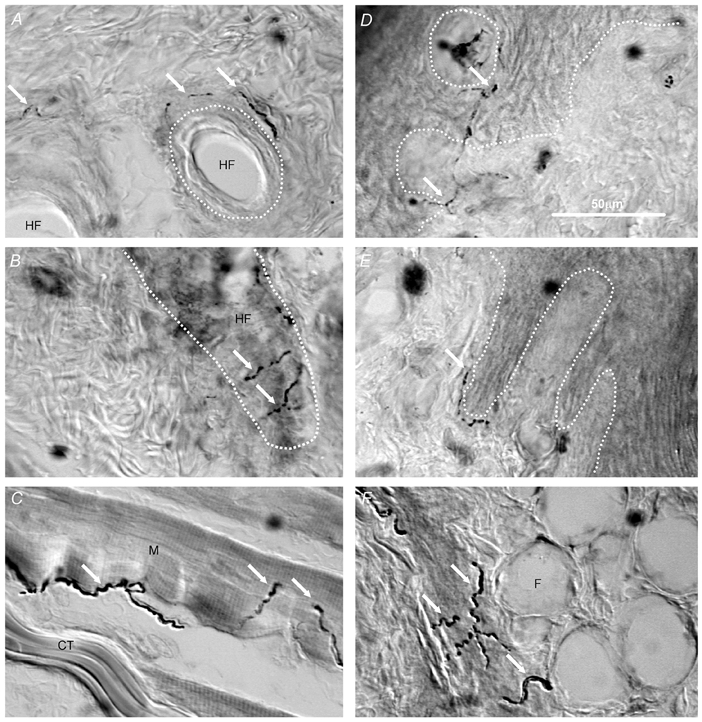
To establish the afferent receptive properties of lumbosacral dorsal root ganglion (DRG) neurones that express calcitonin gene-related peptide (CGRP), intracellular recordings were made with fluorescent dye-filled electrodes in deeply anaesthetised young guinea-pigs. After determination of neuronal functional properties, dye was injected into the soma. CGRP-like immunoreactivity (CGRP-LI) was examined on histological sections of dye-marked neurones. Fourteen of 34 C-fibre neurones showed CGRP-LI. These included 10/21 C-fibre nociceptive neurones. All C-polymodal nociceptors in glabrous (n = 4) but none in hairy skin (n = 4) were positive. Positive C-fibre high threshold mechanoreceptive (HTM) units had receptive fields in dermal or deeper tissue. Four (n = 6) unresponsive or unidentified C-fibre units were positive. Neither C-fibre cooling sensitive (n = 4) nor C-fibre low threshold mechanoreceptive (LTM) units (n = 3) had CGRP-LI. Six of 23 A-fibre nociceptive cells were positive including one Aalpha/beta unit. Three of these positive cells had epidermal and three had dermal/deep receptive fields. Three of 36 A-fibre LTM units exhibited CGRP-LI; all were Aalpha/beta-fibre G hair units. All glabrous skin and muscle spindle units and in hairy skin slowly adapting and field units, and some G-hair units lacked CGRP-LI. CGRP-LI stained fibres were found in tissues containing receptive fields of positive DRG neurones: glabrous skin, near hair follicles and in skeletal muscle. A few substance P-labelled neurones did not exhibit CGRP-LI and vice versa. Thus CGRP expression was detected in under half the nociceptive neurones, was not limited to nociceptive neurones and apart from receptive properties was also related to location/depth in the tissues of a DRG neurone's peripheral terminals.


Figures

Figure 8
Figure 8. Relative intensities of CGRP-LI and SP-LI in the same DRG somata
See Methods for generation of relative intensity measurements on x and y axes. For SP-LI, the values are calculated using image analysis measurements, with 100 % representing maximum intensity and ≥ 20 % relative intensity judged as positive. For CGRP-LI, 5 was maximum and values ≥ 1 were judged as positive. For abbreviations, see Fig. 5 legend.
Figure 5
Figure 5. C-fibre units: intensities of CGRP-LI in relation to afferent receptive properties
A, plot of all units studied. The miscellaneous group (MISC) contained unresponsive or unidentified neurones (see text). B, the nociceptive C-fibre neurones are subdivided according to their responses to noxious mechanical and noxious heat stimuli, and to the apparent depth in the tissues of their receptive fields. Abbreviations as follows: NOCl, nociceptive; COOL, cooling sensitive; MECH, C mechanoreceptor (C LTM); unresp, unresponsive; unid, unidentified.
Figure 1
Figure 1. Cell size and conduction velocity in relation to CGRP-LI
A, distribution of sizes of the largest section through each dye- labelled neurone. Separate plots of C-, Aδ- and Aα/β-fibre neurones are shown. The CGRP-LI positive units are shown as filled bars. The positive G-hair units were the three largest positive Aα/β-fibre units. Below A is a scale converting area to diameter. B, dorsal root CV of the dye-labelled cell is plotted against the relative intensity score for CGRP-LI.
Figure 2
Figure 2. CGRP-LI in identified C-fibre neurones
Left column (A-D) shows fluorescence images prior to immunocytochemistry of Lucifer Yellow (LY) dye-injected neurones; right column shows images of the same neuronal section after immunocytochemistry for CGRP-LI. Cells A, B and D are from L6 and C is from S1. The CVs of the units were (m s−1) A, 0.35; B, 0.36; C, 0.37 and D, 0.76. A, section though a C-fibre heat nociceptor that responded to noxious heat but not innocuous radiant heat on the foot. No mechanically sensitive region could be found. Although this section is through the edge of the cell, it is adjacent to one containing the nucleus in this very small cell. B and C present two examples of CGRP-LI negative H-CPMs (hairy skin C-polymodal nociceptors) with receptive fields on the upper leg/thigh. C, strongly positive unit (arrowhead in C') is just above negative units (shown with two arrows). D, cooling/cold sensitive neurone with spontaneous discharge at room temperature that was promptly inhibited by radiant warming of the sole of the foot. Scale bar in A applies to all images. The subjectively judged scores of CGRP-LI relative intensity are given at the bottom right of each image showing CGRP-LI.
Figure 3
Figure 3. CGRP-LI in identified Aδ-fibre neurones
Layout as in Fig. 2. Left, S1 DRG neurones labelled with LY; right, micrographs of the same DRG sections processed to show CGRP-LI. A, CGRP-LI negative HTM that had a typical superficial, distributed punctate receptive field (CV, 4 m s−1). B, MH cell (mechanical-heat nociceptor) (CV, 1.8 m s−1) that was unambiguously positive for CGRP-LI. The receptive field was superficial and punctate on the side of the upper leg, evoking a response to pressure with needle, pinch by fine forceps and noxious heat. C, section through the nucleus of a D-hair unit (CV, 3.1 m s−1) that was negative for CGRP-LI. The unit had a receptive field on the thigh that gave vigorous responses to moving brush, skin stretch and sudden cooling. Scale bar in A applicable to all.
Figure 4
Figure 4. CGRP-LI in identified Aα/β-fibre neurones
Format as for Fig. 2. In A, B and C, the labelling fluorescent dye was LY and in D was ethidium bromide. A and B are from L6, C is from S1 and D is from L5. A, B and C are through the cell nuclei, whereas D is through the cytoplasm A, Aα/β HTM (CV, 5.5 m s−1) with a superficial punctate receptive field negative for CGRP-LI. B, a slowly adapting type II unit (CV, 5.6 m s−1) with receptive field on the thigh negative for CGRP. C, G2 hair unit (CV, 9.6 m s−1) with receptive field at the top of the thigh showed mottled cytoplasmic CGRP-LI labelling. D, field unit (CV, 6.3 m s−1) negative for CGRP-LI with receptive field on the dorsal surface of a toe. Scale bar in A applicable to all.
Figure 6
Figure 6. A-fibre units: intensities of CGRP-LI in relation to afferent receptive properties
A, nociceptive and LTM units subdivided into Aα/β and Aδ groups. Aα/β cutaneous LTM (cut) and muscle spindle (musc) units shown separately. A subgroup of nociceptors and certain Aα/β LTM units were positive. B, the nociceptive Aδ- and Aα/β-units are separated into those with superficial (probably epidermal) and subepidermal (dermal/deep) receptive fields. Units with superficial fields were divided according to their response to noxious heat into HTM and MH units. C, A-fibre LTMs subdivided according to receptive type. SA, slowly adapting LTM; RA, rapidly adapting LTM; MS, muscle spindle LTM units. Note: none of the Aδ-fibre (D hair) category were positive and G-hair receptors were the sole Aα/β units expressing CGRP-LI.
Figure 7
Figure 7. CGRP-LI in nerve fibres of guinea-pig skin and muscle
Images captured using interference contrast microscopy to show CGRP-LI-labelled nerve fibres in hairy skin (A and B), skeletal muscle (C) and glabrous skin (D, E and F). Some CGRP-LI-labelled nerve fibres are indicated by white arrows. A and B, the approximate outlines of hair follicles are indicated with white dotted lines showing CGRP-LI-positive fibres to be associated with hair follicles. C, CGRP-LI positive fibres are present in skeletal muscle. D and E, the junction between the epidermis and dermis is indicated with the white dotted line. D, the white dotted circle outlines a dermal projection into the epidermis (dermal papilla). CGRP-LI-positive fibres can be seen within dermal pegs and apparently penetrating the epidermis, as well as (E) at the junction between dermis and epidermis. F, fat cells under the foot pad with positive fibres (arrows). CT, connective tissue; M, skeletal muscle fibre; HF, hair follicle; F, fat cell. Scale bar in D is for all images.

Similar articles

Cited by

References

    1. Averill S, Mcmahon SB, Clary DO, Reichardt LF, Priestley JV. Immunocytochemical localization of trkA receptors in chemically identified subgroups of adult rat sensory neurons. European Journal of Neuroscience. 1995;7:1484–1494. - PMC - PubMed
    1. Bennett DL, Dmietrieva N, Priestley JV, Clary D, McMahon SB. TrkA, CGRP and IB4 expression in retrogradely labelled cutaneous and visceral primary sensory neurones in the rat. Neuroscience Letters. 1996;206:33–36. - PubMed
    1. Brain SD, Cambridge H, Hughes SR, Wilsoncroft P. Evidence that calcitonin gene-related peptide contributes to inflammation in the skin and joint. Annals of the New York Academy of Sciences. 1992;657:412–419. - PubMed
    1. Brain SD, Williams TJ. Interactions between the tachykinins and calcitonin gene-related peptide lead to the modulation of oedema formation and blood flow in rat skin. British Journal of Pharmacology. 1989;97:77–82. - PMC - PubMed
    1. Buckley TL, Brain SD, Rampart M, Williams TJ. Time-dependent synergistic interactions between the vasodilator neuropeptide, calcitonin gene-related peptide (CGRP) and mediators of inflammation. British Journal of Pharmacology. 1991;103:1515–1519. - PMC - PubMed

Publication types

Substances

LinkOut - more resources