. 2002 May 6;86(9):1444-8.
doi: 10.1038/sj.bjc.6600282.

Increase in immune cell infiltration with progression of oral epithelium from hyperkeratosis to dysplasia and carcinoma

Affiliations
Free PMC article

Increase in immune cell infiltration with progression of oral epithelium from hyperkeratosis to dysplasia and carcinoma

G Gannot et al. Br J Cancer. .
Free PMC article

Abstract

In the present study, epithelium derived lesions of various pathological manifestations were examined histologically and immunohistochemically for mononuclear cell infiltration. The infiltrate under the transformed epithelium of oral lesions, was examined for differences in the composition of immune mononuclear cells as the epithelium moves from hyperkeratosis through various degrees of dysplasia to squamous cell carcinoma. The study was performed on 53 human tongue tissues diagnosed as hyperkeratosis (11 cases), mild dysplasia (nine cases), moderate and severe dysplasia (14 cases) and squamous cell carcinoma (19 cases). A similar analysis was performed on 30 parotid gland tissues diagnosed as pleomorphic adenoma (14 cases) and carcinoma ex-pleomorphic adenoma (16 cases). Immunohistochemical analysis of various surface markers of the tumour infiltrating immune cells was performed and correlated with the transformation level as defined by morphology and the expression of p53 in the epithelium. The results revealed that, in the tongue lesions, the changes in the epithelium from normal appearance to transformed were accompanied by a corresponding increase in the infiltration of CD4, CD8, CD14, CD19+20, and HLA/DR positive cells. The most significant change was an increase in B lymphocytes in tongue lesions, that was in accordance with the transformation level (P<0.001). In the salivary gland, a significant number of cases did not show an infiltrate. In cases where an infiltrate was present, a similar pattern was observed and the more malignant tissues exhibited a higher degree of immune cell infiltration.


Figures

Figure 1
Figure 1
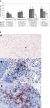
(A) Lymphocyte profile of tongue lesions. Immunohistochemical detection of lymphocyte markers. Sections were reacted with antibodies for CD4, CD8 and CD19+20 using an immunohistochemical protocol. Positive cells were counted out of a total number of infiltrating cells in a 0.04 mm2 area. The bars represent the percentage of positive cells counted out of total number of infiltration cells. In every case four different areas were counted and a mean was established per case. In parentheses are the number of cases for each group. *Significant difference between these groups when compared to hyperkeratosis and mild dysplasia. (B) Immunohistochemical staining of tongue lesions with B cell antibody. The red staining represents positive reaction. (a) Hyperkeratosis, (b) moderate and severe dysplasia, (c) SCC. Demonstration of B cells (stained in red) in cases of hyperkeratosis, moderate and severe dysplasia and SCC. Notice the dramatic elevation in the number of B cells when the epithelium is dysplastic (in the cases of moderate and severe dysplasia and SCC). Magnification of ×200.
Figure 2
Figure 2
CD14 cell profile of tongue lesions. Immunohistochemical detection of CD14 positive cells. Sections were reacted with an antibody for CD14 using an immunohistochemical protocol. Positive cells were counted out of a total number of infiltrating cells in a 0.04 mm2 area. The bars represent the percentage of positive cells counted out of total number of infiltrating cells. In each case four different areas were counted and a mean was established per case.
Figure 3
Figure 3
HLA/DR profile of tongue lesions. Immunohistochemical detection of HLA/DR positive cells. Sections were reacted with an antibody for CD14 using an immunohistochemical protocol. Positive cells were counted out of a total number of infiltrating cells in a 0.04 mm2 area. The bars represent the percentage of positive cells counted out of total number of infiltrating cells. In each case four different areas were counted and a mean was established per case.
Figure 4
Figure 4
p53 profile of epithelium in tongue lesions. Immunohistochemical detection of p53 positive epithelial cells. Sections were reacted with an antibody for p53 using an immunohistochemical protocol. Positive epithelial cells were counted in a 0.04 mm2 area out of a total number of epithelial cells. The bars represent the percentage of positive cells counted out of total number of epithelial cells. In each case four different areas were counted and a mean was established per case.
Figure 5
Figure 5
Profile of infiltrating cells according to p53 staining in SCC tissues of the tongue. Cases of SCC were grouped according to their staining for p53. The cell infiltrate profile of three cases of negative p53 epithelial staining was compared to these of the epithelial p53 positive SCC cases.
Figure 6
Figure 6
A summary of parotid gland results. Sections were reacted with antibodies for CD4 and CD19+20 (B cells) using an immunohistochemical protocol. CD 4 reacted positive with nine out of 14 cases of PA and 12 out of 16 cases of C/PA. Positive staining for CD19+20 was found in eight out of 14 cases of PA and eight out of 16 cases of C/PA. In each case four different areas (0.04 mm2) were counted and a mean was calculated per case. The bars represent the percentage of positive cells counted out of a total number of infiltrating cells in cases positive for the antibody examined.

References

    1. AuclairPLEllisGL1996Atypical features in salivary gland mixed tumors: their relationship to malignant transformation Mod Pathol 9652657 - PubMed
    1. BarreraJLVerasteguiEMenesesAZinserJde la GarzaJHaddenJW2000Combination immunotherapy of squamous cell carcinoma of the head and neck: a phase 2 trial Arch Otolaryngol Head Neck Surg 126345351 - PubMed
    1. EllisGLAuclairPL1996Benign Epithelial NeoplasmsInTumors of the salivary glandspp468Washington, DC: Armed Forces Institute of Pathology
    1. HirotaJUetaEOsakiTOgawaY1990Immunohistologic study of mononuclear cell infiltrates in oral squamous cell carcinoma Head Neck 12118125 - PubMed
    1. IkedaKKatohTHa-KawaSKIwaiHYamashitaTTanakaY1996The usefulness of MR in establishing the diagnosis of parotid pleomorphic adenoma Am J Neuroradiol 17555559 - PMC - PubMed

Publication types

MeSH terms