. 2002 May 3;296(5569):920-2.
doi: 10.1126/science.1068452.

Neurofibromas in NF1: Schwann cell origin and role of tumor environment

Affiliations

Neurofibromas in NF1: Schwann cell origin and role of tumor environment

Yuan Zhu et al. Science. .

Abstract

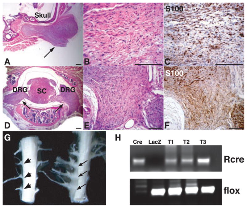
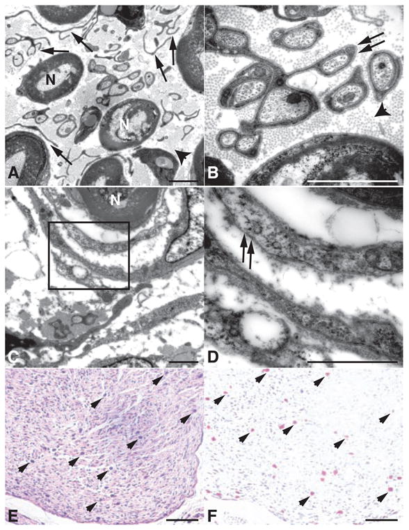
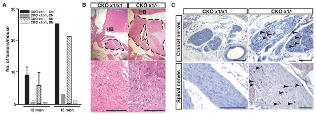
Neurofibromatosis type 1 (NF1) is one of the most prevalent dominantly inherited genetic diseases of the nervous system. NF1 encodes a tumor suppressor whose functional loss results in the development of benign neurofibromas that can progress to malignancy. Neurofibromas are complex tumors composed of axonal processes, Schwann cells, fibroblasts, perineurial cells, and mast cells. Through use of a conditional (cre/lox) allele, we show that loss of NF1 in the Schwann cell lineage is sufficient to generate tumors. In addition, complete NF1-mediated tumorigenicity requires both a loss of NF1 in cells destined to become neoplastic as well as heterozygosity in non-neoplastic cells. The requirement for a permissive haploinsufficient environment to allow tumorigenesis may have therapeutic implications for NF1 and other familial cancers.


Figures

Fig. 1
Fig. 1
Histological and molecular analysis of neurofibromas from NF1 flox/−;Krox20-cre mice. Sections from a neurofibroma (arrow) associated with cranial nerves were stained with hematoxylin and eosin (H&E) (A and B) and S100 antibody (C). Sections from two neurofibromas (arrows) originating from dorsal root ganglia (DRG) were stained with H&E (D and E) and S100 (F). SC, spinal cord. (G) An example of three macroscopic neurofibromas from spinal roots (right, arrows), as compared to control spinal roots (left, arrowheads). (H) Genomic DNAs isolated from three neurofibromas in (G) (T1 to T3) were subjected to polymerase chain reaction analysis, as described in (11). Cre, genomic DNAs from a NF1flox/flox Schwann cell culture infected with Cre-expressing adenovirus. Of note, only the recombined allele (Rcre) remains in this culture. LacZ, genomic DNAs from similar Schwann cell culture infected with LacZ-expressing adenovirus. Scale bar, (A) to (F), 25 μm.
Fig. 2
Fig. 2
Neurofibromas from NF1flox/−;Krox20-cre mice contain multiple cell types. (A) Higher magnification of a neurofibroma demonstrates myelinated axons (N) separated by dissociated Schwann cells (arrows) and collagen bundles (arrowhead). (B) In addition to dissociated Schwann cells, the neurofibroma also contains differentiated Schwann cell processes surrounding individual unmyelinated axons (arrows), suggesting a continuum of dysplastic to overly neoplastic transformation. (C) Electron microscopy analysis demonstrates the presence of perineurial-like cells surrounding axons in a concentric fashion, reminiscent of the multilamellar arrays of perineurial cells seen in human perineuriomas (14). (D) In contrast to Schwann cells elsewhere in the tumor, these perineurial-like cells lack a continuous basal lamina (arrows). Sections from a neurofibroma were stained with H&E (E) and Leder staining, an enzymatic stain for Naphthol AS-D (3-hydroxy-2-naphoic acid-O-toluidine) choloroacetate esterase (F), displaying numerous mast cells (arrowheads) infiltrating the tumors. Scale bar, (A) to (D), 2 μm; (E) and (F), 100 μm.
Fig. 3
Fig. 3
Phenotypic comparison of NF1flox/−;Krox20-cre (CKO×1+/−) and NF1flox/flox;Krox20-cre (CKO×1/×1) mice. (A) Quantification of neurofibromas associated with cranial nerves (CN) and spinal nerves (SN) from 12-month-old (n = 6) and 15-month-old (n = 1) CKO×1/− mutant mice and of hyperplastic lesions from similar areas of 12-month-old (n = 2) and 15-month-old (n = 1) CKO×1/×1 mice. Data are means ±SEM, n = 2 to 6, P < 0.01. (B) Neurofibromas (marked by dotted lines) that develop in the CKO×1/− mice are significantly larger than hyperplastic lesions from CKO×1/×1 mice. HB, hindbrain. (C) A substantial infiltration of mast cells (arrowheads), identified by Leder staining, found in 6-month-old CKO×1/− pre-neoplastic cranial and spinal nerves, whereas few mast cells are present in the hyperplastic nerves of 15-month-old CKO×1/×1 mice. Scale bar, (B), 50 μm; (C), 100 μm.

References

    1. Riccardi VM. Neurofibromatosis: Phenotype, Natural History, and Pathogenesis. 2. Johns Hopkins Univ. Press; Baltimore, MD: 1992.
    1. Cichowski K, Jacks T. Cell. 2001;104:593. - PubMed
    1. Zhu Y, Parada LF. Exp Cell Res. 2001;264:19. - PubMed
    1. Ballester R, et al. Cell. 1990;63:851. - PubMed
    1. Xu GF, et al. Cell. 1990;63:835. - PubMed

Publication types

MeSH terms