Comparative Study
. 2002 Apr;66(2):65-72.

Pathogenic Shiga toxin-producing Escherichia coli in the intestine of calves

Affiliations
Comparative Study

Pathogenic Shiga toxin-producing Escherichia coli in the intestine of calves

K S Sandhu et al. Can J Vet Res. 2002 Apr.

Abstract

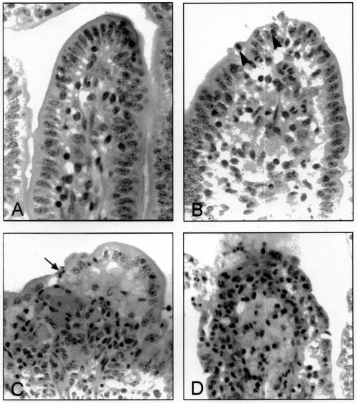
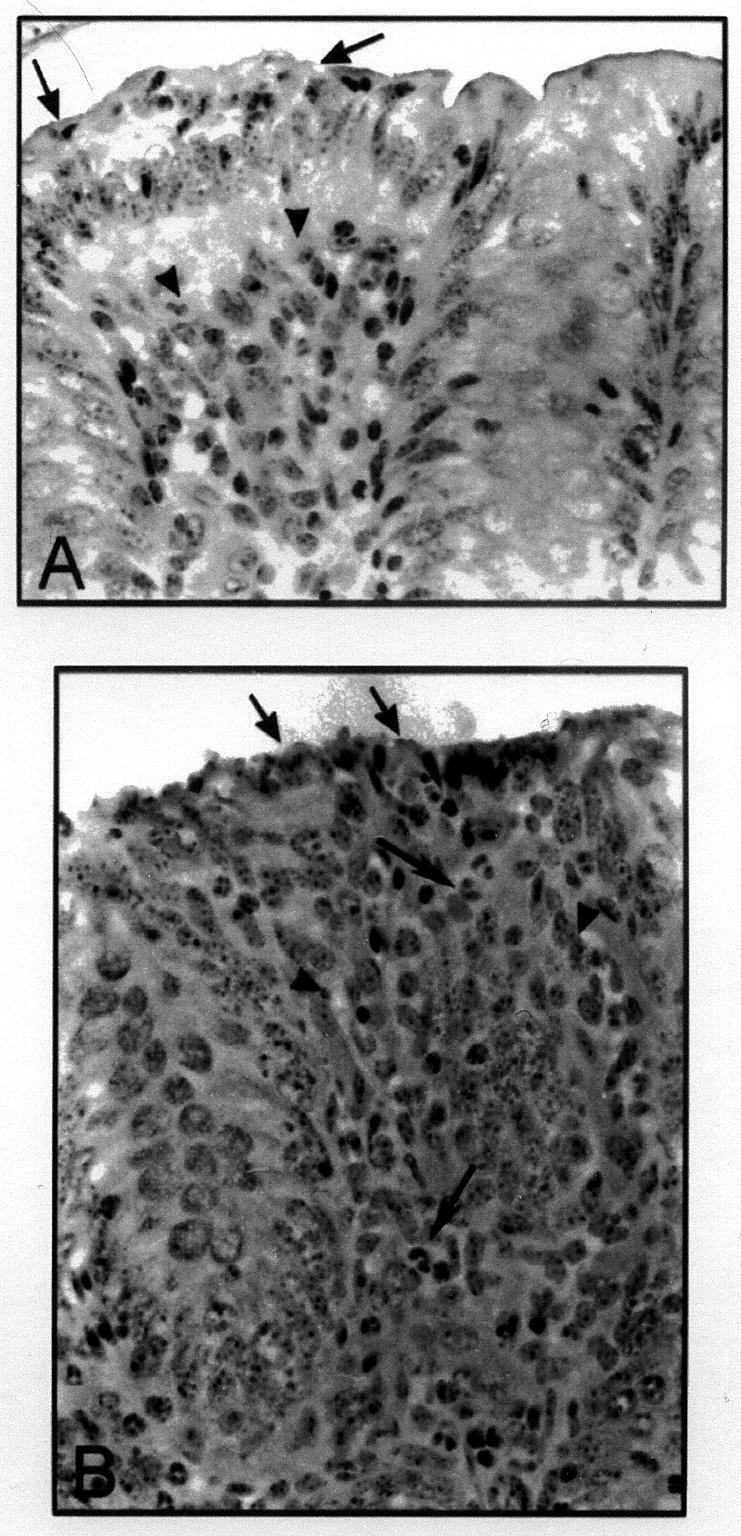
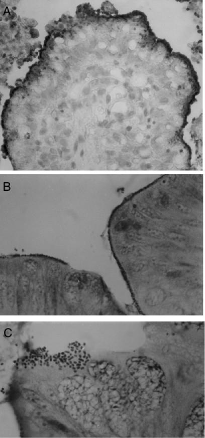
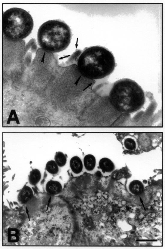
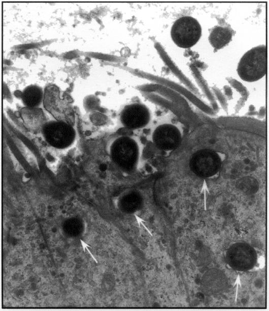
The purpose of this study was to compare the pathological effects of Shiga toxin-producing Escherichia coli (STEC) that vary in their association with bovine and human disease. Shiga toxin-producing E. coli of serotypes associated with both dysentery in calves and hemolytic uremic syndrome (HUS) in humans (O5:H-, O26:H11, O111:H-,O113:H21) were compared with O157:H7 STEC, which are associated with HUS in humans but not with disease in calves. The STEC were administered orally to 80 day-old chicks and into ligated loops in the ileum and colon of four 2- to 6-day-old calves. Examination of the ceca of the chickens 10 d postchallenge showed no adherence or tissue abnormality for any isolate. The calves were euthanized 8 to 10 h postinoculation, and sections of the intestinal loops were examined by light microscopy, transmission and scanning electron microscopy, and immunohistochemistry. All strains showed consistent focal adherence associated with mild lesions in the colon. Attaching and effacing lesions were observed with the eae-positive strains. Ileal lesions were similar to the colonic ones but were sometimes severe, with marked polymorphonuclear leukocyte proliferation in the lamina propria. It is concluded that chickens were unsuitable for studying interaction of STEC with the intestine and that there was no difference in the interaction of the ligated calf intestine with STEC of serotypes associated with disease in calves compared with O157:H7 STEC.


Figures

None
Figure 1. Changes in villi in the ileum of calves in response to Shiga toxin-producing Escherichia coli (STEC). A. Normal villus. The villus is long and slender with intact epithelium, regularly shaped epithelial cells with prominent brush border, and a low concentration of mononuclear leukocytes in the lamina propria. B. Mild reaction (grade 1). Villi are slightly blunted, there are degenerating epithelial cells at the tip, and there is increased cellularity in the lamina propria, with neutrophils migrating into the epithelium (arrowheads). C. Moderate reaction (grade 2). Villi are short and blunt. There is shallow ulceration (arrow), and large numbers of neutrophils are present in the lamina propria and appear to be streaming into the lumen. D. Severe reaction (grade 3). The upper third of the villus is covered with degenerate epithelial cells or is ulcerated, there is massive infiltration of the lamina propria with polymorphonuclear leukocytes, and edema distends the lamina propria.
None
Figure 2. Changes in the appearance of the colon of calves in response to STEC. A. Mild reaction (grade 1). A mild reaction is shown at the left (arrows), adjacent to a normal area of the colon. The area to the left shows marked irregularity of the epithelium, with intraepithelial neutrophils (arrows) and increased cellularity of the lamina propria (arrowheads). B. Moderate reaction (grade 2). There is necrosis of the epithelium (black arrow), increased cellularity of the lamina propria (arrowheads) and a strong neutrophilic reaction (white arrows).
None
Figure 3. Adherence of STEC to ileal and colonic epithelium. A. Immunoperoxidase staining of STEC serotype O26:H11 adherent to villi and in the lumen in the ileum of a calf (40X). B. Bacteria evident as rows of dark-staining organisms at the interphase between epithelium and lumen (Giemsa, 63X). C. A cluster of bacteria adhering in a pattern similar to that seen in localized adherence to HEp-2 cells in vitro (Giemsa, 100X).
None
Figure 4. Transmission electron micrograph of an eae-positive and eae-negative STEC associating with the colonic epithelium. A. The figure shows the classical AE lesion in an area of the colon infected with STEC O5:NM. Note the pedestal formation and the close association of bacteria with the colonic epithelial cells (arrowheads) and the effacement of microvilli (arrows in the lumen). B. Transmission electron micrograph of colonic mucosa infected with an eae-negative STEC of serotype O113:H21 shows that some bacteria are surrounded by an electronlucent zone and are at some distance from the epithelium. However, other bacteria (arrows) appear to be associated with typical AE lesions.
None
Figure 5. Invasion of the ileal epithelium by an O5:NM verotoxin-producing E. coli. Arrows point to an invading organism.

Similar articles

Cited by

References

    1. Karmali MA. Infection by verocytotoxin-producing Escherichia coli. Clin Microbiol Rev 1989;2:15–38. - PMC - PubMed
    1. Nataro J, Kaper JB. Diarrheagenic Escherichia coli. Clin Microbiol Rev 1998;11:142–201. - PMC - PubMed
    1. Gannon VP, Gyles CL. Characteristics of the Shiga-like toxin produced by Escherichia coli associated with porcine edema disease. Vet Microbiol 1990;24:89–100. - PubMed
    1. Chanter N, Hall GA, Bland AP, Hayle AJ, Parsons KR. Dysentery in calves caused by an atypical strain of Escherichia coli (S102-9). Vet Microbiol 1986;12:241–253. - PMC - PubMed
    1. Wray C, McLaren I, Pearson GR. Occurrence of ‘attaching and effacing’ lesions in the small intestine of calves experimentally infected with bovine isolates of verocytotoxic E. coli. Vet Rec 1989;125:365–368. - PubMed

Publication types

MeSH terms

LinkOut - more resources