. 2002 Jun;76(11):5315-25.
doi: 10.1128/jvi.76.11.5315-5325.2002.

Envelope glycoprotein incorporation, not shedding of surface envelope glycoprotein (gp120/SU), Is the primary determinant of SU content of purified human immunodeficiency virus type 1 and simian immunodeficiency virus

Affiliations

Envelope glycoprotein incorporation, not shedding of surface envelope glycoprotein (gp120/SU), Is the primary determinant of SU content of purified human immunodeficiency virus type 1 and simian immunodeficiency virus

Elena Chertova et al. J Virol. 2002 Jun.

Abstract

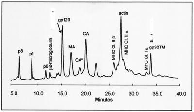
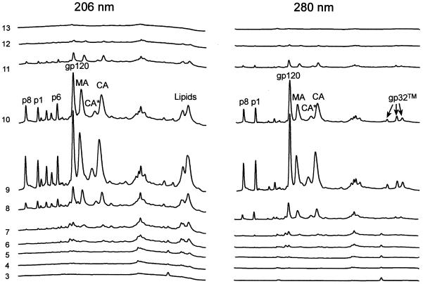
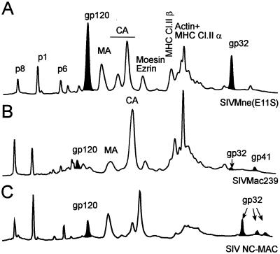
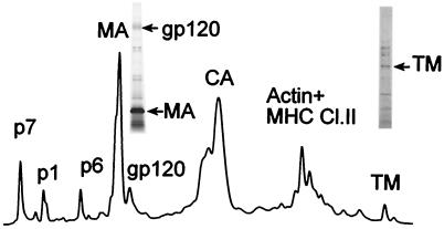
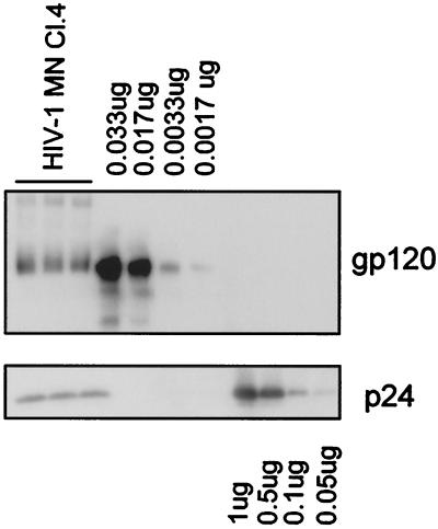
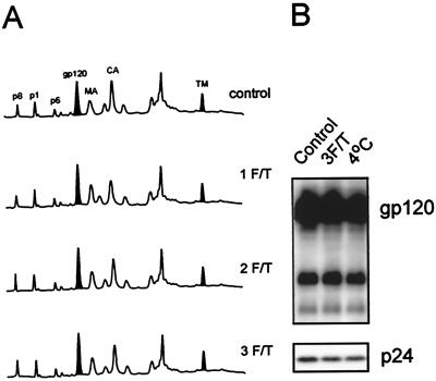
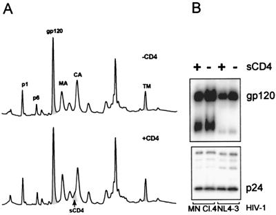
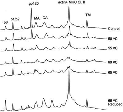
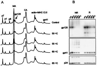
Human immunodeficiency virus type 1 (HIV-1) and simian immunodeficiency virus (SIV) particles typically contain small amounts of the surface envelope protein (SU), and this is widely believed to be due to shedding of SU from mature virions. We purified proteins from HIV-1 and SIV isolates using procedures which allow quantitative measurements of viral protein content and determination of the ratios of gag- and env-encoded proteins in virions. All of the HIV-1 and most of the SIV isolates examined contained low levels of envelope proteins, with Gag:Env ratios of approximately 60:1. Based on an estimate of 1,200 to 2,500 Gag molecules per virion, this corresponds to an average of between 21 and 42 SU molecules, or between 7 and 14 trimers, per particle. In contrast, some SIV isolates contained levels of SU at least 10-fold greater than SU from HIV-1 isolates. Quantification of relative amounts of SU and transmembrane envelope protein (TM) provides a means to assess the impact of SU shedding on virion SU content, since such shedding would be expected to result in a molar excess of TM over SU on virions that had shed SU. With one exception, viruses with sufficient SU and TM to allow quantification were found to have approximately equivalent molar amounts of SU and TM. The quantity of SU associated with virions and the SU:TM ratios were not significantly changed during multiple freeze-thaw cycles or purification through sucrose gradients. Exposure of purified HIV-1 and SIV to temperatures of 55 degrees C or greater for 1 h resulted in loss of most of the SU from the virus but retention of TM. Incubation of purified virus with soluble CD4 at 37 degrees C resulted in no appreciable loss of SU from either SIV or HIV-1. These results indicate that the association of SU and TM on the purified virions studied is quite stable. These findings suggest that incorporation of SU-TM complexes into the viral membrane may be the primary factor determining the quantity of SU associated with SIV and HIV-1 virions, rather than shedding of SU from mature virions.


Figures

FIG. 1.
FIG. 1.
HPLC analysis of sucrose density gradient-purified SIVMne(E11S). After purification, virions were disrupted in 8 M guanidine-HCl and separated by HPLC. Peaks were detected at 280 nm. Proteins were eluted from the column with an acetonitrile gradient as described in Materials and Methods and identified by SDS-PAGE, immunoblot, mass spectrometry, protein sequencing, and amino acid analysis. Protein purity of SU(gp120) and TM(gp32) peaks is shown as insertions on the HPLC profile. Bands were visualized by Coomassie staining. Two UV-absorbing peaks (labeled CA* and CA) were found to contain highly purified monomeric Gag p28CA protein. Subsequent analysis showed that following reduction with 2-mercaptoethanol, the protein in peak CA* eluted as CA, indicating that CA* contained a form of p28CA with at least one internal disulfide bond.
FIG. 2.
FIG. 2.
HPLC analysis of sucrose density gradient fractions from an SIV CP-MAC purification. Cell culture supernatants were processed as described in Materials and Methods. Fractions were collected from the sucrose density gradient after centrifugation. The percent sucrose was determined for each fraction. A 25-ml sample from each fraction was diluted 1:3 with TNE buffer and centrifuged at 100,000 × g to pellet any virus that may have been present. The pellets were resuspended in 8 M guanidine-HCl and analyzed for protein composition by HPLC under nonreducing conditions.
FIG. 3.
FIG. 3.
Comparison of SIV preparations by HPLC analysis of SIVMne(E11S) (A), SIVmac239 (B), and SIV NC-MAC (C). After purification, virions were disrupted in 8 M guanidine-HCl and separated by HPLC. Peaks were detected at 280 nm. Proteins were eluted from the column with an acetonitrile gradient as described in Materials and Methods and analyzed by SDS-PAGE, immunoblot, mass spectrometry, protein sequencing, and amino acid analysis. The peaks corresponding to gp120 (SU) and the peaks identified as gp32 (TM) are denoted black. For SIVmac239 (panel B), both the gp32 and gp41 forms of TM are present, reflecting ongoing selection of truncated forms in the cultures used to produce the virus analyzed. The combined integrated areas of both peaks were used to calculate SU:TM ratios.
FIG. 4.
FIG. 4.
HPLC analysis of HIV-1 MN/H9 cl.4. After purification, virions were disrupted in 8 M guanidine-HCl and separated by HPLC. Peaks were detected at 280 nm. Proteins eluted from the column with an acetonitrile gradient were analyzed by SDS-PAGE, immunoblot, mass spectrometry, protein sequencing, and amino acid analysis. After gp120 and gp41 were identified on the HPLC profile by immunoblot, fractions containing SU and TM were analyzed by SDS-PAGE followed with silver staining (insertions on HPLC picture).
FIG. 5.
FIG. 5.
Analysis of SU and p24CA amounts in HIV-1 MN/H9 cl.4 virus. Virion-associated SU and p24CA levels were analyzed by immunoblot analysis under nonreducing conditions. Bands were visualized with mouse monoclonal antibodies prepared against purified p24 and gp120, followed by enhanced chemiluminescence staining. Band intensities were quantified by densitometry using Scion Image analysis software.
FIG. 6.
FIG. 6.
Freezing and thawing of purified viruses does not result in shedding of SU proteins. Three aliquots of sucrose banded SIVMne(E11S) and HIV-1MN cl.4 (both 1,000-fold) were resuspended and thawed in a 37°C waterbath for 5 min. One aliquot was then immersed in an ice-water bath, completing one freezing and thawing (1 F/T) cycle. Another aliquot was frozen in methanol-dry ice for 10 min, thawed in a 37°C water bath for 5 min, and then immersed in an ice-water bath (2 cycles). The third aliquot was subjected to a third cycle of freezing and thawing (3 cycles). All samples were centrifuged at 100,000 × g for 45 min. The pellets were resuspended and analyzed by the HPLC method (A) for SIVMne(E11S) (280 nm) and by immunoblot analysis (B) for HIV-1 MN cl.4. The control sample was thawed just before HPLC or immunoblot analysis.
FIG. 7.
FIG. 7.
SIVMne(E11S), HIV-1MN/H9 cl.4, and HIV-1 NL4-3/CEMx174 were incubated with or without sCD4 for 2 h at 37°C. Viral samples were then pelleted, and SIVMne(E11S) was analyzed by HPLC (A). Peaks were detected at 280 nm. (B) Virion-associated gp120 and p24CA on HIV-1 isolates were analyzed by SDS-PAGE and immunoblot analysis, followed by densitometry.
FIG. 8.
FIG. 8.
Heat treatment of SIVMne(E11S). SIVMneE11S virus stock (2 ml, 1,000-fold) were thawed for 5 min in a 37°C waterbath, and then five separate 0.3-ml aliquots were each diluted with 0.7 ml of sterile TNE buffer and heated for 1 h at 50, 55, 60, or 65°C; a fifth sample was not heated but incubated at 4°C for 1 h. After incubation, viral samples were centrifuged through a 20% sucrose-TNE buffer pad at 25,000 rpm for 60 min. Pellets were resuspended in TNE buffer and analyzed for gp120 content using HPLC methods under nonreducing and reducing (65°C) conditions. Peaks were detected at 280 nm.
FIG. 9.
FIG. 9.
Heat treatment of HIV-1 MN/H9 cl.4. Virus was incubated at 4, 50, 55, 60, and 65°C for 1 h, and one sample was subjected to freezing and thawing three times. After incubation at various temperatures, viral samples were centrifuged, and pellets were analyzed for gp120 content using HPLC methods (A) under reducing conditions. Peaks were detected at 280 nm. (B) immunoblot analysis of gp120 retention by heat-treated HIV-1MN/H9 cl.4 virus. Virion-associated gp120 and p24CA levels were analyzed by SDS-PAGE under both reducing (R) and nonreducing (NR) conditions, followed by immunoblot analysis. Bands were visualized with mouse monoclonal antibodies prepared against purified p24 and gp120, followed by enhanced chemiluminescence staining.

References

    1. Allan, J. S., E. M. Whitehead, K. Strout, M. Short, P. Kanda, T. K. Hart, and P. J. Bugelski. 1992. Strong association of simian immunodeficiency virus (SIVagm) envelope glycoprotein heterodimers: possible role in receptor-mediated activation. AIDS Res. Hum. Retrovir. 8:2011-2020. - PubMed
    1. Arthur, L. O., J. W. Bess, E. N. Chertova, J. L. Rossio, M. T. Esser, R. E. Benveniste, L. E. Henderson, and J. D. Lifson. 1998. Chemical inactivation of retroviral infectivity by targeting nucleocapsid protein zinc fingers: a candidate SIV vaccine. AIDS Res. Hum. Retrovir. 14(Suppl. 3):S311-S319. - PubMed
    1. Benveniste, R. E., R. W. Hill, L. J. Eron, U. M. Csaikl, W. B. Knott, L. E. Henderson, R. C. Sowder, K. Nagashima, and M. A. Gonda. 1990. Characterization of clones of HIV-1-infected HuT 78 cells defective in gag gene processing and of SIV clones producing large amounts of envelope glycoprotein. J. Med. Primatol. 19:351-366. - PubMed
    1. Bess, J. W., Jr., R. J. Gorelick, W. J. Bosche, L. E. Henderson, and L. O. Arthur. 1997. Microvesicles are a source of contaminating cellular proteins found in purified HIV-1 preparations. Virology 230:134-144. - PubMed
    1. Bess, J. W., P. J. Powell, H. J. Issaq, L. J. Schumack, M. K. Grimes, L. E. Henderson, and L. O. Arthur. 1992. Tightly bound zinc in human immunodeficiency virus type 1, human T-cell leukemia virus type I, and other retroviruses. J. Virol. 66:840-847. - PMC - PubMed

Publication types

MeSH terms