. 2002 Jun;76(11):5357-68.
doi: 10.1128/jvi.76.11.5357-5368.2002.

Modifications of the human immunodeficiency virus envelope glycoprotein enhance immunogenicity for genetic immunization

Affiliations

Modifications of the human immunodeficiency virus envelope glycoprotein enhance immunogenicity for genetic immunization

Bimal K Chakrabarti et al. J Virol. 2002 Jun.

Abstract

In this study, we have investigated the effect of specific mutations in human immunodeficiency virus type 1 (HIV-1) envelope (Env) on antibody production in an effort to improve humoral immune responses to this glycoprotein by DNA vaccination. Mice were injected with plasmid expression vectors encoding HIV Env with modifications in regions that might affect this response. Elimination of conserved glycosylation sites did not substantially enhance humoral or cytotoxic-T-lymphocyte (CTL) immunity. In contrast, a modified gp140 with different COOH-terminal mutations intended to mimic a fusion intermediate and stabilize trimer formation enhanced humoral immunity without reducing the efficacy of the CTL response. This mutant, with deletions in the cleavage site, fusogenic domain, and spacing of heptad repeats 1 and 2, retained native antigenic conformational determinants as defined by binding to known monoclonal antibodies or CD4, oligomer formation, and virus neutralization in vitro. Importantly, this modified Env, gp140 Delta CFI, stimulated the antibody response to native gp160 while it retained its ability to induce a CTL response, a desirable feature for an AIDS vaccine.


Figures
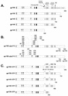
FIG. 1.
FIG. 1.
Schematic representation of functional domains and mutations in HIV-1 Env glycoproteins. Full-length envelope polyprotein, gp160, with the indicated features based on the amino acid residues of HXB2 is shown (top). Functional domains include the gp120/gp41 cleavage site (residues 510 and 511), the fusion domain (residues 512 to 527), the two heptad repeats (residues 546 to 579 and residues 628 to 655), the transmembrane domain (residues 684 to 705), and the cytoplasmic domain (residues 706 to 856). The mutant forms of the envelope proteins are shown below the structure of gp160. COOH deletions were introduced that terminate the envelope protein at positions 752, 704, or 680 to produce gp150, gp145, or gp140, respectively. Two internal deletions that removed the cleavage site, the fusion domain, and the region between the two heptad repeats were introduced into gp160, gp150, gp145, and gp140. A further deletion in the COOH-terminal region at position 592 removed the second heptad repeat and the transmembrane domain to produce gp128ΔCFI. To disrupt potential glycosylation sites, asparagine (N) residues at 11 positions (88, 156, 160, 197, 230, 234, 241, 262, 276, 289, and 295) were replaced with aspartic acid (D) residues in both gp160 and gp150. Versions of both gp160 and gp150 were created with a total of 17 mutated glycosylation sites by including six additional N-to-D substitutions at positions 332, 339, 356, 386, 392, and 448.
FIG. 2.
FIG. 2.
Comparison of the expression of the HIV-1 gp160 with codon-optimized gp160. (A and B) Expression of plasmids encoding Rev-dependent and Rev-independent codon-modified gp160 (lanes 1 and 2). (A) The upper panel shows expression of Rev-dependent viral gp160 (left) and codon-modified gp160 (right) in transfected 293 cells. The lower panel shows comparable expression of β-actin in these transfected cells. (B) Processing of gp160 was detected with a monoclonal antibody to gp41with viral or codon-altered gp160, as indicated (lanes 3 to 9). (C) Expression of mutant CXCR4-tropic HIV Env glycoproteins with COOH-terminal truncations is shown. (D and E) CXCR4-tropic envelope proteins containing mutant glycosylation sites and mutant functional domains are shown. The indicated proteins were detected by immunoblotting as described above. Cell lysates produced by transfection with vector containing no insert were used as controls (vector, first lane in each panel).
FIG. 3.
FIG. 3.
Cytotoxicity of full-length gp160 is eliminated by deletion of the COOH-terminal cytoplasmic domain. Cell rounding and detachment were not observed in control-transfected 293 cells (A), in contrast to full-length gp160 (B), and were observed to a lesser extent in cells transfected with gp150 (C), in contrast to gp145 (D) or gp140 (E).
FIG. 4.
FIG. 4.
Expression of soluble ΔCFI HIV-1 envelope variants. Results of immunoprecipitation and Western blot analysis of supernatants from the indicated transfected cells are shown.
FIG. 5.
FIG. 5.
Interaction of gp140ΔCFI with defined monoclonal antibodies or CD4 and biochemical analysis for oligomerization of gp140ΔCFI. (A) Analysis of the antigenic structure of soluble gp140ΔCFI with monoclonal antibodies. The envelope glycoproteins from the supernatants of the 293 cells transfected with the vector expressing gp140ΔCFI were immunoprecipitated with either 5 μg of monoclonal antibodies 2F5, 2G12, F105, and b12 or with 5 μg of HIV-1 Ig. The proteins were analyzed by SDS-PAGE and detected by Western blotting using the polyclonal antibodies against gp160. The arrow indicates the position of gp140ΔCFI. A nonspecific (ns) band that cross-reacted with the antibody is indicated. (B) Quantification of 2F5 and 2G12 binding to gp140ΔCFI using the indicated concentrations of each antibody as shown compared to a nonreactive Ig isotype (control). The intensity of the gp140ΔCFI band was determined by quantitative phosphorimaging. The arrow indicates the position of gp140ΔCFI. (C) Interaction of soluble gp140ΔCFI protein with CD4. Binding of gp140ΔCFI and gp160, compared to that of controls transfected with vector alone, in an ELISA with CD4 is shown (left panel). The values represent the mean and standard deviation (error bars) for each point. The ability of sCD4 to compete with binding to these envelopes is shown (right panel). (D) Biochemical analysis of soluble gp140ΔCFI oligomerization. Western blot analysis to detect the gp140ΔCFI in different fractions, fractions 1 to 10, after sucrose density gradient. Fraction 1 represents the greatest density, and fraction 10 represents the least density. Equal volumes of the samples from each fraction were analyzed in an SDS-polyacrylamide gel under nonreducing conditions, except the molecular weight marker. The proteins were detected by Western blotting using polyclonal antibody against gp160. The positions of dimer, trimer, and aggregates are shown. The lower panel shows the presence of monomer in these fractions run under reducing conditions. (E) Molecular-exclusion chromatography of soluble of gp140ΔCFI. Membrane-free supernatant containing gp140ΔCFI was analyzed on a Superdex 200 column and compared with a mixture of molecular weight standards; the position of each marker is indicated by arrows. Fractions were analyzed for gp140ΔCFI by immunoprecipitation followed by Western blotting and were quantitated by densitometry.
FIG. 6.
FIG. 6.
Confirmation of cell surface expression of gp140ΔCFI and gp145ΔCFI by flow cytometry. 293 cells were transfected with either a control vector or vectors expressing gp140ΔCFI or gp145ΔCFI, as indicated. The transfection efficiency (∼90%) was calculated based on staining after transfection of the same number of cells under identical conditions with an equal amount of DNA expressing β-galactosidase. Cell surface expression was detected by FACS analysis using Ig purified from the sera of HIV-1-infected individuals (HIV Ig) or different monoclonal antibodies, 2F5 and 2G12. The transfected cells are labeled at the top, and the different antibodies are indicated at right. Light dotted lines indicate FACS analysis with control human Ig, and dark dotted lines indicate FACS analysis with human monoclonal antibodies against HIV-1.
FIG. 7.
FIG. 7.
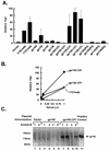
Antibody response against HIV-1 envelope proteins in DNA immunized mice. (A) Comparison of the antibody response in mice immunized with gp140ΔCFI or other Env plasmid expression vectors. Sera were collected 2 weeks after the last immunization and used to immunoprecipitate codon-altered gp160 from lysates of transfected 293 cells as described before. The quantitation of the immunoprecipitated gp160 was done as described for panel B. The average of the normalized data has been presented as a bar diagram. Error bars are indicated. (B) Antibody responses in mice immunized with gp140ΔCFI or gp128ΔCFI relative to a V3-specific monoclonal antibody standard (monoclonal antibody 1727). Antisera from immunized mice were diluted in immunoprecipitation buffer, and 1 μl of each diluted serum was used to immunoprecipitate codon-altered HIV-1 gp160 from lysates of transfected 293 cells as described in the legend to Fig. 3A. The gels were scanned, and the intensity of the gp160 band was determined by densitometry using the program ImageQuant and presented relative to the intensity of gp160 immunoprecipitated with positive control sera (rabbit anti-gp160), which was used to normalize data between experiments. These data are presented graphically to facilitate comparison among groups. Monoclonal antibody 1727 interacts with the V3 loop of HIV IIIb and was kindly provided by the NIH AIDS Research and Reference Reagent Program, from Jon Laman. (C) Antibody responses in mice immunized with gp140 or gp140ΔCFI were determined by immunoprecipitation and Western blotting. Animals received two booster doses (100 μg) of the same plasmid, 2 weeks apart. Sera (1 μl) collected 2 weeks after the last immunization were used to immunoprecipitate codon-optimized HIV-1 gp160 from lysates of transfected 293 cells containing 400 μg of total protein. Each lane corresponds to the serum from an animal immunized with either the control vector (lanes 1 and 2), CXCR4-tropic gp140 (lanes 3 to 6), or plasmid that expresses gp140 with the indicated mutant functional domains (lanes 7 to 10). A mouse monoclonal antibody to gp160 (HIV-1 V3 monoclonal [IIIB-V3-13]; NIH AIDS Research and Reference Reagent Program) was used as a positive control (lane 11).
FIG. 8.
FIG. 8.
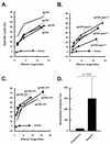
CTL response against HIV-1 envelope proteins in DNA-immunized mice and generation of a neutralizing antibody response in guinea pigs. (A) The CTL response to CXCR4-tropic Env and indicated deletion mutants is shown. (B and C) The CTL responses to CXCR4-tropic envelope with glycosylation site and ΔCFI mutations are shown, respectively. Spleen cells were isolated from immunized mice 2 weeks after the final immunization and stimulated in vitro with irradiated cells expressing gp160 with addition of human interleukin 2 (5 U/ml) at day 4. The cytolytic activity of the restimulated spleen cells was tested after 7 days against V3 peptide-pulsed BC10ME cells. Similar findings were observed with target cells that stably express full-length Env (data not shown). (D) Preimmune sera were collected from four guinea pigs prior to immunization or after DNA priming and ADV boosting with gp140ΔCFI as described in the text. Both preimmune sera and postimmune sera were diluted, and neutralizing activity was measured by reduction of HIV-IIIB virus compared to the untreated control. Neutralizing antibody titers were analyzed as previously described (26). The data represent the dilutions at which the sera can neutralize the virus in the MT2 assay. Standard deviations are indicated.

References

    1. Andre, S., B. Seed, J. Eberle, W. Schraut, A. Bultmann, and J. Haas. 1998. Increased immune response elicited by DNA vaccination with a synthetic gp120 sequence with optimized codon usage. J. Virol. 72:1497-1503. - PMC - PubMed
    1. Binley, J., and J. P. Moore. 1997. HIV-cell fusion. The viral mousetrap. Nature 387:346-348. (Erratum, 389:131.) - PubMed
    1. Binley, J. M., R. W. Sanders, B. Clas, N. Schuelke, A. Master, Y. Guo, F. Kajumo, D. J. Anselma, P. J. Maddon, W. C. Olson, and J. P. Moore. 2000. A recombinant human immunodeficiency virus type 1 envelope glycoprotein complex stabilized by an intermolecular disulfide bond between the gp120 and gp41 subunits is an antigenic mimic of the trimeric virion-associated structure. J. Virol. 74:627-643. - PMC - PubMed
    1. Binley, J. M., R. Wyatt, E. Desjardins, P. D. Kwong, W. Hendrickson, J. P. Moore, and J. Sodroski. 1998. Analysis of the interaction of antibodies with a conserved enzymatically deglycosylated core of the HIV type 1 envelope glycoprotein 120. AIDS Res. Hum. Retrovir. 14:191-198. - PubMed
    1. Bolognesi, D. P., and T. J. Matthews. 1998. HIV vaccines. Viral envelope fails to deliver? Nature 391:638-639. - PubMed

MeSH terms

Substances