. 2002 Jun;76(11):5422-34.
doi: 10.1128/jvi.76.11.5422-5434.2002.

Transduction of interphase cells by avian sarcoma virus

Affiliations

Transduction of interphase cells by avian sarcoma virus

Richard A Katz et al. J Virol. 2002 Jun.

Abstract

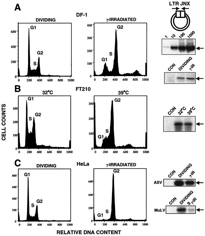
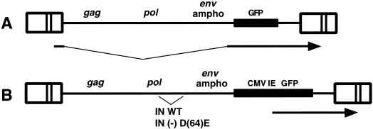
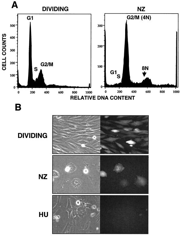
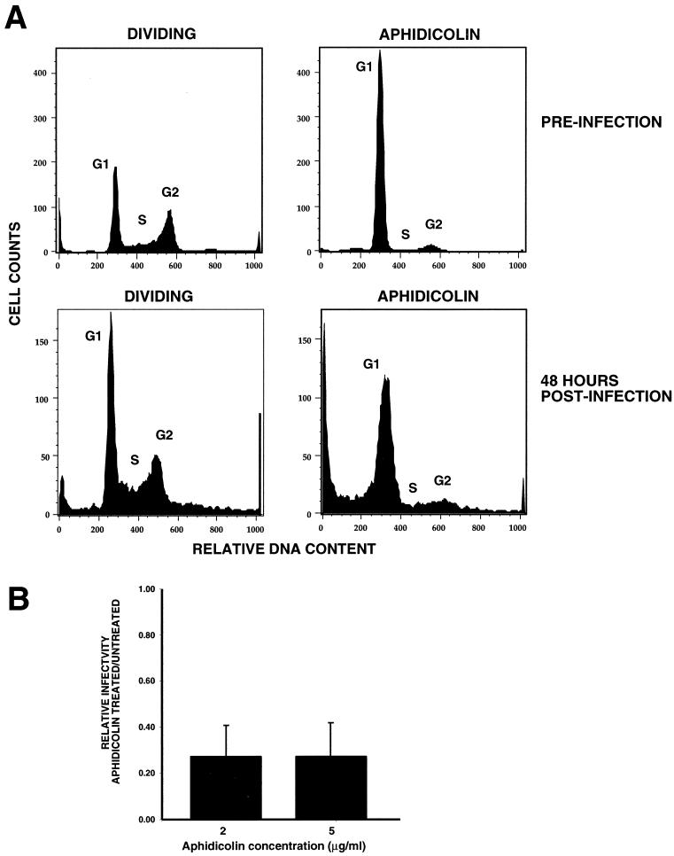
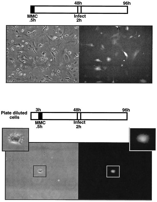
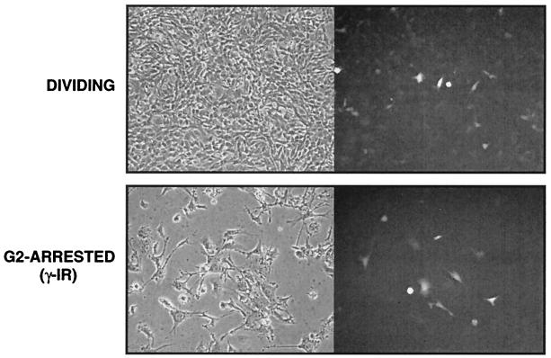
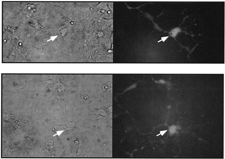
It has been generally believed that oncoretroviruses are dependent on mitosis for efficient nuclear entry of viral DNA. We previously identified a nuclear localization signal in the integrase protein of an oncoretrovirus, avian sarcoma virus (ASV), suggesting an active import mechanism for the integrase-DNA complex (G. Kukolj, R. A. Katz, and A. M. Skalka, Gene 223:157-163, 1998). Here, we have evaluated the requirement for mitosis in nuclear import and integration of ASV DNA. Using a modified ASV encoding a murine leukemia virus amphotropic env gene and a green fluorescent protein (GFP) reporter gene, DNA nuclear import was measured in cell cycle-arrested avian (DF-1) as well as human (HeLa) and mouse cells. The results showed efficient accumulation of nuclear forms of ASV DNA in gamma-irradiation-arrested cells. Efficient transduction of a GFP reporter gene was also observed after infection of cells that were arrested with gamma-irradiation, mitomycin C, nocodazole, or aphidicolin, confirming that nuclear import and integration of ASV DNA can occur in the absence of mitosis. By monitoring GFP expression in individual cells, we also obtained evidence for nuclear import of viral DNA during interphase in cycling cells. Lastly, we observed that ASV can transduce postmitotic mouse neurons. These results support an active nuclear import mechanism for the oncoretrovirus ASV and suggest that this mechanism can operate in both nondividing and dividing cells.


Figures

FIG. 1.
FIG. 1.
Nuclear import of retroviral DNA in arrested cells. Viral DNA LTR junctions signify nuclear entry of viral DNA. In each panel, the cell cycle profiles are shown along with gel analyses of the PCR product corresponding to LTR junctions (LTR JNX [arrows]). FACS analyses for DNA content indicate cell cycle status. Cell cycle profiles shown on the left represent dividing cells, and those on the right are for arrested cells. Dividing and arrested cells were exposed to the ASV amphotropic virus for 2 h, and DNA was isolated 16 to 24 h postinfection for PCR analyses. PCR analysis of mock-infected cells (CON) is shown. Cell cycle profiles were obtained from separate experiments using identical conditions. (A) DF-1 chicken embryo fibroblast line. At top right, a diagram of the PCR strategy is shown; the middle panel presents a standard curve for PCR showing a linear response over 3 to 4 logs. (B) FT210 cell line. (C) HeLa cell line. See Materials and Methods for details.
FIG. 2.
FIG. 2.
Maps of viruses ASVA-GFP (A) and ASVA-CMVEGFP (B). Arrows indicate GFP expression from spliced RNA or CMV-EGFP cassette. LTRs are indicated by boxes. WT, wild type.
FIG. 3.
FIG. 3.
Infection of nocodazole- and hydroxyurea-arrested cells with ASV. (A) Cell cycle profiles of dividing and nocodazole-arrested DF-1 cells obtained after 24 h of treatment (from a separate experiment). (B) DF-1 cells were synchronized by 24 h of treatment with hydroxyurea (HU) and infected with ASVA-GFP in the presence of nocodazole. Cells were maintained in nocodazole (NZ) for an additional 36 h, at which time micrographs were taken. DF-1 cells were arrested with HU for 24 h, infected for 2 h, and maintained in HU for 48 h. Results with control dividing cultures are also shown. Shown are phase-contrast (left) and fluorescence (right) micrographs of dividing and arrested cells 48 h postinfection.
FIG. 4.
FIG. 4.
Infection of aphidicolin-arrested cells with ASV. (A) Cell cycle profiles of dividing and aphidicolin (5 μg/ml)-treated DF-1 cells. In the top panels, DF-1 cells were pretreated with aphidicolin for 24 h prior to infection with ASVA-CMVEGFP (preinfection profile). The bottom panels show the cell cycle profile of aphidicolin-treated cells 48 h postinfection (72 h total exposure). (B) Transduction of aphidicolin-arrested cells relative to dividing cells 48 h postinfection. GFP-expressing cells were measured by FACS analysis. Results at each concentration of aphidicolin were averaged from three separate experiments, and the standard deviation (error bar) is shown.
FIG. 5.
FIG. 5.
Infection of mitomycin C-arrested DF-1 cells with ASV. In the top panels, DF-1 cells were treated with mitomycin C for 0.5 h. After 48 h, cells were infected with ASVA-CMVEGFP and examined for GFP expression 48 h postinfection. Phase-contrast (left) and fluorescence (right) images (GFP) are shown. The bottom panels are the same as the top panels, except that DF-1 cells were monodispersed prior to mitomycin C treatment such that infection of individual cells could be observed. Shown is an arrested premitotic cell expressing GFP.
FIG. 6.
FIG. 6.
Infection of γ-irradiated (γ-IR) HeLa cells with ASV. HeLa cells were arrested in G2 as described in Fig. 1 and were infected with ASVA-CMVEGFP 24 h later. Shown are phase-contrast (left) and fluorescence (right) micrographs of dividing and arrested cells 48 h postinfection.
FIG. 7.
FIG. 7.
Evidence for ASV DNA nuclear import during interphase in dividing cells. HeLa cells were synchronized by mitotic shake-off. Dilute mitotic cells completed cytokinesis, producing two-cell colonies. Three hours after plating, cultures were infected with ASVA-CMVEGFP for 2 h. Cells were observed for GFP expression over the next 24 to 36 h. Phase-contrast (left) and fluorescence (right) images (GFP) are shown. Two fields are shown. The two-cell colony in the lower panel was marked prior to infection.
FIG. 8.
FIG. 8.
Infection of mouse neurons with ASV. Hippocampal neuron explants were prepared from mouse embryos and infected with ASVA-CMVEGFP on day 5 postexplantation. See Results and Materials and Methods for details. Phase-contrast (left) and fluorescence (right) images (GFP) are shown. Arrows identify the cell body. Two fields are shown.

References

    1. Barsov, E. V., and S. H. Hughes. 1996. Gene transfer into mammalian cells by a Rous sarcoma virus-based retroviral vector with the host range of the amphotropic murine leukemia virus. J. Virol. 70:3922-3929. - PMC - PubMed
    1. Blomer, U., L. Naldini, T. Kafri, D. Trono, I. M. Verma, and F. H. Gage. 1997. Highly efficient and sustained gene transfer in adult neurons with a lentivirus vector. J. Virol. 71:6641-6649. - PMC - PubMed
    1. Bouyac-Bertoia, M., J. D. Dvorin, R. A. Fouchier, Y. Jenkins, B. E. Meyer, L. I. Wu, M. Emerman, and M. H. Malim. 2001. HIV-1 infection requires a functional integrase NLS. Mol. Cell 7:1025-1035. - PubMed
    1. Bukrinsky, M. I., S. Haggerty, M. P. Dempsey, N. Sharova, A. Adzhubel, L. Spitz, P. Lewis, D. Goldfarb, M. Emerman, and M. Stevenson. 1993. A nuclear localization signal within HIV-1 matrix protein that governs infection of non-dividing cells. Nature 365:666-669. - PMC - PubMed
    1. Bukrinsky, M. I., N. Sharova, M. P. Dempsey, T. L. Stanwick, A. G. Bukrinskaya, S. Haggerty, and M. Stevenson. 1992. Active nuclear import of human immunodeficiency virus type 1 preintegration complexes. Proc. Natl. Acad. Sci. USA 89:6580-6584. - PMC - PubMed

Publication types

MeSH terms

LinkOut - more resources