. 2002 Jun;76(11):5667-77.
doi: 10.1128/jvi.76.11.5667-5677.2002.

Formation of a human immunodeficiency virus type 1 core of optimal stability is crucial for viral replication

Affiliations

Formation of a human immunodeficiency virus type 1 core of optimal stability is crucial for viral replication

Brett M Forshey et al. J Virol. 2002 Jun.

Abstract

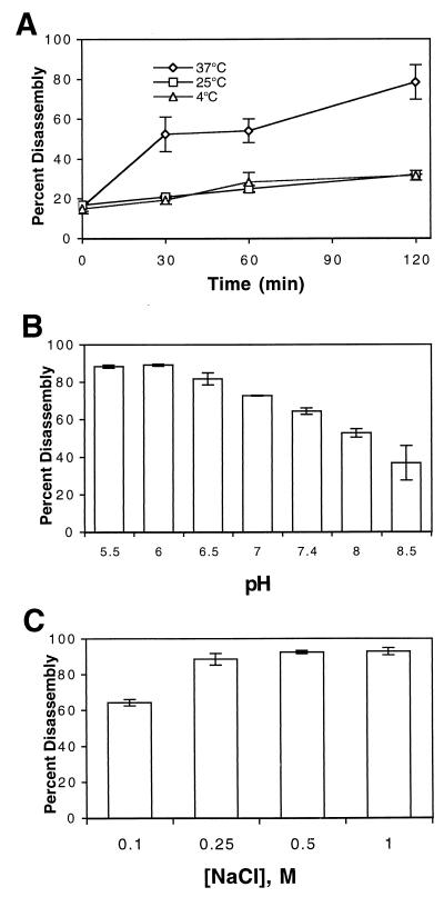
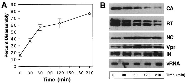
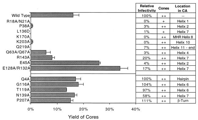
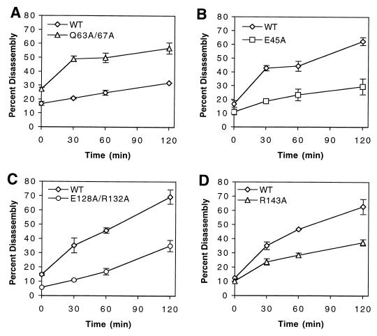
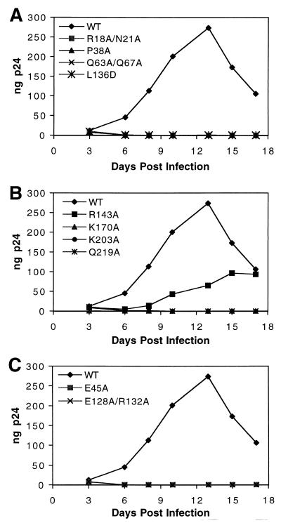
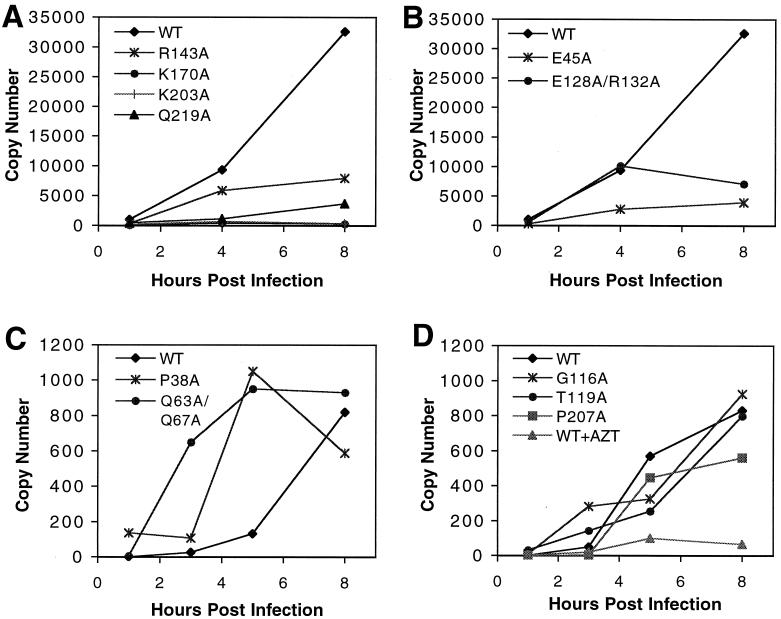
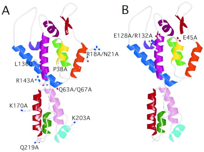
Virions of human immunodeficiency virus type 1 (HIV-1) and other lentiviruses contain conical cores consisting of a protein shell composed of the viral capsid protein (CA) surrounding an internal viral ribonucleoprotein complex. Although genetic studies have implicated CA in both early and late stages of the virus replication cycle, the mechanism of core disassembly following penetration of target cells remains undefined. Using quantitative assays for analyzing HIV-1 core stability in vitro, we identified point mutations in CA that either reduce or increase the stability of the HIV-1 core without impairing conical core formation in virions. Alterations in core stability resulted in severely attenuated HIV-1 replication and impaired reverse transcription in target cells with only minimal effects on viral DNA synthesis in permeabilized virions in vitro. We conclude that formation of a viral core of optimal stability is a prerequisite for efficient HIV-1 infection and suggest that disassembly of the HIV-1 core is a regulated step in infection that may be an attractive target for pharmacologic intervention.


Figures

FIG. 1.
FIG. 1.
Equilibrium density gradient sedimentation of HIV-1 cores and virions. Concentrated virions were layered onto 30 to 70% linear sucrose gradients with (A) or without (B) a layer of Triton X-100. Following ultracentrifugation at 100,000 × g for 16 h at 4°C, fractions (1 ml) were collected from the top of the gradient and were analyzed by p24 ELISA. The density of each fraction was determined by refractometry. Core fractions that were pooled and used for further analysis (Cores) are indicated in panel A.
FIG. 2.
FIG. 2.
Disassembly of purified HIV-1 cores in vitro. Cores were incubated in STE buffer at the indicated temperatures for the times shown (A) or at 37°C for 1 h in STE buffer prepared at the indicated pH values (B) or NaCl concentrations (C). Following incubation, the samples were subjected to ultracentrifugation at 100,000 × g for 15 min at 4°C. The extent of disassembly was determined as the percentage of the total CA protein in the reaction present in the supernatant. (A) Mean values of triplicate determinations, with error bars representing 1 standard deviation. (B and C) Mean values of duplicate determinations, with error bars representing the range of values.
FIG. 3.
FIG. 3.
Kinetic and biochemical analyses of HIV-1 core disassembly in vitro. Purified HIV-1 cores were incubated at 37°C for the indicated times, followed by separation of free and core-associated CA by ultracentrifugation. (A) Dissociation of CA from pelletable cores during incubation at 37°C. Supernatants and pellets were analyzed by p24 ELISA. The extent of disassembly was determined as the percentage of the total CA protein in the reaction detected in the supernatant. (B) Biochemical analysis of particles remaining following disassembly of HIV-1 cores. Pellets from the disassembly reactions shown in panel A were subjected to Western blotting and RNA slot blot analysis. The Western blot membrane was probed sequentially with rabbit anti-CA, anti-RT, anti-NC, and anti-Vpr antibodies. RNA was extracted from the pellets, denatured, and immobilized by vacuum filtration. Relative HIV-1 RNA levels were determined by hybridization with a 32P-labeled HIV-1 probe, followed by detection by autoradiography. In panel A, the values represent means of triplicates, with error bars representing 1 standard deviation. Shown are representative data from one of three independent experiments.
FIG. 4.
FIG. 4.
Identification of CA residues that influence the stability of HIV-1 cores. Concentrated virions were subjected to ultracentrifugation through a detergent layer into a sucrose density gradient. Yield of Cores, percentage of total CA detected in the peak fractions of cores; Relative infectivity, infectivity relative to wild-type HIV-1 as determined by a single-cycle infection assay; Cones, presence of similar quantities (compared to the wild type) of conical cores in virions (as determined by transmission electron microscopy). For R18A/N21A (+), few cones were apparent, and they were frequently aberrantly shaped; for L136D (+), cones were rarely detected. Location refers to the location of the mutation in the structure of HIV-1 CA. Shown are the mean values of at least two determinations, with error bars representing 1 standard deviation of the mean.
FIG. 5.
FIG. 5.
Kinetics of disassembly of wild-type (WT) and CA mutant cores in vitro. Isolated cores were diluted in STE buffer and incubated at 25 (A) or 37°C (B, C, and D). Shown are the mean values of triplicate determinations, with error bars representing 1 standard deviation.
FIG. 6.
FIG. 6.
Replication kinetics of CA mutants in primary T cells. Cultures of activated primary T cells were inoculated with equal quantities (1 ng) of the indicated viruses. Samples were collected on the days indicated and analyzed for p24 content by ELISA. (A and B) Viruses with mutations that lead to decreased core stability in vitro; (C) viruses with mutations that lead to increased core stability in vitro. Shown is a representative growth curve for each virus from duplicate analyses. WT, wild type.
FIG. 7.
FIG. 7.
Kinetics of viral DNA synthesis in vivo. Cultures of CD4-expressing HeLa cells were inoculated with equivalent quantities (100 ng of p24) of the indicated viruses. Individual cultures were harvested at the indicated times, and DNA was isolated for PCR. HIV-1 DNA was quantified by real-time PCR using the SYBR Green protocol. (A and B) Results from analysis using early-stage primers (R and U5); (C and D) DNA products corresponding to a later stage of reverse transcription (U5 and gag). The results are representative of three independent experiments. WT, wild type.
FIG. 8.
FIG. 8.
Location of core stability mutations in proposed complete structure of CA. The structure of the intact CA molecule was based on the work of Gamble et al. (15, 16). (A) Mutations that destabilize the HIV-1 core; (B) mutations that stabilize the core.

References

    1. Accola, M. A., Å hagen, and H. G. Göttlinger. 2000. Isolation of human immunodeficiency virus type 1 cores: retention of Vpr in the absence of p6gag. J. Virol. 74:6198-6202. - PMC - PubMed
    1. Aiken, C., and D. Trono. 1995. Nef stimulates human immunodeficiency virus type 1 proviral DNA synthesis. J. Virol. 69:5048-5056. - PMC - PubMed
    1. Bowzard, J. B., J. W. Wills, and R. C. Craven. 2001. Second-site suppressors of Rous sarcoma virus CA mutations: evidence for interdomain interactions. J. Virol. 75:6850-6856. - PMC - PubMed
    1. Bui, M., G. Whittaker, and A. Helenius. 1996. Effect of M1 protein and low pH on nuclear transport of influenza virus ribonucleoproteins. J. Virol. 70:8391-8401. - PMC - PubMed
    1. Bukrinskaya, A., B. Brichacek, A. Mann, and M. Stevenson. 1998. Establishment of a functional human immunodeficiency virus type 1 (HIV-1) reverse transcription complex involves the cytoskeleton. J. Exp. Med. 188:2113-2125. - PMC - PubMed

Publication types

Substances

LinkOut - more resources