. 2002 Jun;76(11):5720-8.
doi: 10.1128/jvi.76.11.5720-5728.2002.

Measles virus spreads in rat hippocampal neurons by cell-to-cell contact and in a polarized fashion

Affiliations

Measles virus spreads in rat hippocampal neurons by cell-to-cell contact and in a polarized fashion

Markus U Ehrengruber et al. J Virol. 2002 Jun.

Abstract

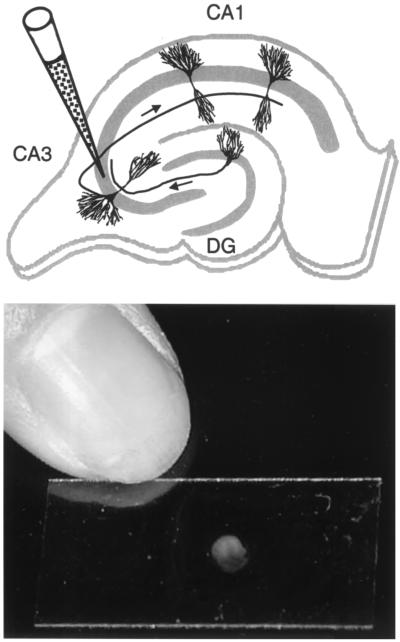
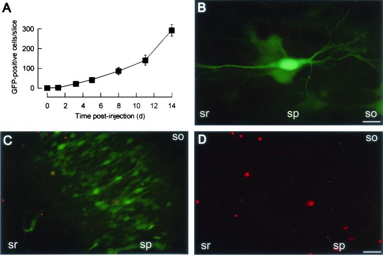
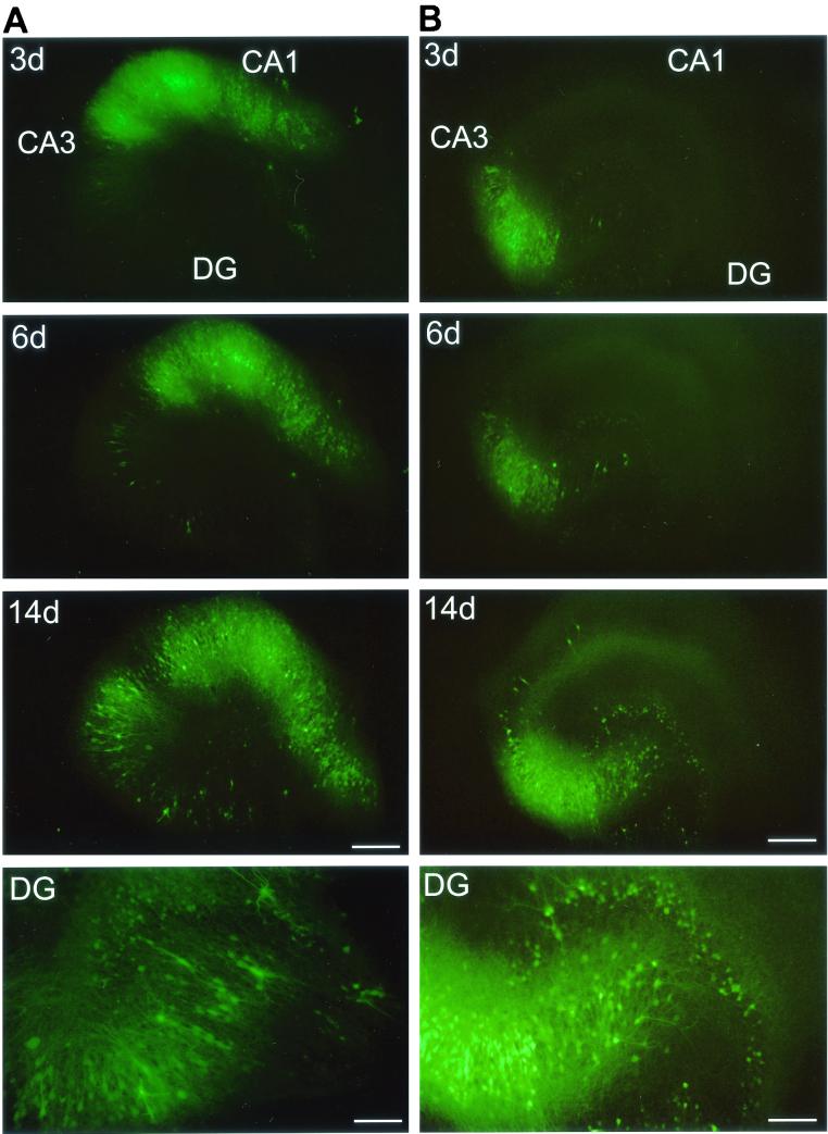
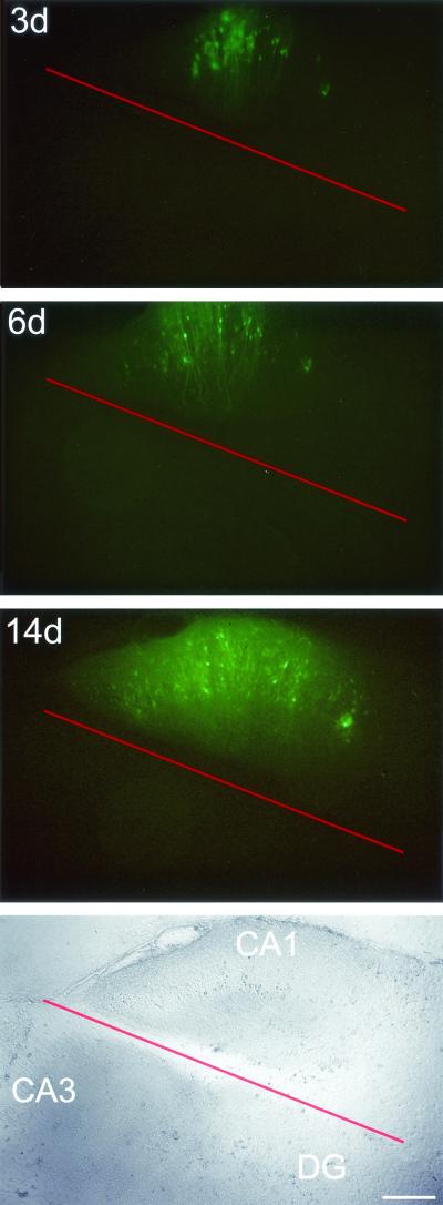
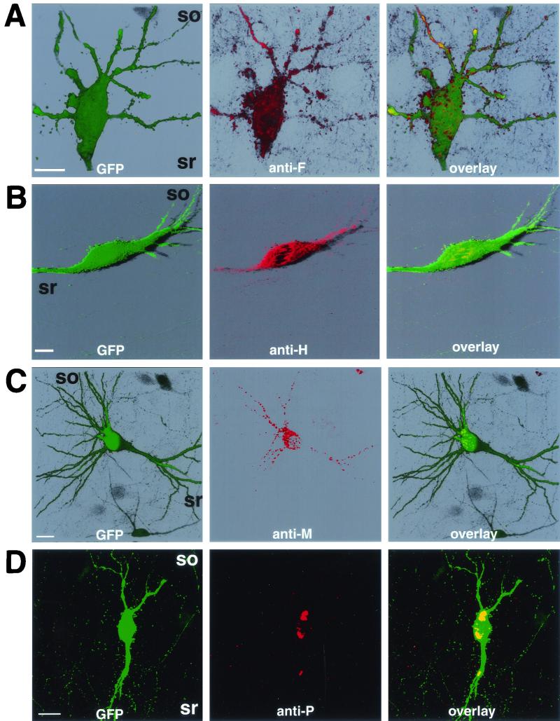
Measles virus (MV) can infect the central nervous system and, in rare cases, causes subacute sclerosing panencephalitis, characterized by a progressive degeneration of neurons. The route of MV transmission in neurons was investigated in cultured rat hippocampal slices by using MV expressing green fluorescent protein. MV infected hippocampal neurons and spread unidirectionally, in a retrograde manner, from CA1 to CA3 pyramidal cells and from there to the dentate gyrus. Spreading of infection depended on cell-to-cell contact and occurred without any detectable release of infectious particles. The role of the viral proteins in the retrograde MV transmission was determined by investigating their sorting in infected pyramidal cells. MV glycoproteins, the fusion protein (F) and hemagglutinin (H), the matrix protein (M), and the phosphoprotein (P), which is part of the viral ribonucleoprotein complex, were all sorted to the dendrites. While M, P, and H proteins remained more intracellular, the F protein localized to prominent, spine-type domains at the surface of infected cells. The detected localization of MV proteins suggests that local microfusion events may be mediated by the F protein at sites of synaptic contacts and is consistent with a mechanism of retrograde transmission of MV infection.


Figures

FIG. 1.
FIG. 1.
Infection of hippocampal slices. (Top) Schematic representation of a hippocampal slice with the locations of CA1 and CA3 pyramidal cells and of a granule cell in the dentate gyrus (DG). The position of a glass micropipette, which contains the viral solution, inserted into the pyramidal cell layer (as indicated by the gray area) of the CA3 region is indicated. Arrows along the axons indicate the unidirectional progression of excitatory pathways, with mossy fibers from granule cells synapsing onto dendrites from CA3 pyramidal cells and Schaffer collaterals from CA3 pyramidal cells connecting to dendrites from CA1 pyramidal cells. Note that granule cells are presynaptic to CA3 pyramidal cells, which themselves are presynaptic to CA1 pyramidal cells, whereas this is normally not the case for the reverse. (Bottom) Photograph of a rat hippocampal slice cultured on a 12- by 24-mm glass coverslip.
FIG. 2.
FIG. 2.
MV spreads in cultured hippocampal slices. (A) Time course of MV transmission. Nine slices were injected with ∼100 infectious MV-GFP particles, and the number of GFP-positive cells per slice was determined at increasing time intervals. (B) High-magnification fluorescence micrograph of a GFP-positive CA1 pyramidal cell from a slice at 9 days after MV-GFP application. (C) Fluorescence micrograph of the CA3 region from a living slice injected with MV-GFP and stained with propidium iodide at 17 dpi. A fluorescein isothiocyanate filter set was used (see Materials and Methods); GFP fluorescence (green) shows MV-infected cells, whereas propidium iodide fluorescence (red) indicates nonviable cells. (D) Propidium iodide fluorescence micrograph of the same region as in panel C with a rhodamine filter set. Abbreviations: so, stratum oriens; sp, stratum pyramidale; sr, stratum radiatum. Bars, 20 μm (B) and 65 μm (C and D).
FIG. 3.
FIG. 3.
Polarity of MV transmission. Injection of MV-GFP into the CA1 region (A) results in the propagation of MV to the CA3 region and dentate gyrus (DG), whereas injection of MV-GFP into the CA3 region (B) results in transmission to the dentate gyrus but only inefficiently to the CA1 region. Fluorescence micrographs at 3, 6, and 14 dpi are shown; bottom panels (DG) show a magnification of the dentate gyrus at ∼14 dpi. The DG panel of the CA1-injected slice (A) shows a magnification that is rotated by 50° counterclockwise with regard to the upper panels and is exposed longer to better visualize GFP-positive granule cells. Bars, 20 μm (3-, 6-, and 14-day panels) and 80 μm (DG panels).
FIG. 4.
FIG. 4.
Separation of the MV-injected CA1 region prevents MV spread to the CA3 region and dentate gyrus (DG). Fluorescence (top) and bright-field (bottom) micrographs at 3, 6, and 14 dpi of a slice for which MV-GFP was applied to the CA1 region and the injected region was separated (red line) immediately thereafter. Bar, 320 μm.
FIG. 5.
FIG. 5.
Localization of F (A), H (B), M (C), and P (D) proteins in infected CA3 pyramidal cells. Slices were fixed at 7 dpi, permeabilized, and incubated with monoclonal antibodies directed against F, H, M, or P proteins (middle panels of A to D, respectively). F, H, and M protein localization is shown by three-dimensional reconstitution (A to C); P protein localization is shown by extended focus of plane images (D). Note that only GFP-positive neurons were reactive toward the antibodies used. Abbreviations: so, stratum oriens; sr, stratum radiatum. Bars, 20 μm.

References

    1. Baczko, K., J. Lampe, U. G. Liebert, U. Brinckmann, V. ter Meulen, I. Pardowitz, H. Budka, S. L. Cosby, S. Isserte, and B. K. Rima. 1993. Clonal expansion of hypermutated measles virus in a SSPE brain. Virology 197:188-195. - PubMed
    1. Baczko, K., U. G. Liebert, M. Billeter, R. Cattaneo, H. Budka, and V. ter Meulen. 1986. Expression of defective measles virus genes in brain tissues of patients with subacute sclerosing panencephalitis. J. Virol. 59:472-478. - PMC - PubMed
    1. Billeter, M. A., and R. Cattaneo. 1991. Molecular biology of defective measles viruses persisting in the human central nervous system, p. 323-345. In D. Kingsbury (ed.), The paromyxoviruses. Plenum Press, New York, N.Y.
    1. Blau, D. M., and R. W. Compans. 1995. Entry and release of measles virus are polarized in epithelial cells. Virology 210:91-99. - PubMed
    1. Burack, M. A., M. A. Silverman, and G. Banker. 2000. The role of selective transport in neuronal protein sorting. Neuron 26:465-472. - PubMed

Publication types

LinkOut - more resources