. 2002 Jun;76(11):5784-92.
doi: 10.1128/jvi.76.11.5784-5792.2002.

Evolution of bacteriophage in continuous culture: a model system to test antiviral gene therapies for the emergence of phage escape mutants

Affiliations

Evolution of bacteriophage in continuous culture: a model system to test antiviral gene therapies for the emergence of phage escape mutants

Björn F Lindemann et al. J Virol. 2002 Jun.

Abstract

The emergence of viral escape mutants is usually a highly undesirable phenomenon. This phenomenon is frequently observed in antiviral drug applications for the treatment of viral infections and can undermine long-term therapeutic success. Here, we propose a strategy for evaluating a given antiviral approach in terms of its potential to provoke the appearance of resistant virus mutants. By use of Q beta RNA phage as a model system, the effect of an antiviral gene therapy, i.e., a virus-specific repressor protein expressed by a recombinant Escherichia coli host, was studied over the course of more than 100 generations. In 13 experiments carried out in parallel, 12 phage populations became resistant and 1 became extinct. Sequence analysis revealed that only two distinct phage mutants emerged in the 12 surviving phage populations. For both escape mutants, sequence variations located in the repressor binding site of the viral genomic RNA, which decrease affinity for the repressor protein, conferred resistance to translational repression. The results clearly suggest the feasibility of the proposed strategy for the evaluation of antiviral approaches in terms of their potential to allow resistant mutants to appear. In addition, the strategy proved to be a valuable tool for observing virus-specific molecular targets under the impact of antiviral drugs.


Figures

FIG. 1.
FIG. 1.
Three-stage continuous culture system for bacteriophages. Host cells were grown in a turbidostat. Host cell suspension and inducer stock solution were transferred into a stirred flow reactor called the induction reactor. The dilution rate was adjusted to reveal a mean residence time of approximately 40 min for host bacteria in this reactor. The suspension of induced host bacteria was finally transferred into as many as six stirred flow reactors called infection reactors. In these infection reactors, host bacteria became infected by bacteriophages.
FIG. 2.
FIG. 2.
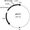
Plasmid map of construct pBL410. The gene of Qβ coat protein was amplified by PCR and cloned into the EcoRI and HindIII sites of vector pKK223-3 (8). The tac promoter and the concomitant rrnB terminator are designated Ptac and rrnB, respectively. ori, origin of replication
FIG. 3.
FIG. 3.
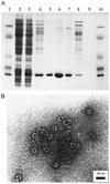
(A) Results of denaturing polyacrylamide gel electrophoresis of the purified coat protein from host cells harboring plasmid pBL410. Lanes: 1 and 10, protein markers (66, 45, 24, 18.4, and 14.3 kDa); 2, cell extract of JM105/pBL410 without induction by IPTG; 3, cell extract of JM105/pBL410 with induction by IPTG; 4 and 8, protein fraction after precipitation with ammonium sulfate; 5, 6, and 7, fractions 5, 6, and 7 from size exclusion chromatography on Sepharose CL-4B, respectively. The coat protein monomer has a molecular mass of 16.9 kDa (52). (B) Electron micrograph of virus-like particles from purified coat protein. Coat protein isolated from E. coli JM105 cells harboring plasmid pBL410 spontaneously formed virus-like capsids that were purified by size exclusion chromatography. The virus-like particles are similar in size and shape to Qβ virions and capsids synthesized in a previous work (27).
FIG. 4.
FIG. 4.
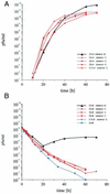
Time courses of phage titers in six representative infection reactors. Phage titers were determined with induced E. coli JM105/pBL410 (A) and plasmid-free JM105 (B). Depicted are the time courses for one population that revealed a variant of the major class (cbsmut 1) of escape mutants (squares), four populations that yielded the cbsmut 2 mutant (triangles), and one population that died out during the time course of the experiment (circles).
FIG. 5.
FIG. 5.
Putative secondary structure of the coat protein binding site. (A) The local secondary structure of Qβ wild-type RNA is depicted. The sequence shown corresponds to the transcripts used for binding studies in vitro, except that an additional U is present at the 3′ terminus. Arrows indicate the point mutations found in the cbsmut 1 and cbsmut 2 variants; the start codon of the replicase gene is also indicated. (B) For the cbsmut 2 variant, an alternative fold can be calculated by standard methods (1).

Similar articles

Cited by

References

    1. Abrahams, J. P., M. van den Berg, E. van Batenburg, and C. Pleij. 1990. Prediction of RNA secondary structure, including pseudoknotting, by computer simulation. Nucleic Acids Res. 18:3035-3044. - PMC - PubMed
    1. Adams, M. H. 1959. Bacteriophages. Wiley Interscience, New York, N.Y.
    1. Arora, R., C. Priano, A. B. Jacobson, and D. R. Mills. 1996. cis-acting elements within an RNA coliphage genome: fold as you please, but fold you must. J. Mol. Biol. 258:433-446. - PubMed
    1. Ball, L. A., and P. Kaesberg. 1973. A polarity gradient in the expression of the replicase gene of RNA bacteriophage Qβ. J. Mol. Biol. 74:547-562. - PubMed
    1. Bauer, G., J. S. McCaskill, H. Otten, and A. Schwienhorst. 1989. Evolution im Laboratorium. Nachrbl. Chem. Tech. Lab. 37:484-488.

Publication types

LinkOut - more resources