

doi: 10.1128/jvi.76.11.5813-5821.2002.
Sequencing of porcine enterovirus groups II and III reveals unique features of both virus groups
Affiliations
- PMID: 11992011
- PMCID: PMC137026
- DOI: 10.1128/jvi.76.11.5813-5821.2002
Item in Clipboard
Sequencing of porcine enterovirus groups II and III reveals unique features of both virus groups
J Virol.
2002 Jun.
Abstract
The molecular classification of the porcine enterovirus (PEV) groups II and III was investigated. The sequence of the almost complete PEV-8 (group II) genome reveals that this virus has unique L and 2A gene regions. A reclassification of this group into a new picornavirus genus is suggested. PEV group III viruses are typical enteroviruses. They differ from other enteroviruses by a prolonged stem-loop D of the 5'-cloverleaf structure.
Figures
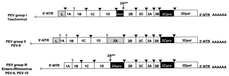
Genome organization of the three PEV groups. The open reading frames are flanked on either side by NTRs. Gene regions (not drawn to scale) are presented. Gene regions encoding proteases are highlighted in black, and those encoding L proteins and the PEV-8 2A gene region are shaded in gray. Viruses of PEV group I, which were recently reclassified as teschoviruses, are characterized by a foot-and-mouth disease virus-like 2A peptidase and a leader protein of unknown function. PEV-8, the only known member of PEV group II, has a leader protein and a 2A protein of unknown function. A 90-nucleotide stretch of the 5′-NTR has a striking homology to the teschovirus 5′-NTR, while the 3′-NTR shares similarities with the PEV group III viruses. PEV-9 and -10 (PEV group III) are typical enteroviruses. Their genome organization is identical to that of entero- and rhinoviruses. The genome-linked 3B peptides at the 5′ end (small circles) and the poly(A) tails at the 3′ ends are indicated. The processing sites of 3C protease (arrowheads) and 2A protease (small arrows) are also shown. A question mark symbolizes unknown proteolytic activities responsible for the maturation cleavage of the 1AB precursor and the release of the PEV-8 leader protein.
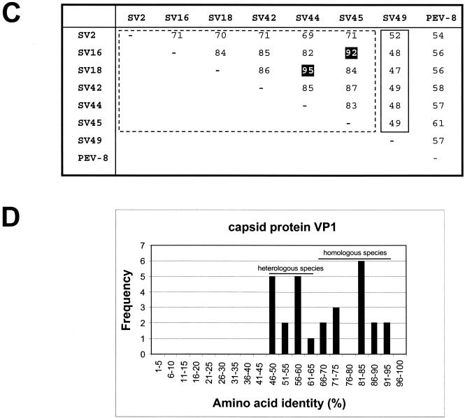
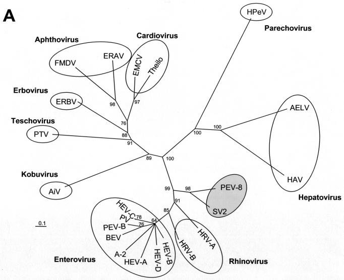
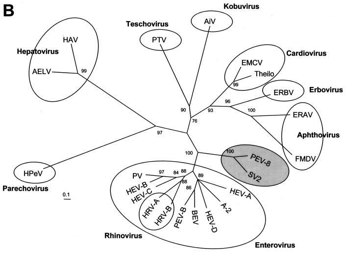
Phylogenetic relationships of PEV-8 to other picornaviruses. Unrooted neighbor-joining trees of the 3D polymerase (A) and the P1 capsid proteins (B) of 22 picornavirus species. Amino acid sequences were aligned with the Clustal W program. Maximum-likelihood branch lengths were calculated by the quartet puzzling method. Branch lengths are proportional to genetic divergence. The scale bar indicates the number of amino acid substitutions per site. Circles indicate picornavirus genera. The proposed genus containing PEV-8 and SV2 is indicated by a shaded circle. Numbers at nodes represent percentages of bipartitions in intermediate trees that have been generated in 10,000 puzzling steps. Note that capsid protein sequences (B) are not suited to differentiate the enterovirus and rhinovirus genera. (C) Pairwise comparison of VP1 sequences of PEV-8 and seven simian picornaviruses. Percent amino acid identities are given. The dotted box indicates amino acid identity values of a major cluster of serotypes that likely represent a new species. The comparison of two strains of the same serotype results in high amino acid identity values, which are highlighted. The small box marks pairwise comparisons of SV49, with the major serotype cluster indicating that SV49 may belong to a different species. (D) Frequency distribution of pairwise amino acid identity scores of the VP1 capsid proteins. The amino acid identity scores of up to 65% are characteristic of comparisons of heterologous species, while amino acid identity scores above 66% indicate comparisons of (i) different strains of homologous serotypes or (ii) heterologous serotypes of homologous species. A-2, A-2 plaque virus; AELV, avian encephalomyelitis-like virus; AiV, aichivirus; BEV, bovine enterovirus; EMCV, encephalomyocarditis virus; ERAV, equine rhinitis A virus (formerly equine rhinovirus 1); ERBV, equine rhinitis B virus (formerly equine rhinovirus 2); FMDV, foot-and-mouth disease virus; HAV, hepatitis A virus; HEV, human enterovirus; HPeV, human parechovirus (formerly echoviruses 22 and 23); HRV, human rhinovirus; PV, poliovirus; SV, simian virus; Theilo, theilovirus.
Phylogenetic relationships of PEV-8 to other picornaviruses. Unrooted neighbor-joining trees of the 3D polymerase (A) and the P1 capsid proteins (B) of 22 picornavirus species. Amino acid sequences were aligned with the Clustal W program. Maximum-likelihood branch lengths were calculated by the quartet puzzling method. Branch lengths are proportional to genetic divergence. The scale bar indicates the number of amino acid substitutions per site. Circles indicate picornavirus genera. The proposed genus containing PEV-8 and SV2 is indicated by a shaded circle. Numbers at nodes represent percentages of bipartitions in intermediate trees that have been generated in 10,000 puzzling steps. Note that capsid protein sequences (B) are not suited to differentiate the enterovirus and rhinovirus genera. (C) Pairwise comparison of VP1 sequences of PEV-8 and seven simian picornaviruses. Percent amino acid identities are given. The dotted box indicates amino acid identity values of a major cluster of serotypes that likely represent a new species. The comparison of two strains of the same serotype results in high amino acid identity values, which are highlighted. The small box marks pairwise comparisons of SV49, with the major serotype cluster indicating that SV49 may belong to a different species. (D) Frequency distribution of pairwise amino acid identity scores of the VP1 capsid proteins. The amino acid identity scores of up to 65% are characteristic of comparisons of heterologous species, while amino acid identity scores above 66% indicate comparisons of (i) different strains of homologous serotypes or (ii) heterologous serotypes of homologous species. A-2, A-2 plaque virus; AELV, avian encephalomyelitis-like virus; AiV, aichivirus; BEV, bovine enterovirus; EMCV, encephalomyocarditis virus; ERAV, equine rhinitis A virus (formerly equine rhinovirus 1); ERBV, equine rhinitis B virus (formerly equine rhinovirus 2); FMDV, foot-and-mouth disease virus; HAV, hepatitis A virus; HEV, human enterovirus; HPeV, human parechovirus (formerly echoviruses 22 and 23); HRV, human rhinovirus; PV, poliovirus; SV, simian virus; Theilo, theilovirus.
Phylogenetic relationships of PEV-8 to other picornaviruses. Unrooted neighbor-joining trees of the 3D polymerase (A) and the P1 capsid proteins (B) of 22 picornavirus species. Amino acid sequences were aligned with the Clustal W program. Maximum-likelihood branch lengths were calculated by the quartet puzzling method. Branch lengths are proportional to genetic divergence. The scale bar indicates the number of amino acid substitutions per site. Circles indicate picornavirus genera. The proposed genus containing PEV-8 and SV2 is indicated by a shaded circle. Numbers at nodes represent percentages of bipartitions in intermediate trees that have been generated in 10,000 puzzling steps. Note that capsid protein sequences (B) are not suited to differentiate the enterovirus and rhinovirus genera. (C) Pairwise comparison of VP1 sequences of PEV-8 and seven simian picornaviruses. Percent amino acid identities are given. The dotted box indicates amino acid identity values of a major cluster of serotypes that likely represent a new species. The comparison of two strains of the same serotype results in high amino acid identity values, which are highlighted. The small box marks pairwise comparisons of SV49, with the major serotype cluster indicating that SV49 may belong to a different species. (D) Frequency distribution of pairwise amino acid identity scores of the VP1 capsid proteins. The amino acid identity scores of up to 65% are characteristic of comparisons of heterologous species, while amino acid identity scores above 66% indicate comparisons of (i) different strains of homologous serotypes or (ii) heterologous serotypes of homologous species. A-2, A-2 plaque virus; AELV, avian encephalomyelitis-like virus; AiV, aichivirus; BEV, bovine enterovirus; EMCV, encephalomyocarditis virus; ERAV, equine rhinitis A virus (formerly equine rhinovirus 1); ERBV, equine rhinitis B virus (formerly equine rhinovirus 2); FMDV, foot-and-mouth disease virus; HAV, hepatitis A virus; HEV, human enterovirus; HPeV, human parechovirus (formerly echoviruses 22 and 23); HRV, human rhinovirus; PV, poliovirus; SV, simian virus; Theilo, theilovirus.
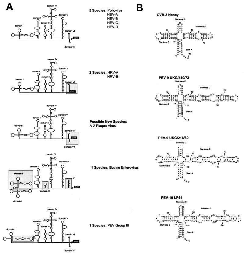
Analysis of enteroviral and rhinoviral 5′-NTRs. (A) Five folding patterns of the 5′-NTRs of the enteroviral and rhinoviral species are schematically illustrated. With respect to the human enteroviruses, the rhinoviruses and the animal enteroviruses (including A-2 plaque virus as a possible new enterovirus species) show significant differences, which are boxed. (B) Comparison of the 5′ cloverleaf of a typical human enterovirus (CVB3) and sequenced virus strains of PEV group III. The stem-loop D of the PEVs is significantly prolonged.
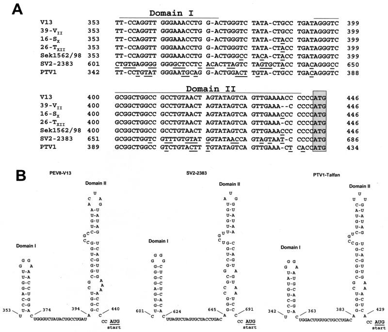
Comparison of partial 5′-NTR sequences of PEV-8, SV2, and PTV-1. (A) Sequence alignment of five PEV-8 strains (V13, 39-VII, 16-SX, 26-TXII, and Sek1562/98), SV2, and PTV-1 Talfan. A homology of this part of the 5′-NTR is obvious. Deviant nucleotides are underlined. The proposed AUG start codon is boxed. (B) The highly conserved nucleotides allow the formation of two putative RNA secondary structures, which are likely to play a role in a specific mechanism of translation initiation of these viruses.
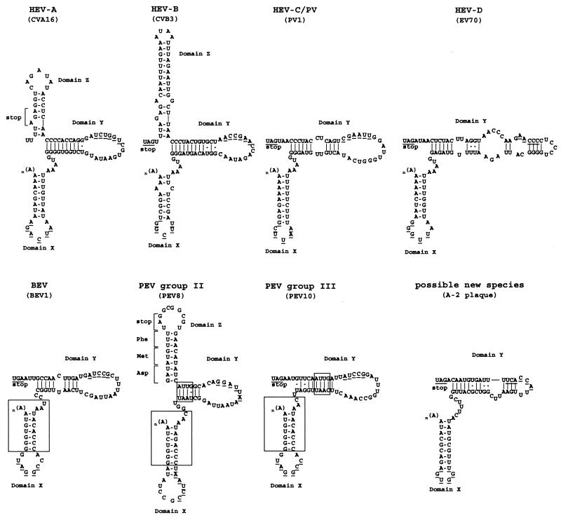
Comparison of the putative 3′-NTR secondary structures of PEV groups II and III with those of other enterovirus species. HEV, human enterovirus. Each species is represented by a typical member (in parentheses). Large boxes indicate conserved sequences of bovine enterovirus (BEV) and PEV species, and small boxes emphasize additional similarities between PEV groups II and III. The putative domains are designated X, Y, and Z starting from the 3′ end. Nucleotides likely to be involved in the formation of a pseudoknot-like element are underlined.
References
-
- Andino, R., G. E. Rieckhoff, and D. Baltimore. 1990. A functional ribonucleoprotein complex forms around the 5′ end of poliovirus RNA. Cell 63:369-380. - PubMed
-
- Dauber, M. 1999. Identification of group I porcine enteroviruses by monoclonal antibodies in cell culture. Vet. Microbiol. 67:1-12. - PubMed
-
- Doherty, M., D. Todd, N. McFerran, and E. M. Hoey. 1999. Sequence analysis of a porcine enterovirus serotype 1 isolate: relationships with other picornaviruses. J. Gen. Virol. 80:1929-1941. - PubMed
-
- Dunne, H. W., J. L. Gobble, J. F. Hokanson, D. C. Kradel, and G. R. Bubash. 1965. Porcine reproductive failure associated with a newly identified “SMEDI” group of picornavirus. Am. J. Vet. Res. 26:1284-1297. - PubMed
MeSH terms
Substances
LinkOut - more resources
Full Text Sources
