

What is the 'true' function of skin?
- PMID: 11994143
- PMCID: PMC7010069
- DOI: 10.1034/j.1600-0625.2002.00112.x
What is the 'true' function of skin?
Abstract
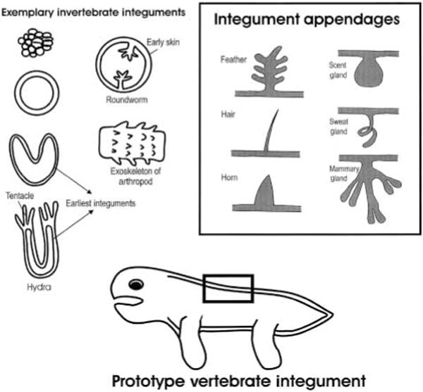
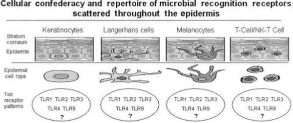
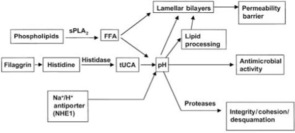
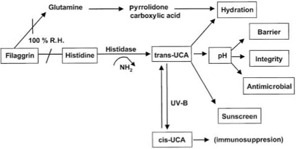
Conventional textbook wisdom portrays the skin as an organ that literally enwraps whatever each of us stands for as a more or less functional, individual member of the mammalian species, and has it that the skin primarily establishes, controls and transmits contacts with the external world. In addition, the skin has long been recognized to protect the organism from deleterious environmental impacts (physical, chemical,microbiological), and is well-known as crucial for the maintenance of temperature, electrolyte and fluid balance. Now, ever more studies are being published that show the skin to also operate as a huge and highly active biofactory for the synthesis,processing and/or metabolism of an astounding range of e.g. structural proteins, glycans, lipids and signaling molecules. Increasingly, it becomes appreciated that the skin, furthermore, is an integral component of the immune, nervous and endocrine systems, with numerous lines of cross-talk between these systems established intracutaneously (e.g. Ann NY Acad Sci Vol 885, 1999; Endocrine Rev 21:457-487, 2000; Physiol Rev 80:980-1020, 2001; Exp Dermatol 10: 349-367, 2001). All these emerging cutaneous functions beyond the classical image of the skin as a barrier and sensory organ are immediately relevant for many of the quandaries that clinical dermatology, dermatopathology, and dermatopharmacology are still struggling with to-date, and offer the practising dermatologist attractive new targets for therapeutic intervention. Yet, many of these skin functions are not even mentioned in dermatology textbooks and await systematic therapeutic targeting. Following a suggestion by Enno Christophers, the current 'Controversies' feature brings together an unusually diverse council of biologists and clinicians, who share their thought-provoking views with the readers and allow us to peek into the future of research in cutaneous biology, not the least by reminding us of the -- often ignored -- evolutionary and embryonal origins of our favorite organ. Hopefully, this unique discussion feature will foster an understanding of the 'true' skin functions that is both more comprehensive and more profound than conventional teaching on this topic, and will stimulate more than 'skin-deep' reflections on the full range of skin functions.
Figures


References
-
- Futuyma DJ. Evolutionary Biology. 3. Sunderland, MA: Sinauer Associates; 1997.
-
- Campbell NA, et al. Biology. 5. New York, NY: Addison-Wesley Longman; 1999.
-
- Bereiter-Hahn J, et al., editors. Biology of the Integument of Vertebrates Berlin. Heidelberg, New York, Tokyo: Springer-Verlag; 1986.
-
- Hebert JM, et al. Cell. 1994;78:1017–1025. - PubMed
Publication types
MeSH terms
Grants and funding
LinkOut - more resources
Full Text Sources
Other Literature Sources
Medical
