. 2002 May;109(9):1231-40.
doi: 10.1172/JCI14698.

Quantitative and functional analysis of PDC-E2-specific autoreactive cytotoxic T lymphocytes in primary biliary cirrhosis

Affiliations

Quantitative and functional analysis of PDC-E2-specific autoreactive cytotoxic T lymphocytes in primary biliary cirrhosis

Hiroto Kita et al. J Clin Invest. 2002 May.

Abstract

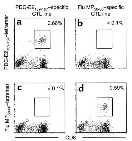
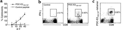
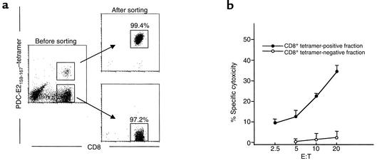
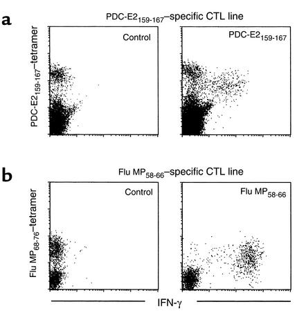
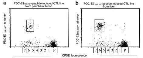
While the pathologic mechanisms responsible for organ-specific tissue damage in primary biliary cirrhosis (PBC) remain an enigma, it has been suggested that the pathology is mediated by autoreactive T cells infiltrating the intrahepatic bile ducts. Previously, we have documented that there is 100-fold enrichment in the frequency of CD4(+) autoreactive T cells in the liver that are specific for peptides encoded by the E2 components of the pyruvate dehydrogenase complexes (PDC-E2). We have also recently characterized the first MHC class I-restricted epitope for PDC-E2, namely amino acid 159-167, a region very similar to the epitope recognized by MHC class II-restricted CD4(+) cells and by autoantibodies. The effector functions of these PDC-E2(159-167)-specific CD8(+) cytotoxic T lymphocytes (CTLs) are not well understood. We have taken advantage of tetramer technology and report herein that there is tenfold increase in the frequency of PDC-E2(159-167)-specific CTLs in the liver as compared with the blood in PBC. In addition, the precursor frequency of the CTLs in blood was significantly higher in early-stage PBC. Of interest was the fact that, upon stimulation with the peptide, the response of PDC-E2(159-167) tetramer-positive cells is heterogeneous with respect to IFN-gamma synthesis. These data, we believe for the first time, document the enrichment of autoantigen-specific CD8(+) T cells in the PBC liver, suggesting that CD8(+) T cells play a significant role in the immunopathogenesis of PBC.


Figures

Figure 1
Figure 1
Verification of the binding specificity of peptide-MHC tetramers. PBMCs from an HLA-A*0201+ PBC patient were cocultured with PDC-E2159-167–loaded APCs (a and c) or Flu MP58-66–loaded APCs (b and d) for 14 days. The cells were stained with the HLA-A*0201 tetramer loaded with PDC-E2159-167 (a and b) or the HLA-A*0201 tetramer loaded with Flu MP58-66 (c and d) in addition to anti-CD4 and anti-CD8 Ab’s, followed by flow cytometric analysis. Displayed in the dot plots are cells gated at lymphocyte population by forward scattering and side scattering and the CD4 population. The cells within the box are considered positive. The number next to the box is the percentage of positively stained cells in the lymphocyte population.
Figure 2
Figure 2
Identification and functional analysis of PDC-E2159-167–specific CD8+ T cells in the peptide-induced CTL line. PBMCs from an HLA-A*0201+ PBC patient were cocultured with PDC-E2159-167–loaded APCs for 14 days to generate a CTL line. (a) The CTL line was tested for cytotoxicity against PDC-E2159-167 peptide– or control peptide–loaded T2 targets at different E/T ratios. HBc18-27, an HLA-A*0201–restricted irrelevant epitope, was used as control. Displayed are the mean specific lysis of triplicate testing. (b) The CTL line was restimulated with PDC-E2159-167 peptide or control peptide in the presence of brefeldin A, followed by intracellular staining for IFN-γ. Displayed in the dot plots are cells gated at lymphocyte population by forward scattering and side scattering and the CD4 population. The number next to the box is the percentage of IFN-γ–producing cells in the lymphocyte population. (c) The CTL line was stained with PDC-E2159-167 tetramer and Ab’s against CD4 and CD8, followed by flow cytometric analysis. Displayed in the dot plots are cells gated at lymphocyte population by forward scattering and side scattering and the CD4 population. The number next to the box is the percentage of positively stained cells in the lymphocyte population.
Figure 3
Figure 3
The specific cytotoxicity of PDC-E2159-167 peptide–induced cell line is mediated by tetramer-positive CD8+ T cells. PBMCs from an HLA-A*0201+ PBC patient were cocultured with PDC-E2159-167–loaded APCs for 14 days to generate a CTL line. (a) Cells were stained with the PDC-E2159-167 tetramer and Ab’s against CD4 and CD8, followed by flow cytometric cell sorting to isolate tetramer-positive CD8+ and tetramer-negative CD8+ populations. (b) The sorted cells were tested for their cytotoxicity against T2 target cells loaded with PDC-E2159-167 peptide or a control peptide HBc18-27 at different E/T ratios. Displayed is the mean specific lysis of triplicate testing.
Figure 4
Figure 4
Antigen specificity of IFN-γ production by CD8+ T cells for PDC-E2 autoantigen or influenza viral antigen. (a) PDC-E2159-167 peptide–induced CTL lines derived from a PBC patient was restimulated with PDC-E2159-167 peptide or a control peptide in the presence of brefeldin A. The cells were first stained with the PDC-E2159-167 tetramer, followed by intracellular staining for IFN-γ. Displayed in the dot plots are cells gated at lymphocyte population by forward scattering and side scattering and the CD8+ population. (b) Flu MP58-66 peptide-induced CTL lines derived from the same PBC patient was restimulated with Flu MP58-66 peptide or a control peptide in the presence of brefeldin A. The cells were first stained with the Flu MP58-66 tetramer, followed by intracellular staining for IFN-γ. Displayed in the dot plots are cells gated at lymphocyte population by forward scattering and side scattering and the CD8+ population.
Figure 5
Figure 5
In vitro expansion of PDC-E2159-167 tetramer–reactive cells. PBMCs (a) and LILs (b) from HLA-A*0201+ PBC patients were stained with CFSE and cocultured with PDC-E2159-167–loaded APCs for 14 days. The cells were stained with the PDC-E2 tetramer and anti-CD8 Ab’s, followed by flow cytometric analysis. Displayed in the dot plots are cells gated at lymphocyte population by forward scattering and side scattering and the CD8+ population. The number next to the box is the percentage of tetramer-stained cells in the lymphocyte population. The horizontal axis is labeled with numbers corresponding to cell divisions and with “p” depicting the undivided parent cell population. This scale was calculated from the distinct CFSE fluorescence peaks produced by polyclonal stimulation with PHA-P and IL-2.

References

    1. Coppel RL, Gershwin ME. Primary biliary cirrhosis: the molecule and the mimic. Immunol Rev. 1995; 144:17–49. - PubMed
    1. Van de Water J, et al. The role of T cells in primary biliary cirrhosis. Semin Liver Dis. 1997; 17:105–113. - PubMed
    1. Colucci G, Schaffner F, Paronetto F. In situ characterization of the cell-surface antigens of the mononuclear cell infiltrate and bile duct epithelium in primary biliary cirrhosis. Clin Immunol Immunopathol. 1986; 41:35–42. - PubMed
    1. Van de Water J, et al. Heterogeneity of autoreactive T cell clones specific for the E2 component of the pyruvate dehydrogenase complex in primary biliary cirrhosis. J Exp Med. 1995; 181:723–733. - PMC - PubMed
    1. Shimoda S, Nakamura M, Ishibashi H, Hayashida K, Niho Y. HLA DRB4 0101-restricted immunodominant T cell autoepitope of pyruvate dehydrogenase complex in primary biliary cirrhosis: evidence of molecular mimicry in human autoimmune diseases. J Exp Med. 1995; 181:1835–1845. - PMC - PubMed

Publication types

MeSH terms