. 2002 May 6;195(9):1155-66.
doi: 10.1084/jem.20011547.

Salmonella pathogenicity island 2 mediates protection of intracellular Salmonella from reactive nitrogen intermediates

Affiliations

Salmonella pathogenicity island 2 mediates protection of intracellular Salmonella from reactive nitrogen intermediates

Dipshikha Chakravortty et al. J Exp Med. .

Abstract

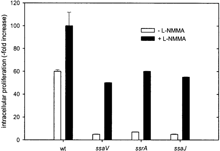
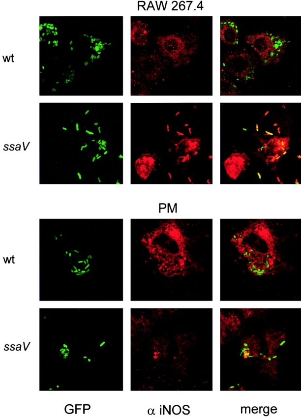
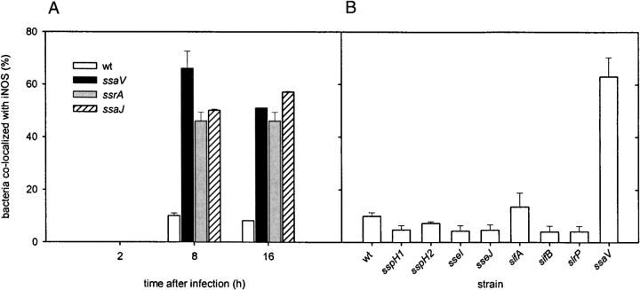
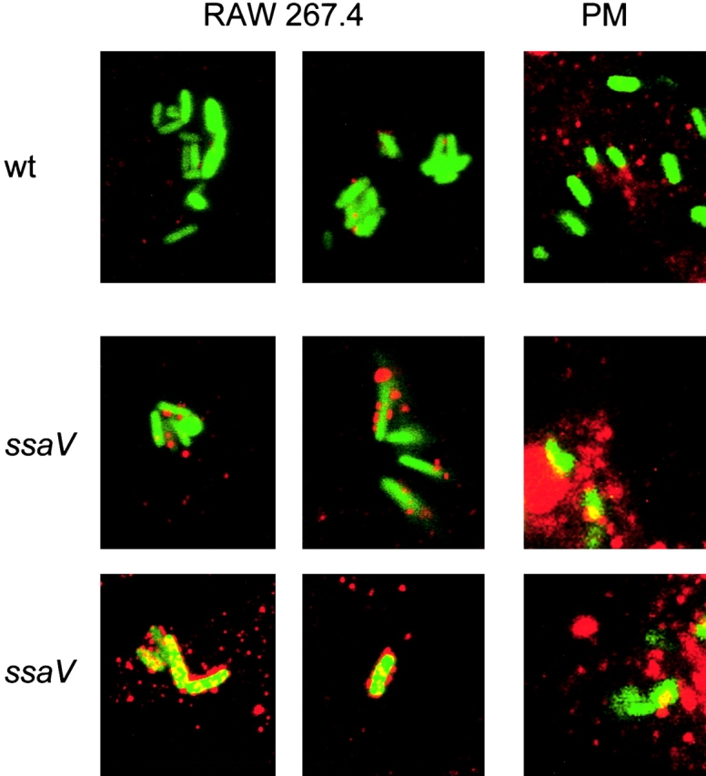
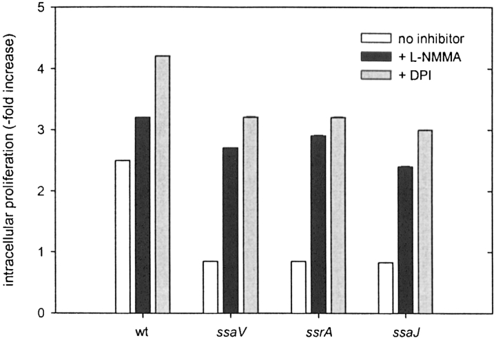
Salmonella typhimurium causes an invasive disease in mice that has similarities to human typhoid. A type III protein secretion system encoded by Salmonella pathogenicity island 2 (SPI2) is essential for virulence in mice, as well as survival and multiplication within macrophages. Reactive nitrogen intermediates (RNI) synthesized by inducible nitric oxide synthase (iNOS) are involved in the control of intracellular pathogens, including S. typhimurium. We studied the effect of Salmonella infection on iNOS activity in macrophages. Immunofluorescence microscopy demonstrated efficient colocalization of iNOS with bacteria deficient in SPI2 but not wild-type Salmonella, and suggests that the SPI2 system interferes with the localization of iNOS and Salmonella. Furthermore, localization of nitrotyrosine residues in the proximity was observed for SPI2 mutant strains but not wild-type Salmonella, indicating that peroxynitrite, a potent antimicrobial compound, is excluded from Salmonella-containing vacuoles by action of SPI2. Altered colocalization of iNOS with intracellular Salmonella required the function of the SPI2-encoded type III secretion system, but not of an individual "Salmonella translocated effector." Inhibition of iNOS increased intracellular proliferation of SPI2 mutant bacteria and, to a lesser extent, of wild-type Salmonella. The defect in systemic infection of a SPI2 mutant strain was partially restored in iNOS(-/-) mice. In addition to various strategies to detoxify RNI or repair damage due to RNI, avoidance of colocalization with RNI is important in adaptation of a pathogen to an intracellular life style.


Figures

Figure 1.
Figure 1.
Induction of iNOS by RAW267.4 macrophages requires live Salmonella. (A) RAW267.4 cells were infected with wild-type Salmonella at various MOI and nitrite in the supernatant was measured by the Griess reaction 4 and 8 h after infection. (B) RAW267.4 macrophages were infected with heat-killed Salmonella and the nitrite was measured 8 and 24 h after infection. The data represent the mean ± SD of three independent experiments done in triplicate. (C) RAW267.4 macrophages were infected with wild-type Salmonella at a MOI of 10 under various experimental conditions as indicated, and nitrite production was measured 8 h after infection. After adding the bacteria to the macrophages and centrifugation, infection took place for 0, 10, 25, or 60 min. In further assays, macrophages were infected for 25 min, but were separated from the bacteria by a 0.4 μM filter, or cytochalasin D was added to the macrophages at 1 μg/ml before infection. not inf., not infected. (D) Effect of bacterial culture conditions on nitrite production. Wild-type S. typhimurium was grown for 16 h in LB media without aeration as standing cultures or with aeration in test tubes in a roller drum. If indicated, the bacteria were opsonized by incubation with 10% normal mouse serum for 30 min. Infection was performed at a MOI of 10 and nitrite production was assayed after 16 h.
Figure 2.
Figure 2.
Nitrite production by RAW267.4 macrophages infected with wild-type Salmonella and SPI2 mutant strains. (A) RAW267.4 macrophages were infected at a MOI of 10 with different SPI2 mutants and the nitrite was measured in the supernatant after 8 h. (B) Infection was performed as before with wild-type S. typhimurium and an isogenic strain deficient in sipC encoded by SPI1. wt, wild-type. (C) Western blot analysis of iNOS expression in noninfected RAW267.4 macrophages, and macrophages infected with wild-type S. typhimurium or strains harboring mutations in SPI2 genes ssaV or ssrA.
Figure 2.
Figure 2.
Nitrite production by RAW267.4 macrophages infected with wild-type Salmonella and SPI2 mutant strains. (A) RAW267.4 macrophages were infected at a MOI of 10 with different SPI2 mutants and the nitrite was measured in the supernatant after 8 h. (B) Infection was performed as before with wild-type S. typhimurium and an isogenic strain deficient in sipC encoded by SPI1. wt, wild-type. (C) Western blot analysis of iNOS expression in noninfected RAW267.4 macrophages, and macrophages infected with wild-type S. typhimurium or strains harboring mutations in SPI2 genes ssaV or ssrA.
Figure 3.
Figure 3.
Intracellular proliferation of Salmonella in the presence or absence of iNOS inhibitor L-NMMA. RAW267.4 macrophages were infected with wild-type (wt) S. typhimurium and various mutant strains in SPI2 genes at a MOI of 10. Non-internalized bacteria were removed by washing and killed by gentamicin. L-NMMA was added at a concentration of 10 μM if indicated. The number of intracellular bacteria was determined by plating serial dilutions of macrophage lysates on LB-agar for counting of cfu. Intracellular replication was expressed as the ratio of cfu at 2 to 16 h after infection.
Figure 4.
Figure 4.
In vitro survival of S. typhimurium under different nitrosative stress conditions. Wild-type (circles) or SPI2 deficient (ssaV mutant, triangles) S. typhimurium were exposed to various concentrations of GSNO (A) or NaNO2 (B) in LB medium (adjusted to pH 5.0) or SIN-1 (C) in LB medium (adjusted to pH 7.0). At 3 h (open symbols) or 9 h (filled symbols) after exposure serial dilutions were plated and cfu were counted. Results are expressed as mean cfu ± SD of two independent experiments in triplicate.
Figure 5.
Figure 5.
Localization of iNOS in Salmonella-infected macrophages. RAW267.4 macrophages and mouse peritoneal macrophages (PM) were infected with GFP-expressing wild-type (wt) S. typhimurium or SPI2 mutant strain defective in ssaV. Non-internalized bacteria were killed by addition of gentamicin. At 12 h after infection, the cells were fixed and permeabilized for immunostaining. iNOS was detected by incubation with a polyclonal antibody against iNOS and a Cy3-labeled secondary antibody. The localization of iNOS (red) and S. typhimurium (green) was analyzed by fluorescence microscopy (not shown) and confocal laser-scanning microscopy. Colocalization of S. typhimurium and iNOS resulted in orange to red staining of the bacterial cells.
Figure 6.
Figure 6.
Colocalization of iNOS with wild-type S. typhimurium and various strains deficient in SPI2 genes or “Salmonella translocated effectors.” (A) RAW267.4 macrophages were infected with wild-type S. typhimurium (wt) and strains deficient in the SPI2-encoded secretion system (ssaV, ssrA, ssaJ). At 2, 8, or 16 h after infection, the percentage of bacteria colocalized with iNOS was scored by examining randomly selected 50 microscopic fields (1,000× magnification) and counted for total green and colocalized yellow or red bacteria. (B) RAW267.4 macrophages were infected with wild-type S. typhimurium, or mutant strains in SPI2 (ssaV) or various STE genes (sspH1, sspH2, sseI, sseJ, sifA, sifB, slrP). 8 h after infection, macrophages were processed for immuno-staining and colocalization was quantified as described in panel A. The data represent two independent experiments.
Figure 7.
Figure 7.
Localization of peroxynitrite in S. typhimurium-infected macrophages. Infection of RAW267.4 macrophages and mouse peritoneal macrophages (PM) was performed for 12 h as described in the legend to Fig. 5. The sites of peroxynitrite formation were detected by immunostaining with an anti-nitrotyrosine antibody and a Cy3-conjugated secondary antibody. Samples were analyzed by confocal laser-scanning microscopy and representative images for the localization of S. typhimurium expressing GFP (green) and peroxynitrite (red) are shown. Various levels of nitrotyrosine formation were observed for the SPI2 mutant strain. wt, wild-type.
Figure 8.
Figure 8.
Intracellular replication in primary macrophages. Peritoneal macrophages were prepared from C57BL/6 mice and infected with S. typhimurium wild-type (wt) or various strains harboring mutations in SPI2 genes. After infection, inhibitors L-NMMA (10 μM) and DPI (10 μM) were added as indicated. The intracellular proliferation was determined as described in the legend of Fig. 3.

Comment in

  • Salmonella selectively stops traffic.
    Fang F, Vazquez-Torres A. Fang F, et al. Trends Microbiol. 2002 Sep;10(9):391-2. doi: 10.1016/s0966-842x(02)02423-x. Trends Microbiol. 2002. PMID: 12217496

References

    1. Shiloh, M.U., J.D. MacMicking, S. Nicholson, J.E. Brause, S. Potter, M. Marino, F. Fang, M. Dinauer, and C. Nathan. 1999. Phenotype of mice and macrophages deficient in both phagocyte oxidase and inducible nitric oxide synthase. Immunity. 10:29–38. - PubMed
    1. Nathan, C., and M.U. Shiloh. 2000. Reactive oxygen and nitrogen intermediates in the relationship between mammalian hosts and microbial pathogens. Proc. Natl. Acad. Sci. USA. 97:8841–8848. - PMC - PubMed
    1. Hueck, C.J. 1998. Type III protein secretion systems in bacterial pathogens of animals and plants. Microbiol. Mol. Biol. Rev. 62:379–433. - PMC - PubMed
    1. Galan, J.E. 2001. Salmonella interactions with host cells: type III secretion at work. Annu. Rev. Cell Dev. Biol. 17:53–86. - PubMed
    1. Wallis, T.S., and E.E. Galyov. 2000. Molecular basis of Salmonella-induced enteritidis. Mol. Microbiol. 36:997–1005. - PubMed

Publication types