. 2002 May 6;195(9):1207-13.
doi: 10.1084/jem.20011783.

The ligand-binding domain of CD22 is needed for inhibition of the B cell receptor signal, as demonstrated by a novel human CD22-specific inhibitor compound

Affiliations

The ligand-binding domain of CD22 is needed for inhibition of the B cell receptor signal, as demonstrated by a novel human CD22-specific inhibitor compound

Soerge Kelm et al. J Exp Med. .

Abstract

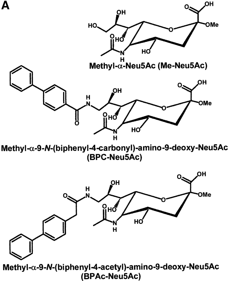
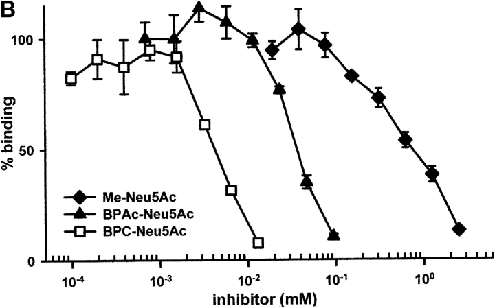
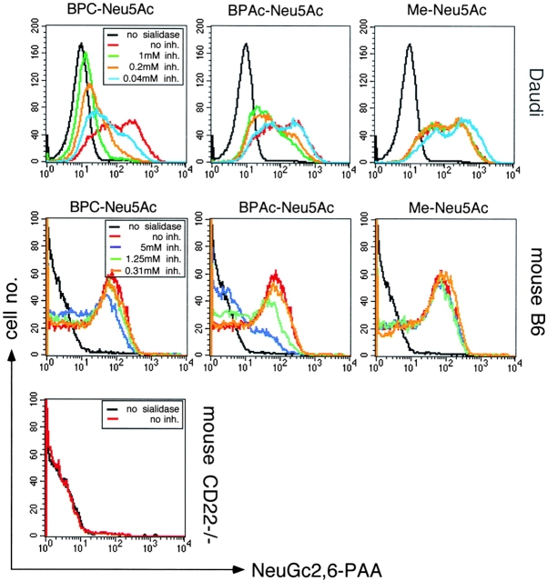
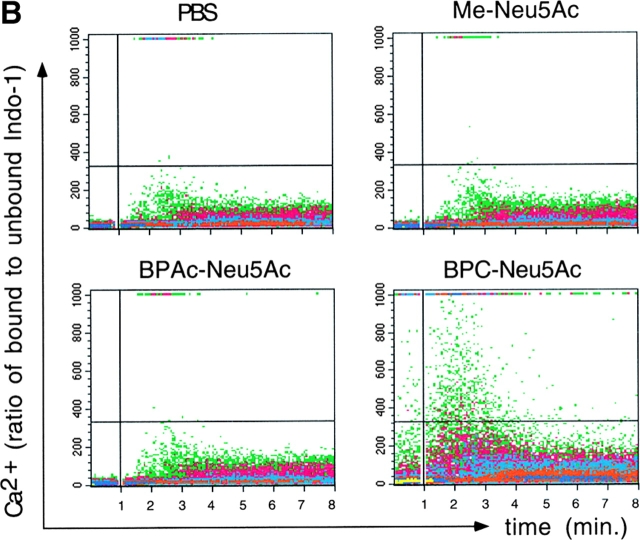
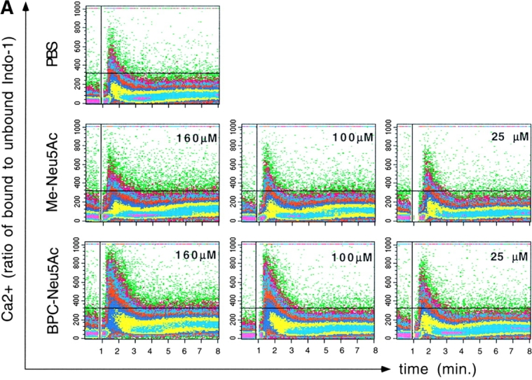
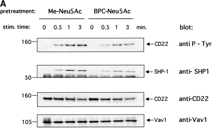
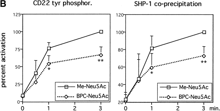
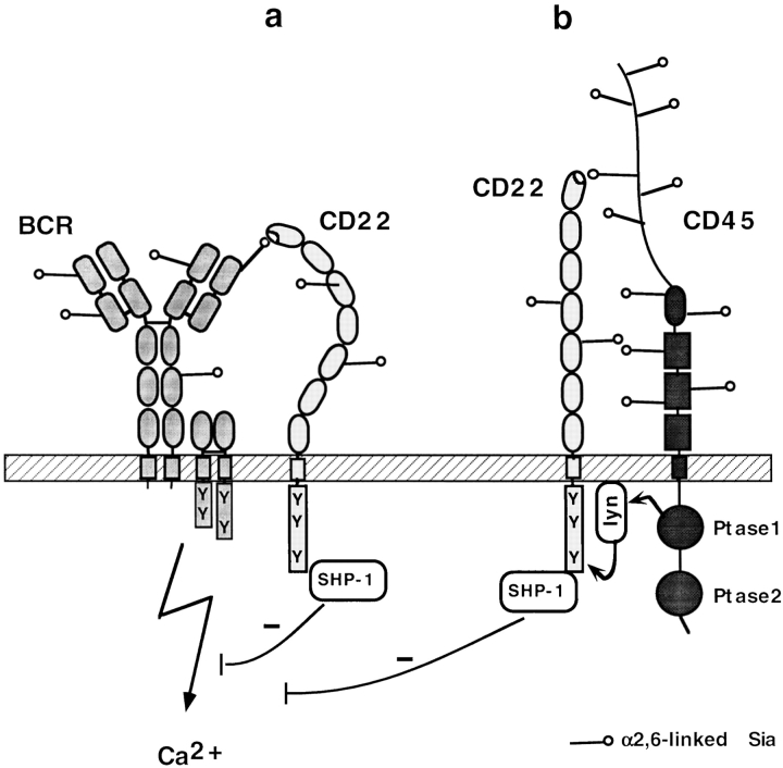
CD22 is a B cell-specific transmembrane protein of the Siglec family. It binds specifically to alpha2,6-linked sialic acid (Sia) residues, which are also present on glycoproteins on the B cell surface. CD22 acts as a negative regulator in B cell receptor-mediated signaling by recruitment of Src homology 2 domain-containing tyrosine phosphatase (SHP)-1 to its intracellular tail. To analyze how ligand-binding of CD22 influences its intracellular signaling domain, we designed synthetic sialosides as inhibitors for the lectin domain of CD22. One of these compounds inhibited binding of human CD22-Fc to target cells over 200-fold better than Sia and was highly selective for human CD22. When Daudi cells or primary B cells were stimulated with anti-immunoglobulin (Ig)M in presence of this sialoside inhibitor, a higher Ca(2+) response was observed, similar to CD22-deficient B cells. Accordingly, a lower tyrosine-phosphorylation of CD22 and SHP-1 recruitment was demonstrated in presence of the sialoside. Thus, by interfering with ligand binding of CD22 on the B cell surface, we have shown for the first time that the lectin domain of CD22 has a direct, positive influence on its intracellular inhibitory domain. Also, we have developed a novel low molecular weight compound which can enhance the response of human B cells.


Figures

Figure 1.
Figure 1.
Inhibition of binding of CD22-Fc to target cells by synthetic sialosides. (A) Structures of sialosides and abbreviations used in this study. (B) Binding assays with human CD22. Binding of hCD22d1–3-Fc to murine AG8 cells was measured as described under Materials and Methods in the presence of sialosides at the concentrations indicated.
Figure 1.
Figure 1.
Inhibition of binding of CD22-Fc to target cells by synthetic sialosides. (A) Structures of sialosides and abbreviations used in this study. (B) Binding assays with human CD22. Binding of hCD22d1–3-Fc to murine AG8 cells was measured as described under Materials and Methods in the presence of sialosides at the concentrations indicated.
Figure 2.
Figure 2.
Specificity of the synthetic sialosides for cellular CD22. Human Daudi cells, mouse C57BL/6 (B6), or mouse CD22−/− B cells were not pretreated (black curves) or sialidase pretreated (all colored curves), then incubated with no inhibitor (no inh., in red) or the indicated inhibitors (same conc. used for one type of cells) and afterwards stained with NeuGc2,6-PAA. The assay shows inhibition of binding of NeuGc2,6-PAA (a synthetic CD22 ligand) to hCD22 and mCD22 by the indicated sialosides. Mouse cells are gated as B220+.
Figure 3.
Figure 3.
Presence of the sialoside BPC-Neu5Ac leads to increased BCR-triggered Ca2+ responses. (A) Daudi B cells were stimulated with anti-IgM in presence of either PBS, Me-Neu5Ac, or BPC-Neu5Ac at the given concentrations. (B) Human blood lymphocytes were stimulated with anti-IgM in presence of either PBS, Me-Neu5Ac, BPAc-Neu5Ac, or BPC-Neu5Ac (250 μM each). CD20+ gated cells are shown. Addition of antibody: at the vertical lines. Horizontal lines are drawn for quantitative comparison. One typical experiment, out of five experiments (for A) or two experiments (for B) is shown.
Figure 3.
Figure 3.
Presence of the sialoside BPC-Neu5Ac leads to increased BCR-triggered Ca2+ responses. (A) Daudi B cells were stimulated with anti-IgM in presence of either PBS, Me-Neu5Ac, or BPC-Neu5Ac at the given concentrations. (B) Human blood lymphocytes were stimulated with anti-IgM in presence of either PBS, Me-Neu5Ac, BPAc-Neu5Ac, or BPC-Neu5Ac (250 μM each). CD20+ gated cells are shown. Addition of antibody: at the vertical lines. Horizontal lines are drawn for quantitative comparison. One typical experiment, out of five experiments (for A) or two experiments (for B) is shown.
Figure 4.
Figure 4.
Lower tyrosine-phosphorylation of CD22 and less SHP-1 recruitment in presence of BPC-Neu5Ac. Daudi cells were stimulated with anti-IgM in presence of Me-Neu5Ac (control) or BPC-Neu5Ac (both 250 μM). Cells were lysed and immunoprecipitated with anti-CD22 plus anti-Vav1. The anti-Vav1 I.P. served as loading control. (A) Immunoprecipitates were separated on a gel, blotted, and probed with the indicated blotting antibodies. The membrane containing the largest fragments was reprobed with anti-CD22. (B) Densitometric analysis of band intensities of the gel. Western blots were scanned and quantified. Intensities of P-tyr bands, or SHP-1 bands were divided by intensities of Vav1 bands. Vav1 was used as loading control, because the anti-CD22 antibody showed interference with the first antibody. The calculated ratio of the highest activation (3 min, Me-Neu5Ac) was set at 100%. Mean results from four experiments are shown (±SD). *P < 0.05; **P < 0.01 in Student's t test.
Figure 4.
Figure 4.
Lower tyrosine-phosphorylation of CD22 and less SHP-1 recruitment in presence of BPC-Neu5Ac. Daudi cells were stimulated with anti-IgM in presence of Me-Neu5Ac (control) or BPC-Neu5Ac (both 250 μM). Cells were lysed and immunoprecipitated with anti-CD22 plus anti-Vav1. The anti-Vav1 I.P. served as loading control. (A) Immunoprecipitates were separated on a gel, blotted, and probed with the indicated blotting antibodies. The membrane containing the largest fragments was reprobed with anti-CD22. (B) Densitometric analysis of band intensities of the gel. Western blots were scanned and quantified. Intensities of P-tyr bands, or SHP-1 bands were divided by intensities of Vav1 bands. Vav1 was used as loading control, because the anti-CD22 antibody showed interference with the first antibody. The calculated ratio of the highest activation (3 min, Me-Neu5Ac) was set at 100%. Mean results from four experiments are shown (±SD). *P < 0.05; **P < 0.01 in Student's t test.
Figure 5.
Figure 5.
Model of possible ligands for CD22 on the B cell surface. (A) Interaction to surface Ig (BCR). (B) Interaction to CD45. Both CD22 interactions would be 2,6 Sia-dependent and could thus be inhibited by BPC-Neu5Ac, leading to impairment of CD22 activation, SHP-1 recruitment and a higher Ca2+ flux.

References

    1. Nitschke, L., R. Carsetti, B. Ocker, G. Köhler, and M.C. Lamers. 1997. CD22 is a negative regulator of B-cell receptor signalling. Curr. Biol. 7:133–143. - PubMed
    1. O'Keefe, T.L., G.T. Williams, S.L. Davies, and M.S. Neuberger. 1996. Hyperresponsive B cells in CD22-deficient mice. Science. 274:798–801. - PubMed
    1. Otipoby, K.L., K.B. Andersson, K.E. Draves, S.J. Klaus, A.G. Farr, J.D. Kerner, R.M. Perlmutter, C.L. Law, and E.A. Clark. 1996. CD22 regulates thymus-independent responses and the lifespan of B cells. Nature. 384:634–637. - PubMed
    1. Sato, S., A.S. Miller, M. Inaoki, C.B. Bock, P.J. Jansen, M.L. Tang, and T.F. Tedder. 1996. CD22 is both a positive and negative regulator of B lymphocyte antigen receptor signal transduction: altered signaling in CD22-deficient mice. Immunity. 5:551–562. - PubMed
    1. Doody, G.M., L.B. Justement, C.C. Delibrias, R.J. Matthews, J. Lin, M.L. Thomas, and D.T. Fearon. 1995. A role in B cell activation for CD22 and the protein tyrosine phosphatase SHP. Science. 269:242–244. - PubMed

Publication types

MeSH terms