

Review
doi: 10.1136/heart.87.5.480.
Apoptosis in the cardiovascular system
Affiliations
- PMID: 11997428
- PMCID: PMC1767116
- DOI: 10.1136/heart.87.5.480
Item in Clipboard
Review
Apoptosis in the cardiovascular system
Heart.
2002 May.
No abstract available
Figures
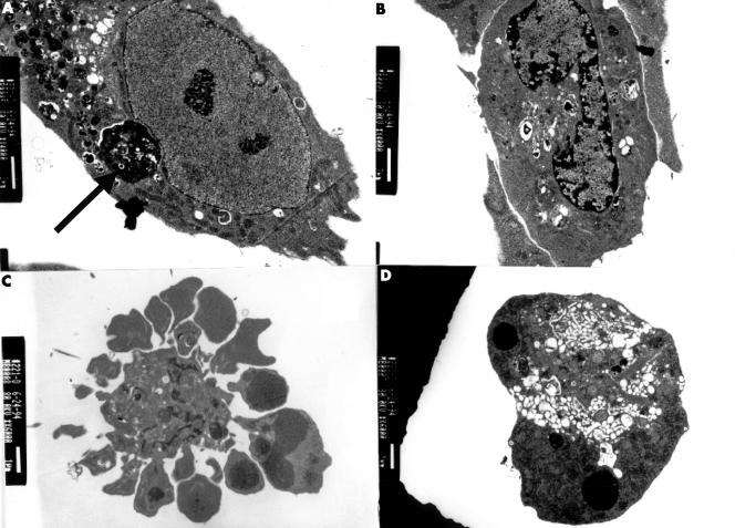
Electron microscopic appearances of a human vascular smooth muscle cell (VSMC) undergoing apoptosis in culture. (A) Normal appearance of a human VSMC. VSMC also contains an apoptotic body (arrow). (B) Peripheral condensation of nuclear chromatin. (C) Intense membrane blebbing and vesicle formation in apoptosis, with condensation of the nuclear chromatin into clumps. (D) An apoptotic body, the end product of apoptosis.
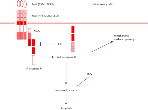
Schematic of Fas death signalling pathways. Fas, the prototypic member of the tumour necrosis factor (TNF) death receptor family, binds to its cognate ligand. Recruitment of the adapter molecule FADD and pro-caspase 8 results in activation of the latter. Caspase 8 activation directly activates downstream caspases, (3, 6, and 7) which results in DNA fragmentation and cleavage of cellular proteins. This pathway is thought to occur in type I cells and does not involve mitochondrial pathways. Caspase 8 activation also results in cleavage of Bid, which translocates and interacts with other Bcl-2 family members (see fig 3).
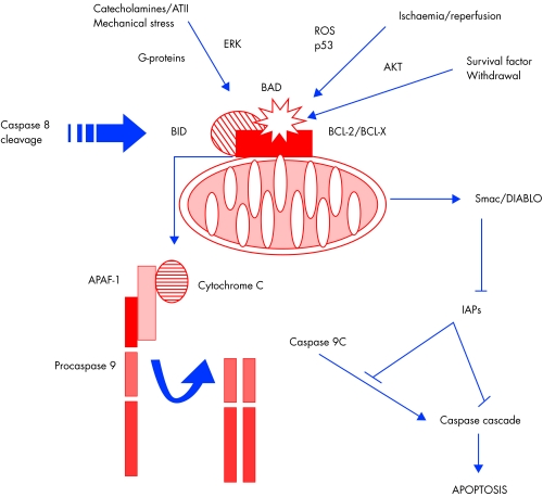
Schematic of mitochondrial death signalling pathways. Anti-apoptotic members of the Bcl-2 family, such as Bcl-2 and Bcl-X, are located on the mitochondrial outer membrane. Here they act to prevent the release of apoptogenic factors from the inner mitochondrial space. Binding of the pro-apoptotic proteins Bid (after cleavage by caspase 8) or Bad (after dephosphorylation) to Bcl-2 mitigates the protective effect of Bcl-2 and triggers release of cytochrome c and Smac/DIABLO. Cytochrome c, in concert with the adapter protein apaf-1 and caspase 9, activates caspase 3 and the downstream caspase cascade. Smac/DIABLO inhibits IAPs (inhibitor of apoptosis proteins), which in turn inhibit caspase activities, thus propagating apoptosis. Stimuli such as growth factor withdrawal or activation of p53 and Fas activation in type II cells act through this mitochondrial pathway.
References
-
- Kerr JF, Wyllie AH, Currie AR. Apoptosis: a basic biological phenomenon with wide-ranging implications in tissue kinetics. Br J Cancer 1972;26:239–57. ▸ The original (morphological) description of apoptosis. The features described are characteristic also of vascular cells, and morphological characterisation remains the “gold standard” for detecting apoptosis. - PMC - PubMed
-
- Saraste A, Pulkki K, Kallajoki M, et al. Apoptosis in human acute myocardial infarction. Circulation 1997;95:320–3. ▸ Detailed description of the timing and spatial characteristics of apoptosis and necrosis after human myocardial infarction. - PubMed
-
- Kajstura J, Cheng W, Reiss K, et al. Apoptotic and necrotic myocyte cell deaths are independent contributing variables of infarct size in rats. Lab Invest 1996;74:86–107. - PubMed
-
- Narula J, Haider N, Virmani R, et al. Apoptosis in myocytes in end-stage heart failure. N Engl J Med 1996;335:1182–9. ▸ This study (and reference 6 below) describe the evidence of cardiomyocyte apoptosis in end stage heart failure in humans, although the quantification of apoptotic index is both studies is now considered impossibly high. - PubMed
Publication types
MeSH terms
LinkOut - more resources
Full Text Sources
Other Literature Sources