. 2002 May 14;99(10):7049-53.
doi: 10.1073/pnas.062160399. Epub 2002 May 7.

Mapping the lethal factor and edema factor binding sites on oligomeric anthrax protective antigen

Affiliations

Mapping the lethal factor and edema factor binding sites on oligomeric anthrax protective antigen

Kristina Cunningham et al. Proc Natl Acad Sci U S A. .

Abstract

Assembly of anthrax toxin complexes at the mammalian cell surface involves competitive binding of the edema factor (EF) and lethal factor (LF) to heptameric oligomers and lower order intermediates of PA(63), the activated carboxyl-terminal 63-kDa fragment of protective antigen (PA). We used sequence differences between PA(63) and homologous PA-like proteins to delineate a region within domain 1' of PA that may represent the binding site for these ligands. Substitution of alanine for any of seven residues in or near this region (R178, K197, R200, P205, I207, I210, and K214) strongly inhibited ligand binding. Selected mutations from this set were introduced into two oligomerization-deficient PA mutants, and the mutants were used in various combinations to map the single ligand site within dimeric PA(63). The site was found to span the interface between two adjacent subunits, explaining the dependence of ligand binding on PA oligomerization. The locations of residues comprising the site suggest that a single ligand molecule sterically occludes two adjacent sites, consistent with the finding that the PA(63) heptamer binds a maximum of three ligand molecules. These results elucidate the process by which the components of anthrax toxin, and perhaps other binary bacterial toxins, assemble into toxic complexes.


Figures
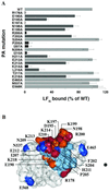
Figure 1
Figure 1
Effects of mutations on ligand binding by cell-bound PA. (A) PA-dependent cell binding of [35S]LFN. Trypsin-nicked wild type (WT)-PA or PA mutant (2.4 × 10⁻⁸ M) was incubated with CHO-K1 cells for 2 h on ice. Cells were washed twice with PBS, incubated for 2 h with [35S]LFN, and lysed. [35S]LFN content was determined by scintillation counting. Nonspecific binding of [35S]LFN to cells (less than 10%) was subtracted from the experimental measurements to determine specific binding. Bars are shaded black, medium gray, and gray to represent samples in which the mutation caused >90% loss of ligand binding, 25–75% loss of ligand binding, and little or no effect on ligand binding, respectively. The error bars represent SE of the mean. (B) Space-filling representation of monomeric PA63 showing the locations of mutated residues. Domain 1′ is colored blue, and domains 2 and 3 are colored gray. Domain 4 was omitted for clarity. The sevenfold axis of symmetry of heptameric PA63 is indicated by a black dot. Residues where mutation caused >90% loss of ligand binding are colored red, those where an intermediate reduction (25–75%) was seen are colored orange, and those where little or no effect was seen are colored dark blue. This figure was generated with MOLSCRIPT (29) and RASTER 3D (30).
Figure 2
Figure 2
PA-dependent cell binding of ligand to PA63 dimer mutants. Trypsin-nicked ligand-binding mutants and their complementary oligomerization-deficient PA mutant pair (1.2 × 10⁻⁸ M of each) were mixed and incubated with CHO-K1 cells for 2 h on ice. Cells were washed twice with PBS and incubated for 2 h with [35S]LFN. Cells were washed and lysed, and the bound [35S]LFN content was determined by scintillation counting. Nonspecific binding of [35S]LFN to cells (less than 10%) was subtracted from the experimental measurements to determine specific binding. Bars shaded gray represent samples in which the ligand-binding mutation was in PA–D512K, and those shaded black represent samples in which the ligand-binding mutation was in PA–K199E/R468A/R470D. The error bars represent SE of the mean. WT, wild type.
Figure 3
Figure 3
Ligand sites on heptameric PA63. (A) A ribbon model of domain 1′ two neighboring PA63 subunits. Residues that participate in ligand binding are highlighted in yellow, red, and green. Residues in red form a complete ligand-binding site spanning the dimer interface. The figure was created with MOLSCRIPT (29) and RASTER 3D (30). The green spheres represent the two Ca2+ in domain 1′. (B) Three sequential ligand-binding sites, each spanning a subunit–subunit interface, are shown on the PA63 heptamer in yellow, red, and green. The heavy black lines mark the boundaries between domain 1′ of the PA63 subunits. Domain 1′ (blue) and domains 2 and 3 (gray) are shown, and domain 4 was omitted for clarity. The figure was created with the INSIGHT(R) II molecular modeling system.

References

    1. Leppla S H. In: Bacterial Toxins and Virulence Factors in Disease. Moss J, Iglewski B, Vaughan M, Tu A T, editors. New York: Dekker; 1995. pp. 543–572.
    1. Leppla S H. Proc Natl Acad Sci USA. 1982;79:3162–3166. - PMC - PubMed
    1. Confer D L, Eaton J W. Science. 1982;217:948–950. - PubMed
    1. Pellizzari R, Guidi-Rontani C, Vitale G, Mock M, Montecucco C. FEBS Lett. 1999;462:199–204. - PubMed
    1. Duesbery N S, Webb C P, Leppla S H, Gordon V M, Klimpel K R, Copeland T D, Ahn N G, Oskarsson M K, Fukasawa K, Paull K D, Woude G F V. Science. 1998;280:734–737. - PubMed

Publication types

LinkOut - more resources