. 2002 May 14;99(10):7054-9.
doi: 10.1073/pnas.092152699. Epub 2002 May 7.

Rescue of a pathogenic Marek's disease virus with overlapping cosmid DNAs: use of a pp38 mutant to validate the technology for the study of gene function

Affiliations

Rescue of a pathogenic Marek's disease virus with overlapping cosmid DNAs: use of a pp38 mutant to validate the technology for the study of gene function

Sanjay M Reddy et al. Proc Natl Acad Sci U S A. .

Abstract

Marek's disease virus (MDV) genetics has lagged behind that of other herpesviruses because of the lack of tools for the introduction of site-specific mutations into the genome of highly cell-associated oncogenic strains. Overlapping cosmid clones have been successfully used for the introduction of mutations in other highly cell-associated herpesviruses. Here we describe the development of overlapping cosmid DNA clones from a very virulent oncogenic strain of MDV. Transfection of these cosmid clones into MDV-susceptible cells resulted in the generation of a recombinant MDV (rMd5) with biological properties similar to the parental strain. To demonstrate the applicability of this technology for elucidation of gene function of MDV, we have generated a mutant virus lacking an MDV unique phosphoprotein, pp38, which has previously been associated with the maintenance of transformation in MDV-induced tumor cell lines. Inoculation of Marek's disease-susceptible birds with the pp38 deletion mutant virus (rMd5 Delta pp38) revealed that pp38 is involved in early cytolytic infection in lymphocytes but not in the induction of tumors. This powerful technology will speed the characterization of MDV gene function, leading to a better understanding of the molecular mechanisms of MDV pathogenesis. In addition, because Marek's disease is a major oncogenic system, the knowledge obtained from these studies may shed light on the oncogenic mechanisms of other herpesviruses.


Figures

Figure 1
Figure 1
Organization of serotype 1 MDV viral genome. (A) The MDV genome consists of a unique long (UL) region flanked by inverted repeats, terminal repeat long (TRL), internal repeat long (IRL), and a unique short region (US) also flanked by two inverted repeats, internal repeat short (IRS) and terminal repeat short (TRS). (B) Schematic representation of the overlapping clones generated to reconstitute an infectious virus from a very virulent (vv) strain of MDV (Md5). The restriction enzymes used to generate the cosmid clones and their positions are indicated. (C) Location of EcoRI restriction sites in cosmid A6 used to delete the MDV-specific pp38 gene.
Figure 2
Figure 2
Southern blot analysis of DNA from wild-type Md5 (lane 1), recombinant rMd5 (lane 2), and pp38 deletion mutants, rMd5Δpp38-19 (lane 3) and rMd5Δpp38-24 (lane 4). Viral DNA isolated from nucleocapsid preparations was digested with EcoRI and probed with all five radiolabeled cosmids (A) or a pp38 probe (B). The pp38 probe hybridizes to the pp38- and pp24-containing band, because both genes share 195 nucleotides. The size of the DNA markers is indicated in kb.
Figure 3
Figure 3
In vitro growth properties of Md5, rMd5, and rMd5Δpp38. DEFs were infected with the indicated viruses, and infected cells were harvested on days 1, 2, 3, 4, and 5 after infection and titered on fresh DEF. Day 0 indicates the titer of the virus in the inoculum. The experiment was performed in duplicate, and the titer (logarithm of the mean number of plaque-forming units per dish) is indicated.
Figure 4
Figure 4
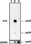
Immunoprecipitation analysis of DEFs uninfected (lane 1) and infected with recombinant rMd5 (lane 2) and pp38 deletion mutant rMd5Δpp38-19 (lane 3). Immunoprecipitation of [35S]methionine-labeled cells was done with mAbs specific to pp38 (H19; Top) or pp40 (2BN90; Bottom). H19 not only immunoprecipitates pp38 but also coprecipitates pp24 in rMd5-infected cells. As expected, in the absence of pp38 (rMd5Δpp38), pp24 is not precipitated by H19. On the other hand, 2BN90 immunoprecipitates pp40 in both virus lysates.
Figure 5
Figure 5
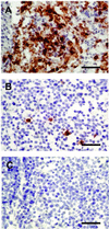
Immunohistochemical analysis of bursa of Fabricius. MDV maternal Ab-negative chickens were inoculated with rMd5 (A), rMd5Δpp38 (B), or mock-inoculated (C). Bursa of Fabricius were harvested 6 days after inoculation and stained with glycoprotein B mAb. Antigen expression is severely impaired in rMd5Δpp38, showing that pp38 is involved in early cytolytic infection in lymphocytes. (Bar = 100 μm.)

References

    1. Churchill A E, Biggs P M. Nature (London) 1967;215:528–530. - PubMed
    1. Nazerian K, Solomon J J, Witter R L, Burmester B R. Proc Soc Exp Biol Med. 1968;127:177–182. - PubMed
    1. Calnek B W, Witter R L. In: Diseases of Poultry. Calnek B W, Barnes H J, Beard C W, McDougald L R, Saif Y M, editors. Ames, IA: Iowa State Univ. Press; 1997. pp. 369–413.
    1. Churchill A E, Chubb R C, Baxendale W. J Gen Virol. 1969;4:557–564. - PubMed
    1. Okazaki W, Purchase H G, Burmester B R. Avian Dis. 1970;14:413–429. - PubMed

LinkOut - more resources