. 2002 Jun;22(11):3610-20.
doi: 10.1128/MCB.22.11.3610-3620.2002.

mtCLIC/CLIC4, an organellular chloride channel protein, is increased by DNA damage and participates in the apoptotic response to p53

Affiliations

mtCLIC/CLIC4, an organellular chloride channel protein, is increased by DNA damage and participates in the apoptotic response to p53

Ester Fernández-Salas et al. Mol Cell Biol. 2002 Jun.

Abstract

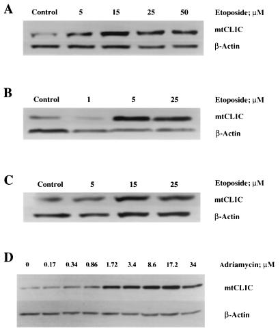
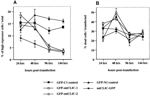
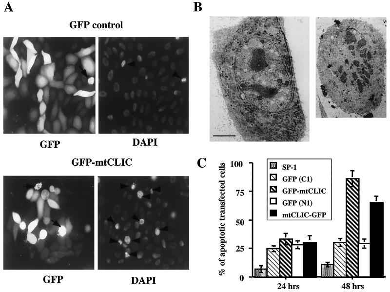
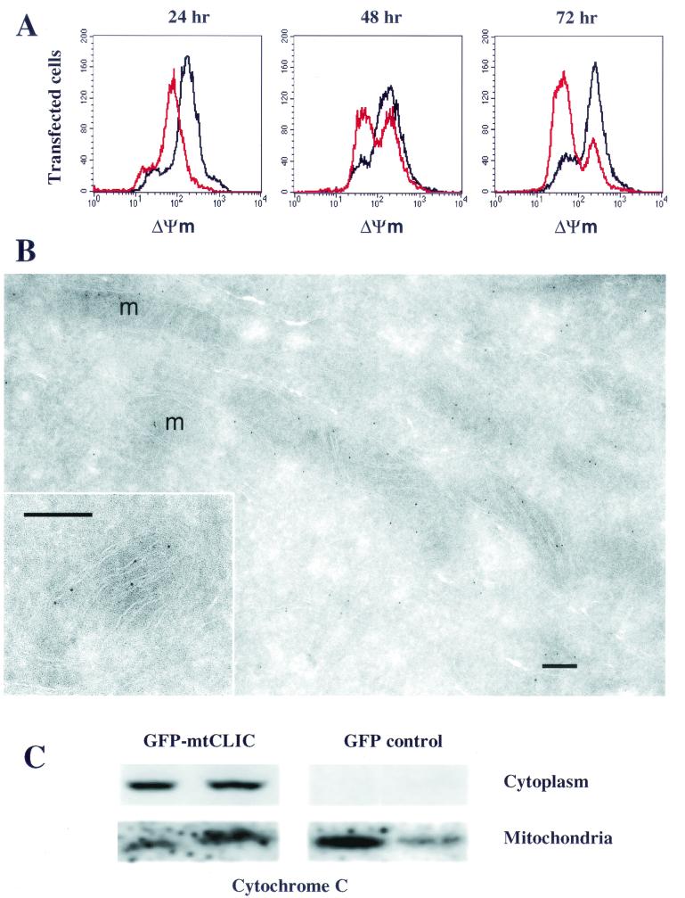
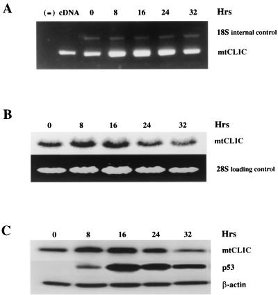
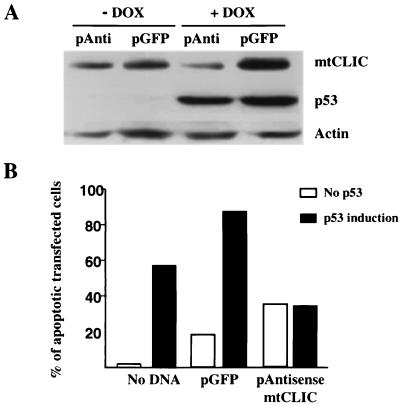
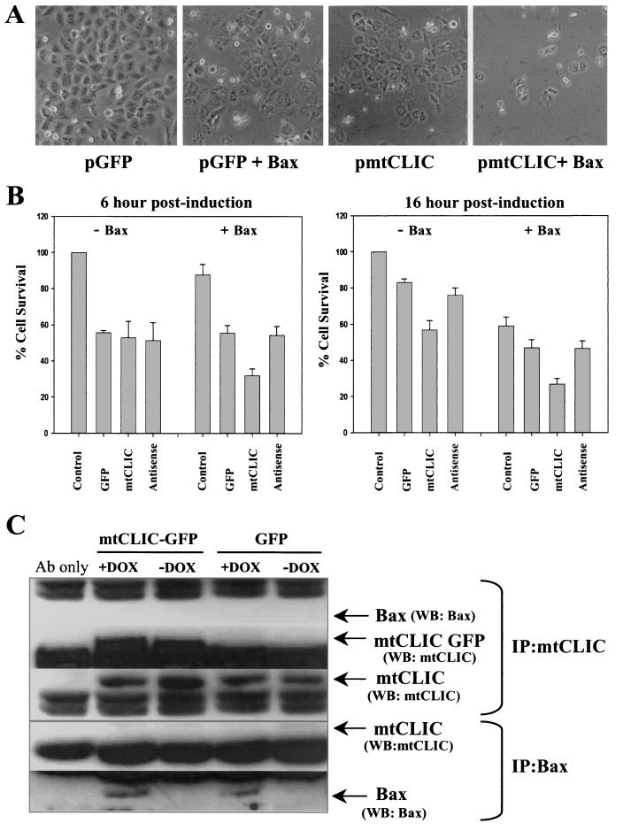
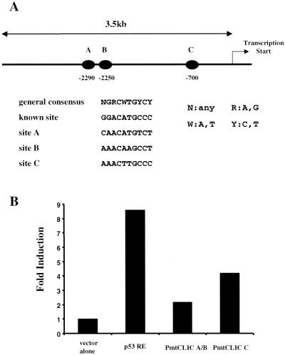
mtCLIC/CLIC4 (referred to here as mtCLIC) is a p53- and tumor necrosis factor alpha-regulated cytoplasmic and mitochondrial protein that belongs to the CLIC family of intracellular chloride channels. mtCLIC associates with the inner mitochondrial membrane. Dual regulation of mtCLIC by two stress response pathways suggested that this chloride channel protein might contribute to the cellular response to cytotoxic stimuli. DNA damage or overexpression of p53 upregulates mtCLIC and induces apoptosis. Overexpression of mtCLIC by transient transfection reduces mitochondrial membrane potential, releases cytochrome c into the cytoplasm, activates caspases, and induces apoptosis. mtCLIC is additive with Bax in inducing apoptosis without a physical association of the two proteins. Antisense mtCLIC prevents the increase in mtCLIC levels and reduces apoptosis induced by p53 but not apoptosis induced by Bax, suggesting that the two proapoptotic proteins function through independent pathways. Our studies indicate that mtCLIC, like Bax, Noxa, p53AIP1, and PUMA, participates in a stress-induced death pathway converging on mitochondria and should be considered a target for cancer therapy through genetic or pharmacologic approaches.


Figures

FIG. 1.
FIG. 1.
mtCLIC protein is upregulated during etoposide- or adriamycin-induced apoptosis. Keratinocytes were treated with different concentrations of etoposide or adriamycin for 24 h. Protein samples were collected for immunoblot analysis for mCLIC, and β-actin was used as a loading control. Data are representative of three separate experiments for etoposide and one experiment for adriamycin. (A and D) Normal keratinocytes (S1 cell line). (B) Neoplastic keratinocytes (SP1 papilloma cell line). (C) p53-null keratinocytes (AK1b cell line).
FIG. 2.
FIG. 2.
Overexpression of mtCLIC in cultured keratinocytes is lethal. Plasmid constructs encoding GFP, GFP-mtCLIC, or mtCLIC-GFP were transiently transfected into SP1 cells. Attached cells were trypsinized 24, 48, 96, and 144 h after transfection; combined with cells that had detached during these times; and incubated with PI. The level of green and red fluorescence was quantitated by FACS analysis. At least 10,000 transfected cells were collected for each quantitation and analyzed without fixation. Values are for duplicate dishes at each time point. Data are representative of two independent experiments. Error bars show standard deviations. GFP-mtCLIC-1 and GFP-mtCLIC-2 were independently constructed plasmids encoding identical sequences. (A) High-expression cells. Cells were classified as high expressors when the amount of green fluorescence was 2 log units higher than that of the cells with the lowest level of expression. Each population shown was quantitated at the indicated times. (B) Dead transfected cells. The percentage of transfected cells that incorporated PI was quantitated by gating the transfected cell population and analyzing green and red fluorescence. PI was added to the suspension of unfixed cells analyzed over 144 h after transfection of GFP-only or GFP fusion plasmids.
FIG. 3.
FIG. 3.
mtCLIC overexpression induces apoptosis in keratinocytes. (A) SP1 keratinocytes were transfected with GFP or GFP-mtCLIC; 48 h later, cells were fixed in neutral buffered formalin and stained with DAPI. Samples were analyzed in a fluorescence microscope. Many cells expressing high levels of mtCLIC displayed morphological features characteristic of apoptotic cells, including cell rounding, condensed nuclei (arrowheads), and bleb formation from the membrane (arrows). A few condensed nuclei were also detected in GFP-transfected cells (GFP control). (B) Transmission electron micrographs of thin sections through a normal S1 keratinocyte transfected with the GFP vector (left panel) or a typical apoptotic S1 cell transfected with mtCLIC-GFP (right panel) and fixed after 48 h. Apoptotic cells are smaller and have condensed and fragmented nuclei, reduced or absent tonofilaments, and ruffling of the plasma membrane. Bar = 5 μm. (C) Apoptotic cells were detected by the presence of annexin V on their membranes. SP1 keratinocytes transfected with GFP control plasmids or mtCLIC-GFP or GFP-mtCLIC fusion proteins were collected 24 and 48 h after transfection. Cells were labeled with annexin V antibody as described in Materials and Methods and analyzed by FACS analysis. A minimum of 20,000 transfected cells were collected for each sample analyzed. The green/transfected population was gated, and the percentage of annexin V-positive cells was calculated. Duplicate dishes were analyzed. The data are representative of two independent experiments; error bars show standard deviations.
FIG. 4.
FIG. 4.
mtCLIC induces apoptosis through dissipation of mitochondrial membrane potential (ΔΨm) and release of cytochrome c. (A) SP1 keratinocytes were transfected with plasmids encoding GFP (black line) or GFP-mtCLIC (red line); 24, 48, and 72 h after transfection, cells were trypsinized and stained with Mitotracker to measure mitochondrial membrane potential. A minimum of 30,000 cells were analyzed by flow cytometry. Data were analyzed by gating the transfected cell population and plotting red fluorescence. Graphs are representative of four separate transfections. (B) Cryosections of HACAT cells were labeled with an affinity-purified antibody against the C-terminal domain of mtCLIC and visualized with protein A-gold. Label is concentrated inside mitochondria. At a higher magnification (inset), gold particles are seen in close association with the inner membrane and cristae. m, mitochondrion. Bar = 0.2 μm. (C) SP1 keratinocytes transfected with GFP or GFP-mtCLIC were collected 48 h after transfection. Cells were fractionated into mitochondrial and cytoplasmic fractions as described previously (15). Samples were analyzed for cytochrome c localization by Western blotting. Each lane represents results from an independent transfection and fractionation experiment.
FIG. 5.
FIG. 5.
Overexpression of p53 upregulates mtCLIC and induces apoptosis. p53 Tet-On Saos-2 cells were treated with doxycycline, and the time course of mtCLIC mRNA induction was determined by RT-PCR detection with primers for mtCLIC RNA and mouse 18S RNA as an internal control (A) or by Northern blotting (B). The negative control (lane −) in panel A contained no DNA, and the positive control (lane cDNA) contained mtCLIC cDNA. (C) p53 Tet-On Saos-2 cells were treated with doxycycline to induce p53 expression and apoptosis. Protein samples were collected at different times after induction and analyzed by Western blotting with β-actin as a loading control.
FIG. 6.
FIG. 6.
mtCLIC activity is an important component of p53-induced apoptosis. (A) Western blot of mtCLIC, p53, and β-actin from lysates of p53 Tet-On Saos-2 cells transfected with the GFP spectrum plasmid (pGFP) or the antisense plasmid (pAnti) for mtCLIC and treated with doxycycline (DOX) to induce p53 expression. Upregulation of mtCLIC but not p53 was blocked by the antisense plasmid. (B) p53 Tet-On Saos-2 cells were transfected with the GFP spectrum plasmid alone or in combination with the antisense mtCLIC plasmid. After 24 h, transfected cells were treated with doxycycline. Cells were collected 24 h later by trypsinization, fixed overnight in 70% ethanol, and stained with PI. DNA content was analyzed by flow cytometry, and sub-G1 cells were quantified by gating green fluorescent cells. A minimum of 10,000 transfected cells were analyzed for each sample. Results shown are representative of three independent experiments.
FIG. 7.
FIG. 7.
mtCLIC cooperates with Bax in the induction of apoptosis without a physical association. The Bax Tet-On Saos-2 cell line was transfected with plasmids encoding the GFP spectrum alone or in combination with either sense mtCLIC or antisense mtCLIC. At 24 h after transfection, cells were treated with doxycycline to induce Bax expression. Cells were analyzed at different time points after Bax induction. (A) Bax or mtCLIC individually induced similar levels of apoptosis, but viable cells were rare in cultures coexpressing Bax and mtCLIC. Photographs shown were taken 22 h after Bax induction and are representative of three independent transfections. (B) Cell survival in the transfected cells was assessed by the MTT assay at 6 h (left panel) and 16 h (right panel) after Bax induction; survival for untransfected-uninduced samples was set at 100%. Data are the means of two independent experiments performed in duplicate. Error bars indicate standard deviations. (C) Immunoprecipitation (IP) and Western blotting (WB) were carried out with transfected cells and affinity-purified antibody (Ab) to mtCLIC or antibody DO-1, which recognizes a p53 sequence tag in the induced Bax protein. mtCLIC and Bax were immunoprecipitated independently, even in the samples overexpressing both proteins, but coimmunoprecipitation was not detected. DOX, doxycycline.
FIG. 8.
FIG. 8.
The region upstream of the human mtCLIC gene contains functional p53-binding sites. (A) Schematic localization of putative p53-binding sites (solid ovals) in a 3.5-kb region upstream of the human mtCLIC transcription start site. Sequence analysis indicates that these sites are more than 90% homologous to known p53-binding consensus sequences. (B) Fragments encompassing elements A and B (PmtCLIC A/B) and C (PmtCLIC C) were cloned into the pSEAP-basic reporter vector system along with a p53 response element (p53 RE) (positive control) (28) and transfected into p53 Tet-On Saos-2 cells. After 12 h, doxycycline was added to the culture medium, and medium was collected 24 h later and assayed for SEAP activity. Results represent the fold increase over the results obtained with the vector alone after subtraction of values from duplicate transfected cultures not induced by doxycycline. All values are corrected for cell numbers. Comparable results were obtained in three separate transfection experiments.

References

    1. Adams, J. M., and S. Cory. 1998. The Bcl-2 protein family: arbiters of cell survival. Science 281:1322-1326. - PubMed
    1. Allen, R. T., W. J. Hunter, I. I. I., and D. K. Agrawal. 1997. Morphological and biochemical characterization and analysis of apoptosis. J. Pharmacol. Toxicol. Methods 37:215-228. - PubMed
    1. Attardi, L. D., E. E. Reczek, C. Cosmas, E. G. Demicco, M. E. McCurrach, S. W. Lowe, and T. Jacks. 2000. PERP, an apoptosis-associated target of p53, is a novel member of the PMP-22/gas3 family. Genes Dev. 14:704-718. - PMC - PubMed
    1. Azzoli, C. G., M. Sagar, A. Wu, D. Lowry, H. Hennings, D. L. Morgan, and W. C. Weinberg. 1998. Cooperation of p53 loss of function and v-Ha-ras in transformation of mouse keratinocyte cell lines. Mol. Carcinog. 21:50-61. - PubMed
    1. Bates, S., and K. H. Vousden. 1996. p53 in signaling checkpoint arrest or apoptosis. Curr. Opin. Genet. Dev. 6:12-18. - PubMed

MeSH terms