. 2002 Jun;22(11):3717-28.
doi: 10.1128/MCB.22.11.3717-3728.2002.

Cyclic AMP blocks cell growth through Raf-1-dependent and Raf-1-independent mechanisms

Affiliations

Cyclic AMP blocks cell growth through Raf-1-dependent and Raf-1-independent mechanisms

Nicolas Dumaz et al. Mol Cell Biol. 2002 Jun.

Abstract

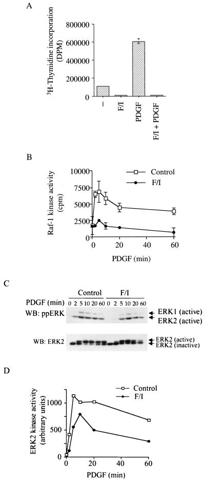
It is widely accepted that cyclic AMP (cAMP) can block cell growth by phosphorylating Raf-1 on serine 43 and inhibiting signaling to extracellular signal-regulated protein kinase. We show that the suppression of Raf-1 by cAMP is considerably more complex than previously reported. When cellular cAMP is elevated, Raf-1 is phosphorylated on three residues (S43, S233, and S259), which work independently to block Raf-1. Both Ras-dependent and Ras-independent processes are disrupted. However, when cAMP-insensitive versions of Raf-1 are expressed in NIH 3T3 cells, their growth is still strongly suppressed when cAMP is elevated. Thus, although Raf-1 appears to be an important cAMP target, other pathways are also targeted by cAMP, providing alternative mechanisms that lead to suppression of cell growth.


Figures

FIG. 1.
FIG. 1.
Activity and phosphorylation of Raf-1 in NIH 3T3 cells. (A) mRaf-1 kinase activity. mRaf-1 (Raf) or mA43Raf-1 (A43) was transiently expressed in NIH 3T3 cells as indicated. The cells were serum starved (24 h) and left untreated or treated with 8-bromo-cAMP (cAMP) (1 mM) as indicated. Ten minutes later, the cells were treated with PDGF (10 min) as indicated, cell extracts were prepared, and Raf kinase activity was measured using 9E10 to capture the myc-epitope-tagged proteins. Background counts (∼5,800 cpm) were subtracted. The results are for one experiment assayed in triplicate, with error bars representing standard deviations. Similar results were obtained in two independent experiments. (B) Phosphorylation of endogenous Raf-1. NIH 3T3 cells were serum starved (24 h) and left untreated (Control) or pretreated with forskolin and IBMX (F/I) as indicated. Ten minutes later, the cells were treated with PDGF for the indicated times and cell extracts were prepared. Endogenous Raf-1 was immunoprecipitated with polyclonal antibody C20 for blotting with phospho-specific antibodies to S43 (p43; upper panel), S338 (p338; second panel) and S259 (p259; third panel). Blots were reprobed with monoclonal antibody M40091.G to reveal total Raf-1, and one representative reprobed blot is shown (lower panel). Similar results were obtained in three independent experiments. WB, Western blot.
FIG. 2.
FIG. 2.
Activity and phosphorylation of Raf-1 in COS cells. (A) Phosphorylation of endogenous Raf-1. COS cells were serum starved (24 h) and left untreated (Control) or pretreated with forskolin and IBMX (F/I) as indicated. Ten minutes later, the cells were treated with EGF for the indicated times and cell extracts were prepared. Endogenous Raf-1 was immunoprecipitated with polyclonal antibody C20 for blotting with phospho-specific antibodies to S43 (p43; upper panel), S338 (p338; second panel) and S259 (p259; third panel). Western blots (WB) were reprobed with monoclonal antibody M40091.G to reveal total Raf-1, and one representative reprobed blot is shown (lower panel). Similar results were obtained in three independent experiments. (B) mRaf-1 kinase activity. COS cells were transiently transfected with mRaf-1 (Raf), mA43Raf-1 (A43), mA259Raf-1 (A259), and mA43, A259Raf-1 (A43, A259). The cells were serum starved (for 24 h) and left untreated or pretreated with forskolin and IBMX (F/I). Ten minutes later, the cells were treated with EGF (20 min) as indicated and extracts were prepared. The kinase activity of the mRaf-1 proteins was measured using 9E10 monoclonal antibody to capture the myc-tagged proteins. Background counts (∼5,000 cpm) were subtracted; the results are for one experiment assayed in triplicate, with error bars to indicate standard deviations from the mean. Similar results were obtained in two independent assays.
FIG. 3.
FIG. 3.
Role of Ras in cAMP-mediated suppression of Raf-1. (A and B) Activation of endogenous Ras. NIH 3T3 cells (A) or COS cells (B) were serum starved (for 24 h) and left untreated (Control) or pretreated with forskolin and IBMX (F/I). Ten minutes later, the cells were treated with PDGF (A) or EGF (B) as indicated, and cell extracts were prepared at the indicated times for Ras activation assays. Total Ras protein in 10% of the extracts is shown in the upper panels, and activated Ras (Ras.GTP) is shown in the lower panels. Similar results were obtained in two independent assays. (C) Ras-Raf-1 binding. Serum-starved COS cells were treated as described for panels A and B, and extracts were prepared for Ras-Raf-1 binding assays. The levels of Raf-1 and Ras in 10% of the cell extracts are shown in the upper two panels and the immunoprecipitated Ras is shown in the lower panel. Raf-1 coprecipitated with Ras.GTP is shown in the third panel. Raf-1 was revealed with antibody M40091.G, and Ras was revealed with the pan-Ras antibody. Similar results were obtained in two independent experiments. WB, Western blot.
FIG. 4.
FIG. 4.
Membrane-targeted Raf-1 is still subject to cAMP-mediated suppression. mRaf-1 (Raf), m89LRafCAAX (89L-CX), and mΔRafCAAX (Δ-CX) were transiently transfected into COS cells and treated as described for Fig. 2B, for the Raf-1 kinase assay. The results are for one experiment assayed in triplicate, with error bars indicating standard deviations from the mean. Similar results were obtained in two independent experiments.
FIG. 5.
FIG. 5.
Binding of antibody R19120 to Raf-1. (A) Binding to endogenous Raf-1 protein. NIH 3T3 cells were treated as described in Fig. 1B, and Western blots (WB) were performed using monoclonal antibodies R19120 (upper panel) or M40091.G (lower panel). Similar results were obtained in at least three independent experiments. (B) Details of the R19120 epitope. The sequence of human Raf-1, amino acids 228 to 241, is shown. The R19120 minimal epitope is indicated by the open box, and the PKA consensus and the sequence of synthetic peptide Raf228-238 are indicated by the short and long lines, respectively. S233 is highlighted in the oblong, and the potential chymotrypsin cleavage sites are indicated by the arrowheads. (C) Effects of mutants on R19120 binding. COS cells transiently expressing mRaf-1 (Raf) or mA233Raf-1 (A233) were serum starved (for 24 h) and left untreated (C) or treated with forskolin and IBMX (F/I) (10 min). Myc-epitope-tagged proteins were immunoprecipitated with 9E10 and immunoblotted with R19120 (upper panel) or M40091.G (lower panel). Vector, vector control transfection. Similar results were obtained in two independent experiments.
FIG. 6.
FIG. 6.
S233 is phosphorylated in vitro by PKA and in vivo following F/I treatment. (A) Two-dimensional phosphopeptide mapping. COS cells were transfected with mRaf-1 (Raf-1) or mA233Raf-1 (A233) and labeled with [32P]orthophosphate. Cells were either untreated or treated for 10 min with forskolin and IBMX (F/I), and mRaf-1 proteins were immunoprecipitated for two-dimensional phosphopeptide mapping analysis. For each sample, ∼1,000 cpm (Cerenkov counting) were loaded, except for the mix, where ∼2,000 cpm were loaded. The peptides whose phosphorylation was enhanced by F/I treatment are indicated with arrows, and the peptides absent in the map from mA233Raf-1 are indicated with open arrows. A scheme of the peptide identity is presented, with peptides being labeled from A to G. O, origin. Similar results were obtained in two experiments. (B) Phosphorylation of Raf228-238 by PKA. The peptides Raf228-238 and pRaf228-238 were incubated with PKA for in vitro phosphorylation, followed by digestion with chymotrypsin as indicated, and separated by electrophoresis at pH 1.9 on phospho-cellulose plates. O, origin. Similar results were obtained in two experiments. (C) Two-dimensional phosphopeptide mapping. Upper panel, phosphopeptide map of Raf-1 treated with forskolin and IBMX as described for panel A (∼2,000 cpm). Middle panel, the PKA phosphorylated chymotryptic fragment of peptide Raf228-238 from panel B (peptide; ∼100 cpm). Lower panel, mixture of Raf-1 treated with forskolin and IBMX (∼2,000 cpm) and the fragment from Raf228-238 (∼100 cpm). The peptides which comigrate are indicated by an arrow. (D) Phosphoamino acid analysis. The peptides indicated by the arrows in Fig. 6C above were subjected to phosphoamino acid analysis. The circles indicate the positions of migration of phosphoserine (P-S) and phosphothreonine plus phosphotyrosine (P-T/P-Y) standards. Pi, inorganic phosphate.
FIG. 6.
FIG. 6.
S233 is phosphorylated in vitro by PKA and in vivo following F/I treatment. (A) Two-dimensional phosphopeptide mapping. COS cells were transfected with mRaf-1 (Raf-1) or mA233Raf-1 (A233) and labeled with [32P]orthophosphate. Cells were either untreated or treated for 10 min with forskolin and IBMX (F/I), and mRaf-1 proteins were immunoprecipitated for two-dimensional phosphopeptide mapping analysis. For each sample, ∼1,000 cpm (Cerenkov counting) were loaded, except for the mix, where ∼2,000 cpm were loaded. The peptides whose phosphorylation was enhanced by F/I treatment are indicated with arrows, and the peptides absent in the map from mA233Raf-1 are indicated with open arrows. A scheme of the peptide identity is presented, with peptides being labeled from A to G. O, origin. Similar results were obtained in two experiments. (B) Phosphorylation of Raf228-238 by PKA. The peptides Raf228-238 and pRaf228-238 were incubated with PKA for in vitro phosphorylation, followed by digestion with chymotrypsin as indicated, and separated by electrophoresis at pH 1.9 on phospho-cellulose plates. O, origin. Similar results were obtained in two experiments. (C) Two-dimensional phosphopeptide mapping. Upper panel, phosphopeptide map of Raf-1 treated with forskolin and IBMX as described for panel A (∼2,000 cpm). Middle panel, the PKA phosphorylated chymotryptic fragment of peptide Raf228-238 from panel B (peptide; ∼100 cpm). Lower panel, mixture of Raf-1 treated with forskolin and IBMX (∼2,000 cpm) and the fragment from Raf228-238 (∼100 cpm). The peptides which comigrate are indicated by an arrow. (D) Phosphoamino acid analysis. The peptides indicated by the arrows in Fig. 6C above were subjected to phosphoamino acid analysis. The circles indicate the positions of migration of phosphoserine (P-S) and phosphothreonine plus phosphotyrosine (P-T/P-Y) standards. Pi, inorganic phosphate.
FIG. 7.
FIG. 7.
Raf-1 regulation by cAMP involves multiple phosphorylation events. (A) mRaf-1 kinase activity. mRaf-1 (Raf), mA233Raf-1 (A233), mA43,A233Raf-1 (A43, A233), mA233,A259Raf-1 (A233, A259) or mA43,A233,A259Raf-1 (A43,A233,A259) were transiently expressed in COS cells and treated as for Fig. 2B for the Raf-1 kinase assay. The results are for one experiment assayed in triplicate, with error bars indicating standard deviations from the mean. Similar results were obtained in two independent experiments. (B) S43 phosphorylation. mRaf-1 (Raf), mA233Raf-1 (A233), mA259Raf-1 (A259), mA233,A259Raf-1 (A233,A259) or mA43,A233,A259Raf-1 (A43,A233,A259) were transiently expressed in COS cells which were either untreated or treated for 10 min with forskolin and IBMX as indicated. An equal amount of myc-tagged Raf-1 protein was immunoprecipitated and probed with a phospho-specific antibody to S43. Similar results were obtained in two independent experiments.
FIG. 8.
FIG. 8.
cAMP regulates of Raf-1/ERK signaling and cell growth in NIH 3T3 cells. (A) DNA synthesis. NIH 3T3 cells were serum starved and left untreated or treated with forskolin and IBMX (F/I) as indicated. Ten minutes later, the cells were treated with PDGF as indicated and DNA synthesis was measured. The results presented are for one experiment assayed in triplicate with error bars representing standard deviations. Similar results were obtained in three independent experiments. (B) Endogenous Raf-1 activity. NIH 3T3 cells were serum starved (for 24 h) and left untreated (Control) or pretreated with forskolin and IBMX (F/I). Ten minutes later, the cells were treated with PDGF as indicated, and cell extracts were prepared at the indicated times. Endogenous Raf-1 was immunoprecipitated with M40091.G, and kinase activity was measured. The results presented are for one experiment assayed in triplicate with error bars representing standard deviations. Background counts (∼5,000 cpm) were subtracted, and similar results were obtained in three independent assays. (C) Phosphorylation of endogenous ERK. NIH 3T3 cells were serum starved (for 24 h) and left untreated (Control) or pretreated with forskolin and IBMX (F/I). Ten minutes later, the cells were stimulated with PDGF for the indicated times. ERK phosphorylation is shown in the upper blot, and total ERK is shown in the lower blot. Results are for one assay, and similar results were obtained in two independent experiments. WB, Western blot. (D) Endogenous ERK activity. The kinase activity of ERK2 was measured from the same samples as in panel C. Results are for one assay, and similar results were obtained in two independent experiments.
FIG. 9.
FIG. 9.
mA43,A233,A259Raf-1 rescues ERK activation but not cell proliferation. (A) Raf-1 kinase activity. NIH 3T3 cells stably expressing mRaf-1 (Raf; upper panel) or mA43,A233,A259Raf-1 (A43,A233,A259; lower panel) were treated as described for Fig. 8B, and extracts were prepared. Myc-tagged Raf proteins were immunoprecipitated using 9E10 for Raf kinase assay determination. The results are for one experiment assayed in triplicate, with error bars representing standard deviations from the mean. Similar results were obtained with two independently derived clones for each line, each assayed on two separate occasions. (B) Endogenous ERK activation. NIH 3T3 cells stably expressing mRaf-1 (Raf) or mA43,A233,A259Raf-1 (A43,A233,A259) were serum starved and treated with forskolin and IBMX (F/I) for 10 min followed by PDGF for a further 2 min as indicated. Extracts were prepared, and immunoblots were performed for ppERK (upper blot) or total ERK (lower blot). Similar results were obtained in two independent assays. WB, Western blot. (C) DNA synthesis. NIH 3T3 cells stably expressing mRaf-1 (Raf) or mA43,A233,A259Raf-1 (A43,A233,A259) were treated as described for Fig. 8A and analyzed for DNA synthesis. The results presented are for one experiment assayed in triplicate with error bars representing standard deviations. Similar results were seen with two individual clones of each line, each assayed at least twice.

References

    1. Abraham, D., K. Podar, M. Pacher, M. Kubicek, N. Welzel, B. A. Hemmings, S. M. Dilworth, H. Mischak, W. Kolch, and M. Baccarini. 2000. Raf-1-associated protein phosphatase 2A as a positive regulator of kinase activation. J. Biol. Chem. 275:22300-22304. - PubMed
    1. Ally, S., T. Clair, D. Katsaros, G. Tortora, H. Yokozaki, R. A. Finch, T. L. Avery, and Y. S. Cho-Chung. 1989. Inhibition of growth and modulation of gene expression in human lung carcinoma in athymic mice by site-selective 8-Cl-cyclic adenosine monophosphate. Cancer Res. 49:5650-5655. - PubMed
    1. Avruch, J., A. Khokhlatchev, J. M. Kyriakis, Z. Luo, G. Tzivion, D. Vavvas, and X. F. Zhang. 2001. Ras activation of the Raf kinase: tyrosine kinase recruitment of the MAP kinase cascade. Recent Prog. Horm. Res. 56:127-155. - PubMed
    1. Bos, J. L., J. de Rooij, and K. A. Reedquist. 2001. Rap1 signalling: adhering to new models. Nat. Rev. Mol. Cell Biol. 2:369-377. - PubMed
    1. Boyle, W. J., P. van der Geer, and T. Hunter. 1991. Phosphopeptide mapping and phosphoamino acid analysis by two-dimensional separation on thin-layer cellulose plates. Methods Enzymol. 201:110-149. - PubMed

Publication types