. 2002 Jun;22(11):3744-56.
doi: 10.1128/MCB.22.11.3744-3756.2002.

Induced expression and association of the Mona/Gads adapter and Gab3 scaffolding protein during monocyte/macrophage differentiation

Affiliations

Induced expression and association of the Mona/Gads adapter and Gab3 scaffolding protein during monocyte/macrophage differentiation

Caroline Bourgin et al. Mol Cell Biol. 2002 Jun.

Abstract

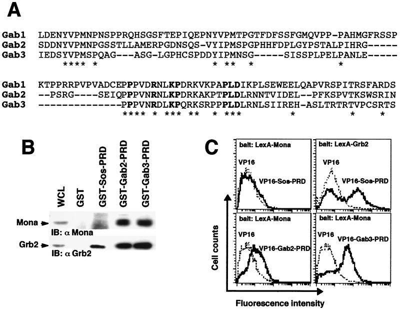
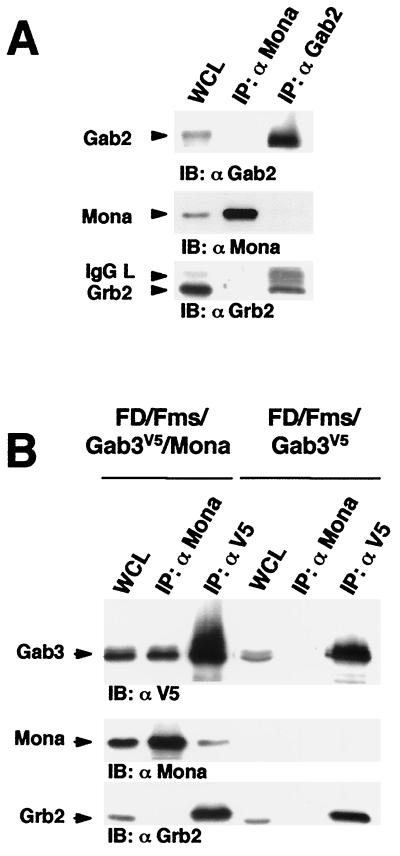
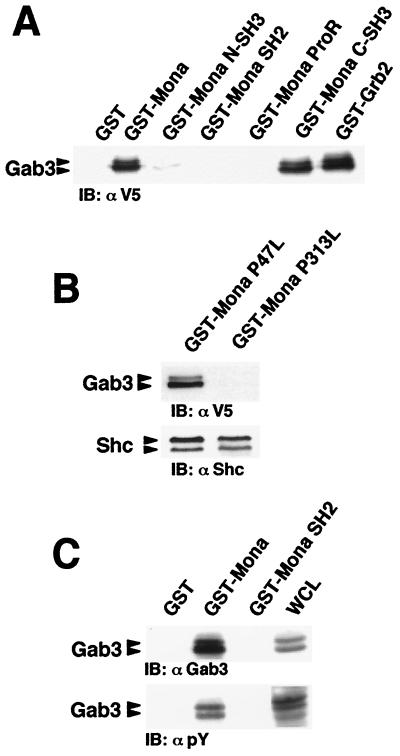
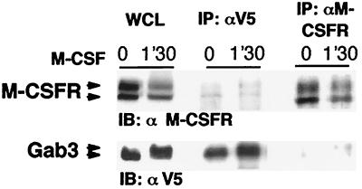
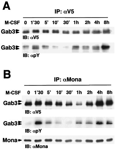
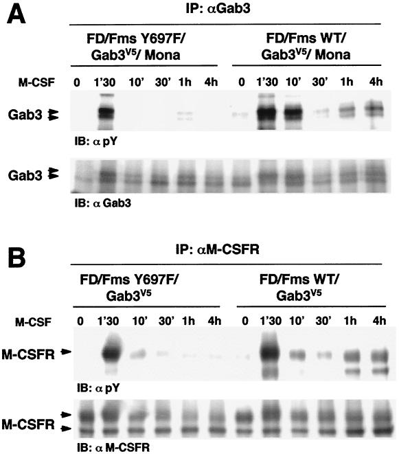
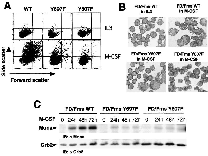
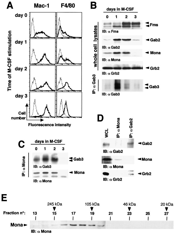
Mona/Gads is a Grb2-related, Src homology 3 (SH3) and SH2 domain-containing adapter protein whose expression is restricted to cells of hematopoietic lineage (i.e., monocytes and T lymphocytes). During monocyte/macrophage differentiation, Mona is induced and interacts with the macrophage colony-stimulating factor receptor, M-CSFR (also called Fms), suggesting that Mona could be involved in developmental signaling downstream of the M-CSFR by recruiting additional signaling proteins to the activated receptor. Our present results identify Mona as a specific partner protein for the DOS/Gab family member Gab3 in monocytic/macrophage development. Mona does not interact with Gab2; however, Gab3 also forms a complex with the Mona-related adapter Grb2. Glutathione S-transferase pull-down experiments demonstrate that the Mona and Gab3 interaction utilizes the carboxy-terminal SH3 domain of Mona and the atypical proline-rich domain of Gab3. Mona is known to interact with the phosphorylated Y697 site of the M-CSFR. The M-CSFR mutation Y697F exhibited qualitative and quantitative abnormalities in receptor and Gab3 tyrosine phosphorylation, and Mona induction was greatly reduced. The Y807F M-CSFR mutation is defective in differentiation signaling, but not growth signaling, and also fails to induce Mona protein expression. During M-CSF-stimulated macrophage differentiation of mouse bone marrow cells, Mona and Gab3 expression is coinduced, these proteins interact, and Mona engages in multimolecular complexes. These data suggest that association of Mona and Gab3 plays a specific role in mediating the M-CSFR differentiation signal.


Figures

FIG. 1.
FIG. 1.
Gab2 and Gab3 are potential Mona partners. (A) Alignment of the carboxy-terminal PRDs of Gab family members. Bold characters show amino acid residues featuring the atypical SH3-binding domain as defined by Lock et al . (32). (B) The PRD is sufficient to mediate binding of Grb2 and Mona to either Gab2 or Gab3. Lysates from FD/Fms/Mona cells were incubated with purified, immobilized GST fused to protein segments encompassing Gab2- or Gab3-PRD. Bound proteins were run on an SDS-12% PAGE gel and transferred onto nitrocellulose membranes that were immunoblotted (IB) with anti-Mona (1:1,000) or anti-Grb2 (1:5,000) antibody (left panel). The GST-Sos-PRD fusion protein was used as a specificity control, known to bind Grb2 only (right panel). (C) Interaction between Mona and Gab proteins assessed in the yeast two-hybrid system. Bait and target proteins were expressed in yeast as described in Materials and Methods. Positive interactions of bait and target proteins were detected by fluorescence due to transcriptional activation of the GFP reporter gene. Data are representative of three independent experiments (except for Grb2, for which n = 2). For LexA-Mona experiments, average fluorescence intensities were 7.6 ± 1.0 (empty VP16), 8.0 ± 0.6 (VP16-Sos-PRD), 14.7 ± 1.3 (VP16-Gab2-PRD), and 44.2 ± 3.0 (VP16-Gab3-PRD). For LexA-Grb2 experiments, average fluorescence intensities were 9.0 (empty VP16) and 125.2 (VP16-Sos-PRD). Values are given as means ± standard errors of the means.
FIG. 2.
FIG. 2.
Gab3, but not Gab2, interacts with Mona in vivo. (A) Cell lysates from FD/Fms/Mona cells were immunoprecipitated (IP) using anti-Mona or anti-Gab2 antibody. Whole cell lysates (WCL; 50 μg) and immunoprecipitates were separated by SDS-12% PAGE and immunoblotted (IB) with anti-Gab2 (1:2,000), anti-Mona (1:1,000), or anti-Grb2 (1:5,000) antibody. (B) Cell lysates from FD/Fms/Gab3V5/Mona and FD/Fms/Gab3V5 cells were immunoprecipitated using antibodies against Mona or the V5 epitope detecting Gab3. WCL (50 μg) and immunoprecipitates were separated by SDS-12% PAGE and then immunoblotted with anti-V5 (1:5,000), anti-Mona, or anti-Grb2 antibody.
FIG. 3.
FIG. 3.
The carboxy-terminal Mona SH3 domain mediates binding to Gab3. (A) Lysates from FD/Fms/Gab3V5 cells were incubated with purified, immobilized GST fused to the full-length Mona or Grb2 protein or to the individual Mona domains, including amino-terminal SH3 domain (N-SH3), SH2 domain, ProR, and carboxy-terminal SH3 domain (C-SH3). Bound proteins were run on an SDS-12% PAGE gel and immunoblotted (IB) with anti-V5 (1:5,000) antibody. (B) Mona C-SH3 domain mutant fails to bind Gab3. Purified, immobilized GST fusion proteins containing full-length Mona protein harboring the P47L mutation in the amino-terminal SH3 domain or the P313L mutation in the carboxy-terminal SH3 domain were mixed with FD/Fms/Gab3V5 cell lysates. Bound proteins were analyzed on an SDS-12% PAGE gel by immunoblotting with anti-V5 or anti-Shc (1:2,000) antibody. (C) Mona SH2 domain does not bind to tyrosine phosphorylated Gab3. Cell lysates from M-CSF-stimulated FD/Fms/Gab3V5 cells (1 min 30 s, 37°C) were incubated with purified, immobilized GST fused with full-length Mona or Mona SH2 domain. WCL (50 μg) and protein complexes were separated by SDS-7.5% PAGE and immunoblotted with anti-Gab3 (1:100) or antiphosphotyrosine (αpY; 1:10,000) antibody.
FIG. 4.
FIG. 4.
Examination of M-CSFR and Gab3 association. FD/Fms/Gab3V5 cells were stimulated, where indicated, for 1 min 30 s with 2,500 U of M-CSF per ml. Proteins in cell lysates were immunoprecipitated (IP) using anti-M-CSFR or anti-V5 antibody as specified. WCL (50 μg) and immunoprecipitates were separated by SDS-7.5% PAGE and proteins were visualized by immunoblotting (IB) using anti-M-CSFR (1:500) or anti-V5 (1:5,000) antibody.
FIG. 5.
FIG. 5.
M-CSF regulates Gab3 phosphorylation and association with Mona. (A) FD/Fms/Gab3V5/Mona cells were washed in IMDM, starved, and then stimulated (except the zero time point) with 2,500 U of M-CSF per ml for the indicated times at 37°C. Cell lysates from each time were subjected to immunoprecipitation (IP) using anti-V5 antibody. Immunoprecipitates were separated by SDS-12% PAGE and proteins were visualized by immunoblotting (IB) using antiphosphotyrosine (αpY; 1:10,000) or anti-V5 (1:5,000) antibody. (B) As for panel A, but proteins in cell lysates were immunoprecipitated with anti-Mona antibody. Immunoprecipitates were separated by SDS-12% PAGE and visualized by immunoblotting using anti-V5, antiphosphotyrosine, or anti-Mona (1:1,000) antibody.
FIG. 6.
FIG. 6.
The Y697F mutation of M-CSFR results in altered M-CSFR and Gab3 phosphorylation. (A) Effects of Y697F mutation on Gab3 phosphorylation. FD/Fms WT/Gab3V5/Mona and FD/Fms Y697F/Gab3V5/Mona cells were stimulated with 2,500 U of M-CSF per ml at 37°C for the indicated times. Proteins in cell lysates were immunoprecipitated (IP) using anti-Gab3 antibody. Immunoprecipitates were separated by SDS-7.5% PAGE and visualized by immunoblotting (IB) using antiphosphotyrosine (αpY; 1:10,000) or anti-Gab3 (1:100) antibody. (B) Effects of Y697F mutation on M-CSFR phosphorylation. FD/Fms WT/Gab3V5 and FD/Fms Y697F/Gab3V5 cells were stimulated with M-CSF as described above, and proteins in cell lysates were immunoprecipitated using anti-M-CSFR antibody. Immunoprecipitates were separated by SDS-7.5% PAGE and visualized by immunoblotting using antiphosphotyrosine (1:10,000) or anti-M-CSFR (1:500) antibody.
FIG. 7.
FIG. 7.
Comparison of Mona expression in FD/Fms WT, FD/Fms Y697F, and FD/Fms Y807F cells following M-CSF stimulation. (A) Fluorescence-activated cell sorter analysis of cell morphology. Cells were either maintained in IL-3 or incubated with 2,500 U of M-CSF per ml for 3 days and then analyzed by flow cytometry for light scatter intensity to monitor cellular size (forward scatter) and cellular granularity (side scatter). (B) As for panel A, but cells were stained with May-Grünwald Giemsa reagent. (C) Induction of Mona expression in FD/Fms cells is dependent on macrophage differentiation. Cells were cultivated as indicated above for indicated times. Cell lysates from each time point were analyzed for Mona and Grb2 expression on an SDS-12% PAGE gel by immunoblotting (IB) using anti-Mona (1:1,000) or anti-Grb2 (1:5,000) antibody.
FIG. 8.
FIG. 8.
Mona/Gab3 complexes are induced during differentiation of normal macrophage precursors. (A) Differentiation of bone marrow-derived macrophage precursors. Day 6 FL cells (day 0) were obtained following cultivation of normal mouse bone marrow cells for 6 days in the presence of FL and then shifted to M-CSF-containing medium (1,000 U/ml) for various times as specified in the figure. Differentiation was detected by flow cytometry based on expression of the macrophage markers Mac-1 and F4/80. Dotted lines represent control cells labeled with secondary antibody alone. Bold lines show cells labeled for the differentiation markers specified. (B) Upregulated expression of Fms, Mona, Gab2, and Gab3. Bone marrow cells were cultivated in the presence of FL for 6 days (day 6 FL, day 0 in M-CSF) and then in the presence of 1,000 U of M-CSF per ml for 1, 2, or 3 days. Fms, Gab2, Mona, and Grb2 expression was detected in WCL following SDS-12% PAGE and immunoblotting (IB) using anti-Fms (1:500), anti-Gab2 (1:2,000), anti-Mona (1:1,000), or anti-Grb2 (1:5,000) antibody. The same lysates (1 mg) were used for Gab3 immunoprecipitation, run on an SDS-7.5% PAGE gel, and immunoblotted with anti-Gab3 antibody. (C) Mona associates with Gab3 during macrophage differentiation. Cell lysates were prepared from cells cultivated as described above, immunoprecipitated using anti-Mona antibody, separated on an SDS-12% PAGE gel, and immunoblotted with anti-Gab3 or anti-Mona antibody. (D) Mona does not associate with Gab2 during macrophage differentiation. Cell lysates (day 6 FL, day 2 in M-CSF) were obtained as described above, and proteins were immunoprecipitated using anti-Mona or anti-Gab2 antibodies, run on an SDS-12% PAGE gel, and immunoblotted with anti-Gab2, anti-Mona, or anti-Grb2 antibody. (E) Mona is found in multimolecular complexes in monocytes/macrophages. Day 6 FL cells were cultivated for 1 day in the presence of 1,000 U of M-CSF per ml. Protein complexes in WCL were separated on a Superose 12 HR10/60 gel filtration column. Protein complexes from fractions 13 to 31 were then separated by SDS-12% PAGE and analyzed for Mona expression by immunoblotting with anti-Mona antibody. Estimated molecular masses were calculated based on the standard curve produced from the molecular size markers as indicated in Materials and Methods.

Similar articles

Cited by

References

    1. Asada, H., N. Ishii, Y. Sasaki, K. Endo, H. Kasai, N. Tanaka, T. Takeshita, S. Tsuchiya, T. Konno, and K. Sugamura. 1999. Grf40, a novel Grb2-family member, is involved in T-cell signaling through interaction with SLP76 and LAT. J. Exp. Med. 189:1383-1390. - PMC - PubMed
    1. Asakura, E., T. Yamauchi, A. Umemura, T. Hanamura, and T. Tanabe. 1997. Intravenously administered macrophage colony-stimulating factor (M-CSF) specifically acts on the spleen, resulting in the increasing and activating spleen macrophages for cytokine production in mice. Immunopharmacology 37:7-14. - PubMed
    1. Austyn, J. M., and S. Gordon. 1981. F4/80, a monoclonal antibody directed specifically against the mouse macrophage. Eur. J. Immunol. 11:805-815. - PubMed
    1. Bardelli, A., P. Longati, D. Gramaglia, M. C. Stella, and P. M. Comoglio. 1997. Gab1 coupling to the HGF/Met receptor multifunctional docking site requires binding of Grb2 and correlates with the transforming potential. Oncogene 15:3103-3111. - PubMed
    1. Berry, D. M., S. J. Benn, A. M. Cheng, and C. J. McGlade. 2001. Caspase-dependent cleavage of the hematopoietic specific adapter Gads alters signaling from the T cell receptor. Oncogene 20:1203-1211. - PubMed

Publication types

MeSH terms