. 2002 Jun;22(11):3864-74.
doi: 10.1128/MCB.22.11.3864-3874.2002.

The PERK eukaryotic initiation factor 2 alpha kinase is required for the development of the skeletal system, postnatal growth, and the function and viability of the pancreas

Affiliations

The PERK eukaryotic initiation factor 2 alpha kinase is required for the development of the skeletal system, postnatal growth, and the function and viability of the pancreas

Peichuan Zhang et al. Mol Cell Biol. 2002 Jun.

Abstract

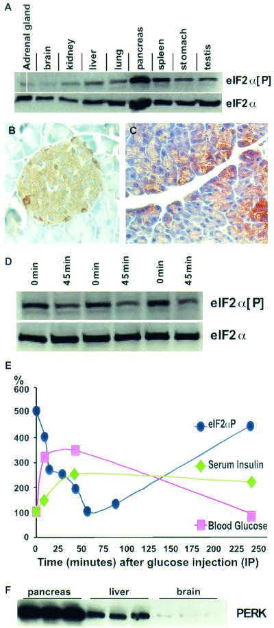
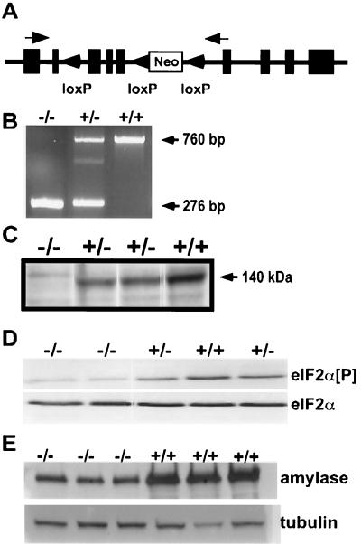
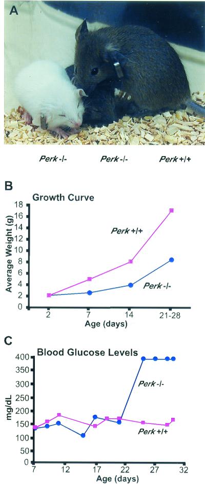
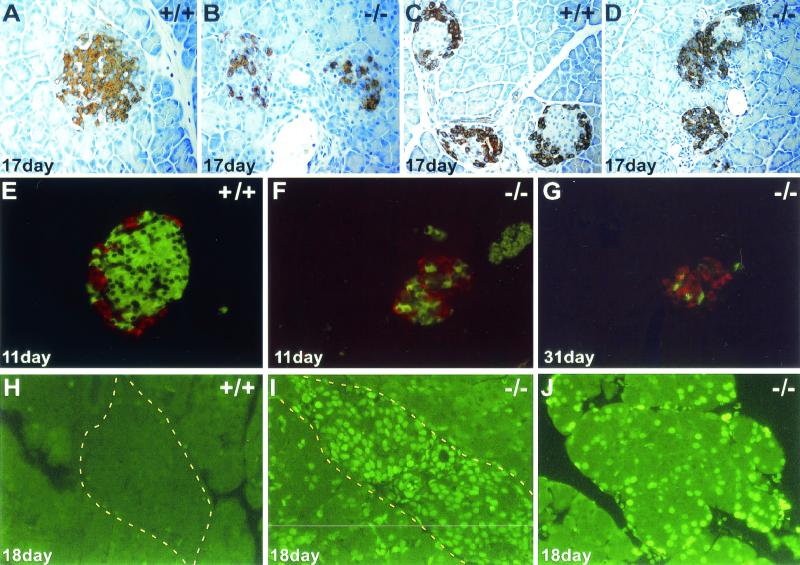
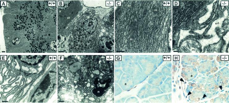
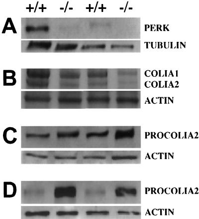
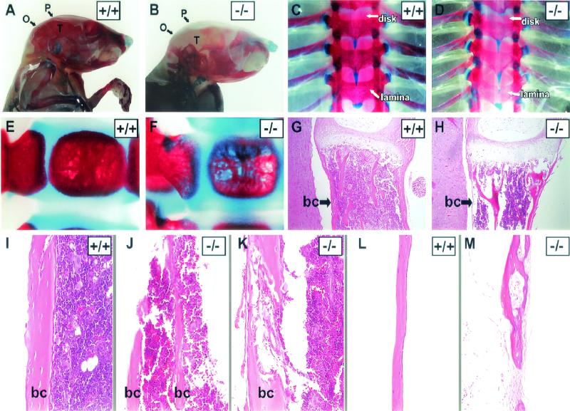
Phosphorylation of eukaryotic initiation factor 2 alpha (eIF-2 alpha) is typically associated with stress responses and causes a reduction in protein synthesis. However, we found high phosphorylated eIF-2 alpha (eIF-2 alpha[P]) levels in nonstressed pancreata of mice. Administration of glucose stimulated a rapid dephosphorylation of eIF-2 alpha. Among the four eIF-2 alpha kinases present in mammals, PERK is most highly expressed in the pancreas, suggesting that it may be responsible for the high eIF-2 alpha[P] levels found therein. We describe a Perk knockout mutation in mice. Pancreata of Perk(-/-) mice are morphologically and functionally normal at birth, but the islets of Langerhans progressively degenerate, resulting in loss of insulin-secreting beta cells and development of diabetes mellitus, followed later by loss of glucagon-secreting alpha cells. The exocrine pancreas exhibits a reduction in the synthesis of several major digestive enzymes and succumbs to massive apoptosis after the fourth postnatal week. Perk(-/-) mice also exhibit skeletal dysplasias at birth and postnatal growth retardation. Skeletal defects include deficient mineralization, osteoporosis, and abnormal compact bone development. The skeletal and pancreatic defects are associated with defects in the rough endoplasmic reticulum of the major secretory cells that comprise the skeletal system and pancreas. The skeletal, pancreatic, and growth defects are similar to those seen in human Wolcott-Rallison syndrome.


Figures

FIG. 1.
FIG. 1.
Physiological regulation of eIF-2α[P] and correlated expression of PERK in the pancreas. (A) Immunoblot analysis of eIF-2α[P] shows moderate levels of expression in the spleen and high levels in the pancreas of wild-type C57BL6 mice, while most tissues exhibit a low level of eIF-2α[P]. Although the pancreas also exhibits a relatively high level of total eIF-2α (phosphorylated and nonphosphorylated), quantitative analysis indicates that the pancreas shows approximately twice the amount of eIF-2α[P] compared to other tissues after normalizing to total eIF-2α. (B and C) Cellular localization of eIF-2α[P] in the islets of Langerhans and acinar cells of wild-type mouse pancreas. (D) Pancreatic eIF-2α[P] and total eIF-2α (nonphosphorylatedand phosphorylated) in pancreata from fasted mice (0 min) versus mice 45 min after the intraperitoneal injection of glucose (45 min). (E) Blood glucose (▪), serum insulin (♦), and eIF-2α[P] (•) of fasted mice injected at time zero with glucose. Insulin and glucose values are normalized to time zero, and eIF-2α[P] values are normalized to total eIF-2α and to the 1-h time point. Each data point represents the average of three to five individual mice. Representative data for the fasted and glucose-injected mice are shown in panel D. Four other similar experiments were conducted over a 2-year period and resulted in a similar pattern of glucose-stimulated dephosphorylation of eIF-2α. (F) Immunoblot analysis of wild-type mice shows high levels of PERK in the pancreas, moderate levels in the liver, and low levels in the brain. C57BL/6 (Jackson Lab) male mice, 6 to 10 weeks old, were used for all glucose injection experiments described above.
FIG. 2.
FIG. 2.
Generation of Perk−/− knockout mice. (A) Schematic of targeting construct designed for generation of Perk−/− mice. Tandemly oriented loxP sites were inserted into a 14.0-kb genomic clone of the Perk gene and then cloned into the pGK-neo-loxP targeting vector. Black boxes represent PERK-encoding exons, and arrows indicate priming sites for RT-PCR. Perk−/− mice described in the text have suffered a deletion of all genomic DNA between the first and third loxP sites. (B) RT-PCR analysis of total RNA using primers flanking the Cre-excised exons. Both Perk+/− and Perk−/− mice produced the expected 276-bp truncated mRNA band, while both the Perk+/− and Perk+/+ mice produced the expected 760-bp band, which was confirmed by sequence analysis. (C) Immunoblot of PERK from pancreatic lysates isolated from Perk+/+, Perk+/−, and Perk−/− mice with antisera recognizing the kinase domain of PERK. PERK is not expressed in the pancreas of Perk−/− mice, and expression is reduced in the Perk+/− mice. (D) Immunoblot analysis of eIF-2α[P] and eIF-2α from pancreatic lysates isolated from 12-day-old Perk+/+, Perk+/−, and Perk−/− mice. Note the greatly reduced levels of eIF-2α[P] in the Perk−/− mice. (E) Immunoblot analysis of amylase from pancreatic lysates isolated from 26- to 28-day-old Perk+/+ and Perk−/− mice, using antisera for amylase.
FIG. 3.
FIG. 3.
Postnatal growth retardation and the development of hyperglycemia in Perk−/− mice. (A) Perk−/− mice (two smaller mice, left) are growth retarded compared to a Perk+/+ littermate (far right). (B) Growth curve illustrating the average weight for Perk+/+ and Perk−/− littermates plotted against age. (C) Blood glucose levels (in milligrams per deciliter) of Perk+/+ and Perk−/− mice. After postnatal day 22, blood glucose of the Perk−/− mice often exceeded the detection capability of the blood glucose monitor.
FIG. 4.
FIG. 4.
Apoptotic loss of pancreatic islet of Langerhans and acinar cells in Perk−/− mice. (A and B) Anti-insulin immunostaining of Perk+/+ and Perk−/− islets. (C and D) Antiglucagon immunostaining of Perk+/+ and Perk−/− islets. Redistribution of the beta cells from the periphery into the central region of the islet has occurred before hyperglycemia has even become apparent. The alpha cells outnumber the beta cells in the mutant islets at this stage, and the entire islet size is drastically reduced. (E to G) Fluorescent double-labeling using Cy2-conjugated donkey anti-guinea pig IgG secondary antibody against guinea pig anti-bovine insulin (green) and Cy3-conjugated donkey anti-rabbit IgG secondary antibody against rabbit antiglucagon (red). (G) Decrease in overall size of the islet and the loss of the alpha and beta cells in a 31-day-old diabetic Perk−/− mouse. (H to J) Apoptosis in an islet (I) and acinar cells (J) detected by TUNEL labeling on 18-day-old Perk−/− mice. Yellow dashed line outlines edges of islets.
FIG. 5.
FIG. 5.
Cellular and subcellular anomalies in pancreata and bone tissue in Perk−/− mice. Pancreatic acinar cells exhibit fragmented and distended ER (er) in Perk−/− mice (B and D) compared to that in wild-type mice (A and C). Note the crowding of the zymogen granules (z) around the nucleus in Perk−/− acinar cells (B) compared to that in the wild-type cells (A) due to the breakdown of normal ER structure. a, acini; n, nucleus; er, RER; erl, ER lumen; m, mitochondria. Bars: A and B, 2.0 μm; C, D, and E, 0.2 μm. (E) A wild-type osteoblast showing a prototypical RER structure. (F) An osteoblast cell in compact bone tissue of a Perk−/− mouse exhibiting highly fragmented and distended ER with densely stained material in the cisternae (erl). (G and H) A large fraction of the pancreatic acinar cells seen in a 32-day-old Perk−/− mouse are severely vacuolated and lack zymogen granules (H), compared to a Perk+/+ pancreas (G). Magnification, ×60. Black arrows in panel H indicate periacinar cells that have proliferated in the Perk−/− pancreas, and white arrows indicate vacuolated acinar cells that have apparently succumbed to apoptosis. This tissue section was immunostained (brown) for eIF-2α[P] by using an antisera specific for eIF-2α[P]. The presence of a high level of eIF-2α[P] is highly correlated with abnormally vacuolated acinar cells.
FIG. 6.
FIG. 6.
PERK expression in bone tissue and misregulation of PROCOLIA2 and COLIA2 in Perk−/− mice. (A) Immunoblot of PERK in bone tissues (5 μg). Lanes 1 and 2, thoracic vertebrae; lanes 3 and 4, tibial long bones. The faint band present in the Perk−/− mutants is due to cross-reactivity with an unrelated protein. (B) COLIA1 and COLIA2 in tibia long bone tissue (lanes 1 and 2) and thoracic vertebrae (lanes 3 and 4) (10 μg). (C) Immunoblot of PROCOLIA2 of tibia long bone tissue (10 μg). (D) Immunoblot of PROCOLIA2 of cultured osteoblasts (10 μg). Quantitative Phosphorimager analysis was performed on these data and on other replicate samples. The large differences in COLIA1, COLIA2, and PROCOLIA2 between Perk−/− mice and their wild-type littermates shown here were consistently observed across several litters. PROCOLIA1 is also reduced in Perk−/− mice (data not shown), proportionate to the reduction observed in PROCOLIA2.
FIG. 7.
FIG. 7.
Perk−/− mice display multiple skeletal dysplasias. (A to F) Alizarin Red (mineralized bone) and Alcian Blue (cartilage) skeletal staining of 18-day-old (A to D) and 2-day-old (E and F) mice. The mineralization of the flat bones of the skull (P, parietal; O, occipital; T, temporal) is greatly reduced in the Perk−/− mouse (B). (C and D) The Perk−/− dorsal thoracic vertebral column shows lack of mineralization across the spine, but especially in the lamina compared to the wild type, as indicated by arrows. Asymmetrical formation and compression of the vertebral column are indicated by arrows pointing to the intervertebral disks. (E and F) Single vertebrae (ventral bodies) of the Perk+/+ and Perk−/− mice (2 days old). Notice the obvious porosity of the vertebrae body in the Perk−/− mouse. (G to M) Hematoxylin and eosin staining of Perk+/+ and Perk−/− bones. (G and H) Longitudinal sections of single vertebrae bodies. Arrows point to the three layers of the bone collar (bc), which are extremely thin in the Perk−/− vertebrae. (I to K) Tibial sections show the porosity and abnormal formation of the bone collars of long bone. (L and M) The weakness, lack of organization, and porosity of the mutant bones are also apparent in the parietal bones of the skull. Panels B and M are at higher magnification.

References

    1. Abraham, N., D. F. Stojdl, P. I. Duncan, N. Methot, T. Ishii, M. Dube, B. C. Vanderhyden, H. L. Atkins, D. A. Gray, M. W. McBurney, A. E. Koromilas, E. G. Brown, N. Sonenberg, and J. C. Bell. 1999. Characterization of transgenic mice with targeted disruption of the catalytic domain of the double-stranded RNA-dependent protein kinase, PKR. J. Biol. Chem. 274:5953-5962. - PubMed
    1. Berlanga, J. J., J. Santoyo, and C. De Haro. 1999. Characterization of a mammalian homolog of the GCN2 eukaryotic initiation factor 2α kinase. Eur. J. Biochem. 265:754-762. - PubMed
    1. Bertolotti, A., Y. Zhang, L. M. Hendershot, H. P. Harding, and D. Ron. 2000. Dynamic interaction of BiP and ER stress transducers in the unfolded-protein response. Nat. Cell Biol. 2:326-332. - PubMed
    1. Casini, A., A. Galli, P. Pignalosa, L. Frulloni, C. Grappone, S. Milani, P. Pederzoli, G. Cavallini, and C. Surrenti. 2000. Collagen type I synthesized by pancreatic periacinar stellate cells (PSC) co-localizes with lipid peroxidation-derived aldehydes in chronic alcoholic pancreatitis. J. Pathol. 192:81-89. - PubMed
    1. Chen, J. J., and I. M. London. 1995. Regulation of protein synthesis by heme-regulated eIF-2 alpha kinase. Trends Biochem. Sci. 20:105-108. - PubMed

Publication types

MeSH terms