A human immunoglobulin G1 antibody originating from an in vitro-selected Fab phage antibody binds avidly to tumor-associated MUC1 and is efficiently internalized
- PMID: 12000712
- PMCID: PMC1850867
- DOI: 10.1016/S0002-9440(10)61107-8
A human immunoglobulin G1 antibody originating from an in vitro-selected Fab phage antibody binds avidly to tumor-associated MUC1 and is efficiently internalized
Abstract
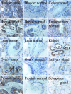
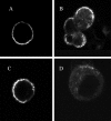
We describe the engineering and characterization of a whole human antibody directed toward the tumor-associated protein core of human MUC1. The antibody PH1 originated from the in vitro selection on MUC1 of a nonimmune human Fab phage library. The PH1 variable genes were reformatted for expression as a fully human IgG1. The resulting PH1-IgG1 human antibody displays a 160-fold improved apparent kd (8.7 nmol/L) compared to the kd of the parental Fab (1.4 micromol/L). In cell-binding studies with flow cytometry and immunohistochemistry, PH1-IgG1 exhibits staining patterns typical for antibodies recognizing the tumor-associated tandem repeat region on MUC1, eg, it binds the tumor-associated glycoforms of MUC1 in breast and ovarian cancer cell lines, but not the heavily glycosylated form of MUC1 on colon carcinoma cell lines. In many tumors PH1-IgG1 binds to membranous and cytoplasmic MUC1, with often intense staining of the whole-cell membrane (eg, in adenocarcinoma). In normal tissues staining is either absent or less intense, in which case it is found mostly at the apical side of the cells. Finally, fluorescein isothiocyanate-labeled PH1-IgG1 internalizes quickly after binding to human OVCAR-3 cells, and to a lesser extent to MUC1 gene-transfected 3T3 mouse fibroblasts. The tumor-associated binding characteristics of this antibody, its efficient internalization, and its human nature, make PH1-IgG1 a valuable candidate for further studies as a cancer-targeting immunotherapeutic.
Figures


Similar articles
-
PH1-derived bivalent bibodies and trivalent tribodies bind differentially to shed and tumour cell-associated MUC1.Protein Eng Des Sel. 2010 Sep;23(9):721-8. doi: 10.1093/protein/gzq044. Epub 2010 Jul 8. Protein Eng Des Sel. 2010. PMID: 20616115
-
Human single-chain Fv antibodies to MUC1 core peptide selected from phage display libraries recognize unique epitopes and predominantly bind adenocarcinoma.Cancer Res. 1998 Oct 1;58(19):4324-32. Cancer Res. 1998. PMID: 9766660
-
Characterization of a new MUC1 monoclonal antibody (VU-2-G7) directed to the glycosylated PDTR sequence of MUC1.Tumour Biol. 2000 Jul-Aug;21(4):197-210. doi: 10.1159/000030126. Tumour Biol. 2000. PMID: 10867613
-
Expression of antibody Fab fragments and whole immunoglobulin in mammalian cells.Methods Mol Biol. 2002;178:389-95. doi: 10.1385/1-59259-240-6:389. Methods Mol Biol. 2002. PMID: 11968508 Review. No abstract available.
-
Mechanisms of antitumor and immune-enhancing activities of MUC1/sec, a secreted form of mucin-1.Immunol Res. 2013 Dec;57(1-3):70-80. doi: 10.1007/s12026-013-8451-6. Immunol Res. 2013. PMID: 24222275 Review.
Cited by
-
Generation and characterization of the human neutralizing antibody fragment Fab091 against rabies virus.Acta Pharmacol Sin. 2011 Mar;32(3):329-37. doi: 10.1038/aps.2010.209. Epub 2011 Jan 31. Acta Pharmacol Sin. 2011. PMID: 21278782 Free PMC article.
-
Recombinant nanobody against MUC1 tandem repeats inhibits growth, invasion, metastasis, and vascularization of spontaneous mouse mammary tumors.Mol Oncol. 2022 Jan;16(2):485-507. doi: 10.1002/1878-0261.13123. Epub 2021 Nov 19. Mol Oncol. 2022. PMID: 34694686 Free PMC article.
-
Efficient production of soluble recombinant single chain Fv fragments by a Pseudomonas putida strain KT2440 cell factory.Microb Cell Fact. 2011 Feb 21;10:11. doi: 10.1186/1475-2859-10-11. Microb Cell Fact. 2011. PMID: 21338491 Free PMC article.
-
Recombinant full-length human IgG1s targeting hormone-refractory prostate cancer.J Mol Med (Berl). 2007 Oct;85(10):1113-23. doi: 10.1007/s00109-007-0208-z. Epub 2007 Jun 7. J Mol Med (Berl). 2007. PMID: 17554518
-
PankoMab: a potent new generation anti-tumour MUC1 antibody.Cancer Immunol Immunother. 2006 Nov;55(11):1337-47. doi: 10.1007/s00262-006-0135-9. Epub 2006 Feb 17. Cancer Immunol Immunother. 2006. PMID: 16485130 Free PMC article.
References
-
- Grillo-Lopez AJ, White CA, Varns C, Shen D, Wei A, McClure A, Dallaire BK: Overview of the clinical development of rituximab: first monoclonal antibody approved for the treatment of lymphoma. Semin Oncol 1999, 26:66-73 - PubMed
-
- Riethmuller G, Holz E, Schlimok G, Schmiegel W, Raab R, Hoffken K, Gruber R, Funke I, Pichlmaier H, Hirche H, Buggisch P, Witte J, Pichlmayr R: Monoclonal antibody therapy for resected Dukes’ C colorectal cancer: seven-year outcome of a multicenter randomized trial. J Clin Oncol 1998, 16:1788-1794 - PubMed
-
- Baselga J: Clinical trials of Herceptin (trastuzumab). Eur J Cancer 2001, 37(Suppl 1):S18-S24 - PubMed
-
- Gendler S, Taylor-Papadimitriou J, Duhig T, Rothbard J, Burchell J: A highly immunogenic region of a human polymorphic epithelial mucin expressed by carcinomas is made up of tandem repeats. J Biol Chem 1988, 263:12820-12823 - PubMed
-
- Taylor-Papadimitriou J, Burchell J, Miles DW, Dalziel M: MUC1 and cancer. Biochim Biophys Acta 1999, 1455:301-313 - PubMed
Publication types
MeSH terms
Substances
LinkOut - more resources
Full Text Sources
Other Literature Sources
Research Materials
Miscellaneous

