. 2002 May;160(5):1619-28.
doi: 10.1016/s0002-9440(10)61109-1.

Interaction between the transforming growth factor-beta type II receptor/Smad pathway and beta-catenin during transforming growth factor-beta1-mediated adherens junction disassembly

Affiliations

Interaction between the transforming growth factor-beta type II receptor/Smad pathway and beta-catenin during transforming growth factor-beta1-mediated adherens junction disassembly

Ya Chung Tian et al. Am J Pathol. 2002 May.

Abstract

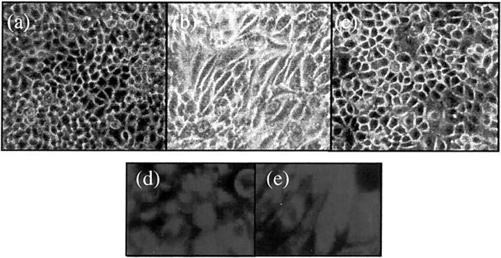
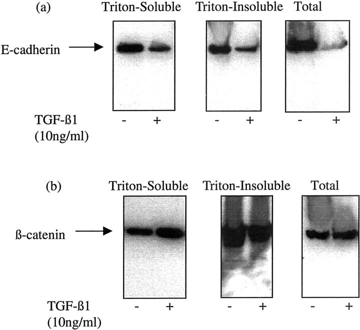
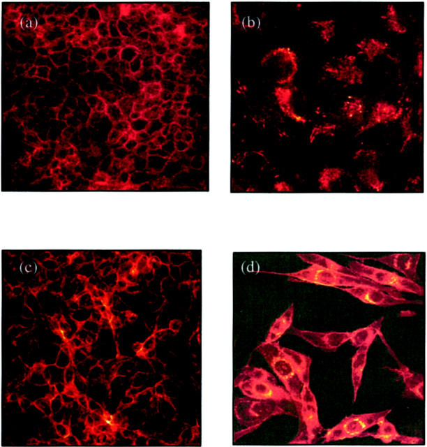
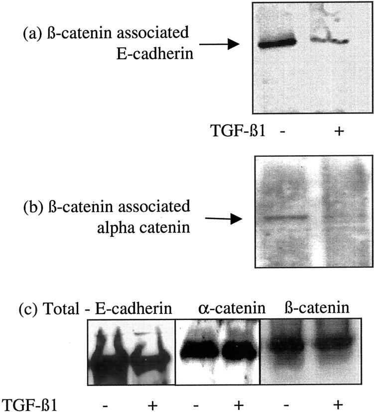
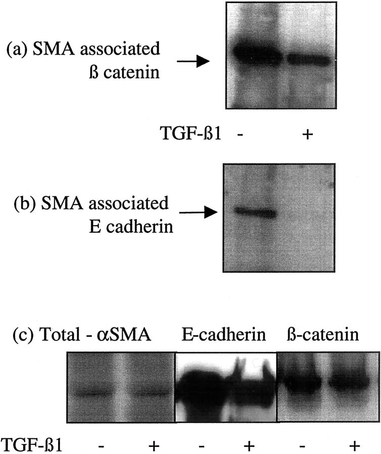
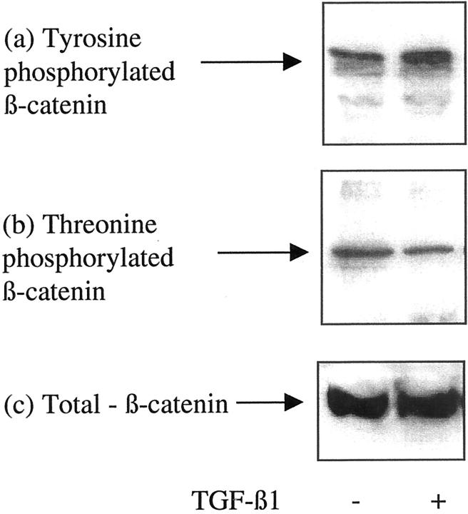
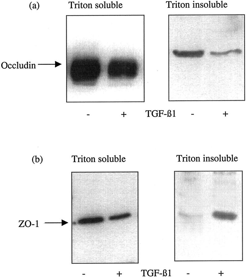
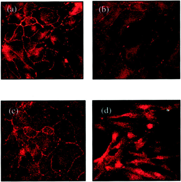
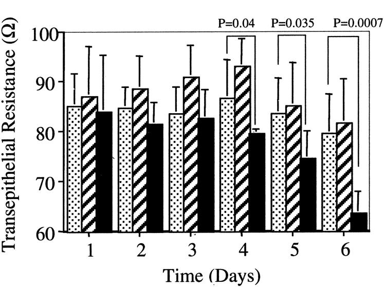
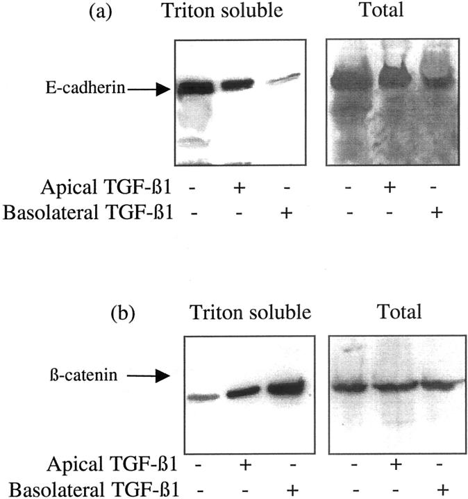
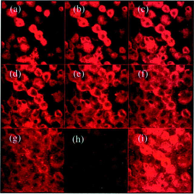
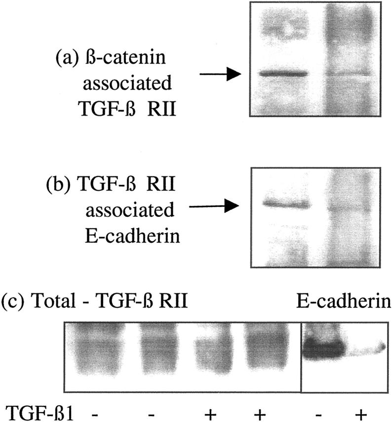
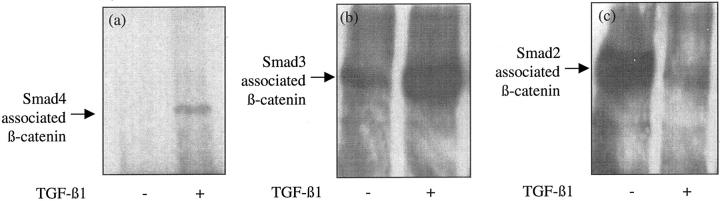
The aim of the current study was to examine the influence of transforming growth factor (TGF)-beta 1 on proximal tubular epithelial cell-cell interaction, with particular emphasis on the regulation of adherens junction complex formation. Stimulation of the proximal tubular cell line HK-2 cells by TGF-beta 1 led to loss of cell-cell contact and disassembly of both adherens and tight junctional complexes. Adherens junction disassembly was associated with reduction of both Triton-soluble and Triton-insoluble E-cadherin, and an increase in detergent-soluble beta-catenin. Under these conditions, immunoprecipitation and Western analysis demonstrated decreased association of beta-catenin, both with E-cadherin, alpha-catenin, and the cell cytoskeleton. Confocal microscopy after immunostaining, showed decreased intensity of peripheral E-cadherin staining, and redistribution of beta-catenin expression to a perinuclear location. Tight junction disassembly was manifest by a reduction in the expression of Triton-soluble occludin and ZO-1 by Western analysis and their disassociation manifested by immunostaining and confocal microscopy. Loss of cell-cell contact and disassembly of adherens junctions were seen after addition of TGF-beta 1 to the basolateral aspect of the cells. Immunoprecipitation experiments demonstrated co-localization of E-cadherin, beta-catenin, and TGF-beta 1 RII in unstimulated cells. After TGF-beta 1 stimulation, the TGF-beta 1 RII no longer associated with either E-cadherin or beta-catenin. Dissociation of the adherens junction protein from the TGF-beta 1 receptor was associated with increased beta-catenin tyrosine phosphorylation and decreased threonine phosphorylation. Furthermore after receptor ligand binding, beta-catenin became associated with the TGF-beta 1-signaling molecules Smad3 and Smad4.


Figures

Figure 1.
Figure 1.
Morphological alterations after TGF-β1 stimulation. Appearance of control HK-2 cells grown on a glass surface without any treatment after serum deprivation for 7 days (a). Cells grow as a typical cobblestone monolayer. Stimulation of confluent monolayers of HK-2 cells with recombinant TGF-β1 (10 ng/ml) under serum-free conditions results in marked changes in cell morphology at day 2 (b). Cells lose their regular cuboidal appearance becoming elongated and spindle shaped. Reversibility of incorporation of α-SMA into stress fibers was determined by removal of supernatant containing TGF-β1 after 72 hours, before washing the cells with phosphate-buffered saline. Subsequently medium supplemented with 10% fetal calf serum was added to the cells for a further 48 hours (c). By immunohistochemistry, both unstimulated (d) and TGF-β1-treated (e) HK-2 cells stain positively for the epithelial cell marker cytokeratin.
Figure 2.
Figure 2.
TGF-β1-induced alteration in E-cadherin and β-catenin expression. Recombinant TGF-β1 (10 ng/ml) was added to confluent monolayers of growth-arrested HK-2 cells under serum-free conditions. On day 3, cells were either lysed by the addition of SDS sample buffer (total lysates), or extracted by the sequential addition of 1% Triton alone (Triton-soluble extract) and 1% Triton/0.5% deoxycholate and 0.1% SDS (Triton-insoluble extract) as described in Materials and Methods. Subsequently Western blot analysis for either E-cadherin (a) or β-catenin (b), of each of the extract samples was performed by standard methodologies. In control experiments serum-free medium alone was added to the confluent monolayers before lysis or sequential extraction. One representative experiment of at least four individual replicate experiments is shown.
Figure 3.
Figure 3.
Immunohistochemistry for E-cadherin (a and b) and β-catenin (c and d). Confluent monolayers of HK-2 cells were stimulated with TGF-β1 for 4 days before fixation and analysis by immunohistochemistry as described in Materials and Methods. Under control conditions (serum-free medium alone) both E-cadherin (a) and β-catenin (c) clearly outline the cell contour. After stimulation with recombinant TGF-β1 E-cadherin staining intensity becomes weaker and discontinuous (b). β-catenin staining after addition TGF-β1 demonstrates a relocation from the cell periphery to a perinuclear location (d).
Figure 4.
Figure 4.
Association of β-catenin with E-cadherin and α-catenin. Recombinant TGF-β1 (10 ng/ml) was added to confluent monolayers of growth-arrested HK-2 cells under serum-free conditions. On day 3 β-catenin was immunoprecipitated as described in Materials and Methods, and E-cadherin (a) or α-catenin (b) expression in the precipitate examined by Western analysis. For comparison total E-cadherin, α-catenin, and β-catenin expression is shown (c). In control experiments serum-free medium was added to the confluent monolayers on cells. One representative experiment of at least four individual replicate experiments is shown.
Figure 5.
Figure 5.
Association of β-catenin with the actin cytoskeleton. Recombinant TGF-β1 (10 ng/ml) was added to confluent monolayers of growth-arrested HK-2 cells under serum-free conditions. On day 3 α-SMA was immunoprecipitated as described in Materials and Methods, and β-catenin (a) or E-cadherin (b) expression in the precipitate examined by Western analysis. For comparison total α-SMA, E-cadherin, and β-catenin expression is shown (c). In control experiments serum-free medium was added to the confluent monolayers on cells. One representative experiment of at least four individual replicate experiments is shown.
Figure 6.
Figure 6.
Influence of TGF-β1 on β-catenin phosphorylation status. Recombinant TGF-β1 (10 ng/ml) was added to confluent monolayers of growth-arrested HK-2 cells under serum-free conditions. On day 3 phosphotyrosine (a) or phosphothreonine (b) antibodies were used for immunoprecipitation as described in Materials and Methods, and β-catenin expression in the immunoprecipitate examined by Western analysis. For comparison, total β-catenin expression is shown (c). In control experiments serum-free medium was added to the confluent monolayers on cells. One representative experiment of at least four individual replicate experiments is shown.
Figure 7.
Figure 7.
TGF-β1-induced alteration in occludin and ZO-1 expression. Recombinant TGF-β1 (10 ng/ml) was added to confluent monolayers of growth-arrested HK-2 cells under serum-free conditions. On day 3, cell proteins were extracted by the sequential addition of 1% Triton alone (Triton-soluble extract) and 1% Triton/0.5% deoxycholate and 0.1% SDS (Triton-insoluble extract) as described in Materials and Methods. Subsequently Western blot analysis for either occludin (a) or ZO-1 (b), of each of the extract samples was performed by standard methodologies. In control experiments serum-free medium alone was added to the confluent monolayers before lysis or sequential extraction. One representative experiment of at least four individual replicate experiments is shown.
Figure 8.
Figure 8.
Immunohistochemistry for occludin (a and b) and ZO-1 (c and d). Confluent monolayers of HK-2 cells were stimulated with TGF-β1 for 4 days before fixation and analysis by immunohistochemistry as described in Materials and Methods. Under control conditions (serum-free medium alone) both occludin (a) and ZO-1 (c) clearly outline the cell contour. After stimulation with recombinant TGF-β1, occludin staining intensity becomes weaker and discontinuous (b). ZO-1 staining after the addition TGF-β1 demonstrates relocation from the cell periphery into the cell cytoplasm (d).
Figure 9.
Figure 9.
Polarity of TGF-β1-mediated alteration in transepithelial resistance. HK-2 cells were grown to confluence on porous tissue culture inserts as described in Materials and Methods. Subsequently TGF-β1 was added to either the apical (hatched bars) or basolateral (black bars) compartment on day 1. In control experiments serum-free medium alone was added to both compartments (dotted bars). Subsequently transepithelial resistance was determined daily. Each reading represents triplicate determinants of six independent experiments.
Figure 10.
Figure 10.
Polarity of TGF-β1-mediated alteration in E-cadherin and β-catenin expression. HK-2 cells were grown to confluence on porous tissue-culture inserts as described in Materials and Methods. Subsequently TGF-β1 was added to either the apical or basolateral compartment and either the total cell protein or Triton-soluble protein extracted as described in Materials and Methods before analysis of either E-cadherin (a) or β-catenin (b) by Western analysis. One representative experiment of at least four individual replicate experiments is shown.
Figure 11.
Figure 11.
Immunhistochemical localization of TGF-β type II receptor expression. Confluent growth-arrested monolayers of HK-2 cells were fixed before analysis of TGF-β type II receptor by immunohistochemistry as described in Materials and Methods. Subsequently localization of fluorescence was examined in eight 0.8-μm serial sections from the apical to the basolateral aspect of the cell using the confocal microscope (a–g). In addition a composite image of the superimposed images was generated after immunostaining in the presence (h) or absence (i) of blocking peptide.
Figure 12.
Figure 12.
Association of β-catenin with the TGF-β1 RII. Recombinant TGF-β1 (10 ng/ml) was added to confluent monolayers of growth-arrested HK-2 cells under serum-free conditions. On day 3 β-catenin was immunoprecipitated as described in Materials and Methods, and TGF-β1 RII expression in the precipitate was examined by Western analysis. For comparison total TGF-β RII and E-cadherin expression is shown (c) In control experiments serum-free medium was added to the confluent monolayers on cells. One representative experiment of at least four individual replicate experiments is shown.
Figure 13.
Figure 13.
Association of β-catenin with Smad proteins. Recombinant TGF-β1 (10 ng/ml) was added to confluent monolayers of growth-arrested HK-2 cells under serum-free conditions. On day 3 Smad4 (a), Smad3 (b), or Smad2 (c) were immunoprecipitated as described in Materials and Methods, and β-catenin expression in the precipitates examined by Western analysis. In control experiments serum-free medium was added to the confluent monolayers on cells. One representative experiment of at least four individual replicate experiments is shown.

Similar articles

Cited by

References

    1. Bohle A, Mackensen-Haen S, Gise H: Significance of tubulointerstitial changes in the renal cortex for the excretory function and concentration ability of the kidney: a morphometric contribution. Am J Nephrol 1987, 6:421-433 - PubMed
    1. Mackensen-Haen S, Bader R, Grund KE, Bohle A: Correlations between renal cortical interstitial fibrosis, atrophy of the proximal tubules and impairment of the glomerular filtration rate. Clin Nephrol 1981, 15:167-172 - PubMed
    1. Phillips AO, Steadman R, Topley N, Williams JD: Elevated D-glucose concentrations modulate TGF-β1 synthesis by human cultured renal proximal tubular cells: the permissive role of platelet derived growth factor. Am J Pathol 1995, 147:362-374 - PMC - PubMed
    1. Phillips AO, Topley N, Steadman R, Morrisey K, Williams J: Induction of TGF-β1 synthesis in D-glucose primed human proximal tubular cells: differential stimulation by the macrophage derived pro-inflammatory cytokines IL-1β and TNFα. Kidney Int 1996, 50:1546-1554 - PubMed
    1. Phillips AO, Topley N, Morrisey K, Williams JD, Steadman R: Basic fibroblast growth factor stimulates the release of pre-formed TGF-β1 from human proximal tubular cells in the absence of de-novo gene transcription or mRNA translation. Lab Invest 1997, 76:591-600 - PubMed

Publication types

MeSH terms

LinkOut - more resources