. 2002 May;160(5):1683-93.
doi: 10.1016/S0002-9440(10)61115-7.

Suppression of diabetic retinopathy with angiopoietin-1

Affiliations

Suppression of diabetic retinopathy with angiopoietin-1

Antonia M Joussen et al. Am J Pathol. 2002 May.

Abstract

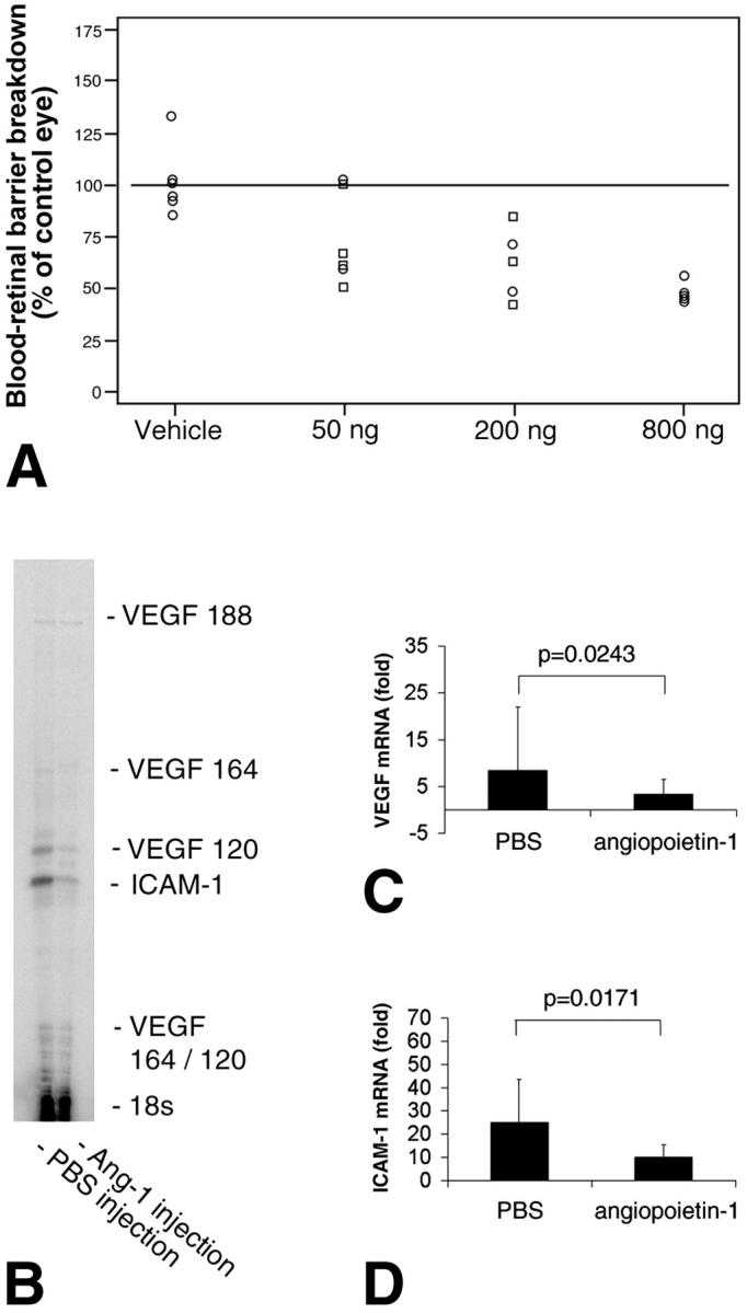
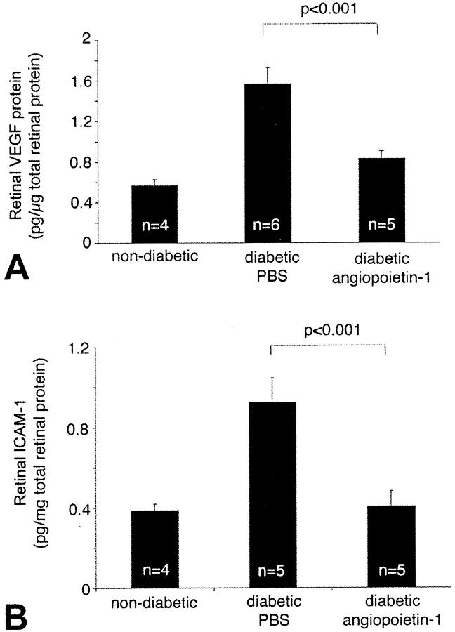
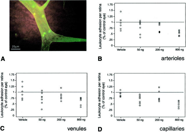
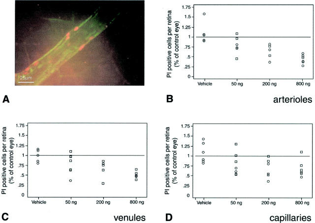
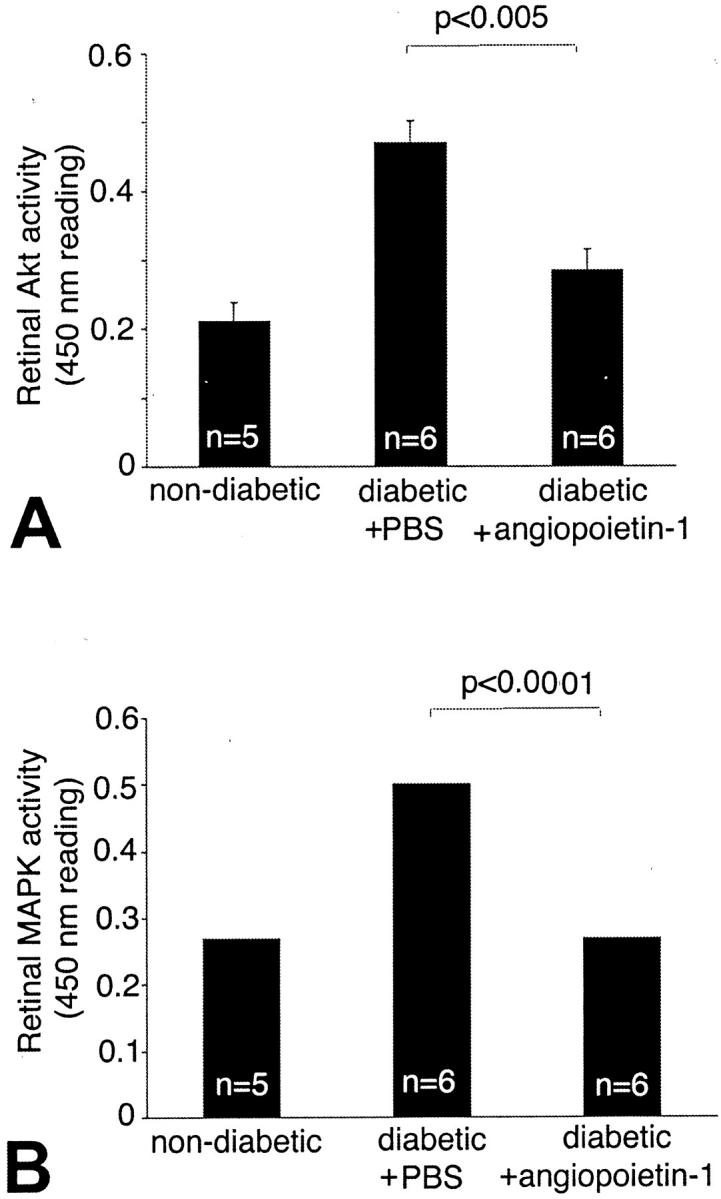
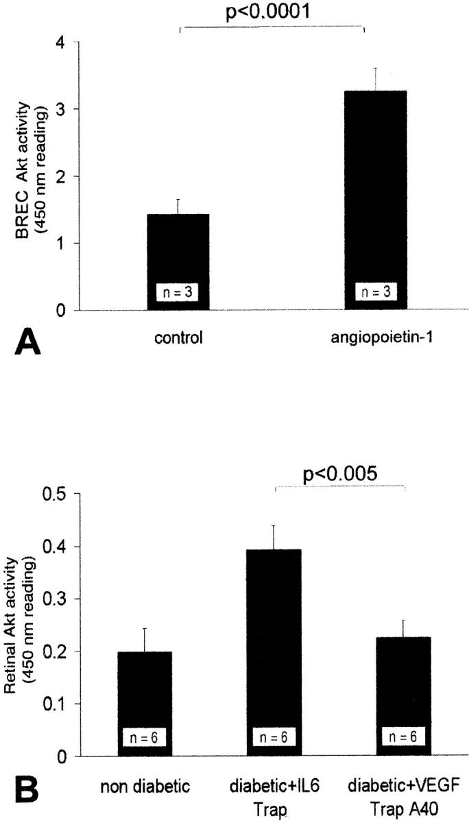
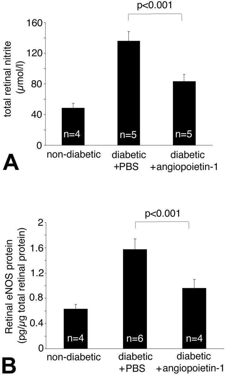
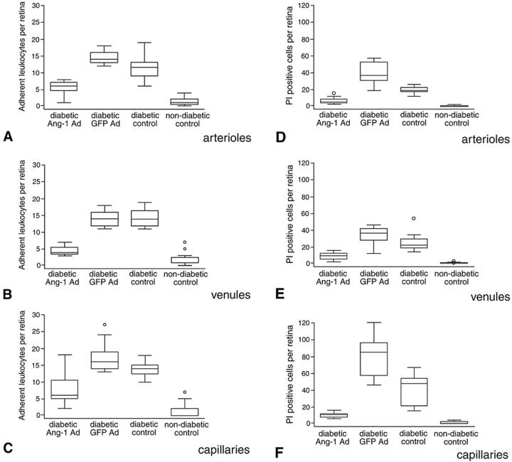
Diabetic retinopathy remains a leading cause of irreversible blindness. A critical early pathology in the disease is the adhesion of leukocytes to the retinal vasculature, a process that occurs, in part, via intercellular adhesion molecule-1. Once leukocyte adhesion occurs, endothelial cell injury ensues, as does blood-retinal barrier breakdown. Here we show that angiopoietin-1 can prevent and reverse these diabetic retinal vascular changes in both new and established diabetes. Angiopoietin-1, when given intravitreally to newly diabetic rats, normalized retinal vascular endothelial growth factor (VEGF) and intercellular adhesion molecule-1 mRNA and protein levels, leading to reductions in leukocyte adhesion, endothelial cell injury, and blood-retinal barrier breakdown. When an adenovirus coding for angiopoietin-1 was given systemically to mice with established diabetes, it similarly inhibited leukocyte adhesion and endothelial cell injury and blood-retinal barrier breakdown. These changes coincided with reductions in retinal eNOS, nitric oxide, Akt (protein kinase B), and MAP kinase activity, known mediators of VEGF bioactivity and leukocyte adhesion. When endogenous VEGF bioactivity was inhibited with a soluble Flt-1/Fc chimera, retinal Akt kinase activity was significantly reduced in vivo. Taken together, these data document new vascular and anti-inflammatory bioactivities for angiopoietin-1 and identify it as the first naturally occurring protein that directly protects the retinal vasculature in diabetes.


Figures

Figure 1.
Figure 1.
Ang-1 suppresses blood-retinal barrier breakdown and reduces VEGF and ICAM-1 expression in the retina. A: Locally administered Ang-1 suppresses diabetic retinal vascular permeability. Five μl of 160 μg/ml, 40 μg/ml, or 10 μg/ml of Ang-1 (800 ng, 200 ng, or 50 ng total) or vehicle was injected into one eye of 1-week diabetic rats in a masked manner. The contralateral eye of each rat received an equivalent volume of PBS alone as a control. The animals were studied 24 hours later. Evans blue dye was used to measure vascular permeability 24 hours after intra-ocular injection. Ang-1 dose-dependently reduced the retinal vascular permeability induced by experimental diabetes (P < 0.0001). B–D: Locally administered Ang-1 (200 ng) suppresses VEGF and ICAM-1 mRNA levels in diabetic retina. Retinae were harvested for RNA isolation 24 hours after intra-ocular injection with either Ang-1 or vehicle alone. B: A representative VEGF/ICAM-1 ribonuclease protection assay of eyes treated with PBS (left lane) or Ang-1 (right lane). C: When normalized to 18S RNA, diabetic retinal VEGF mRNA levels were reduced by 61.5% with Ang-1 (P < 0.03, n = 5). The graph shows the combined signal of the VEGF120, VEGF164, and VEGF188 transcripts, compared with diabetic eyes treated with PBS. D: When normalized to 18S RNA, retinal ICAM-1 mRNA levels were reduced by 59.5% with Ang-1 (P < 0.02, n = 5).
Figure 2.
Figure 2.
Ang-1 decreases retinal VEGF and ICAM-1 protein levels. VEGF and ICAM-1 protein levels in the retina were measured using an ELISA technique. A: Ang-1 reduced diabetic retinal VEGF protein levels by 39.6% (0.95 ± 0.14 pg/μg versus 1.57 ± 0.17 pg/μg; n = 4 to 6, P < 0.001). B: Ang-1 reduced diabetic retinal ICAM-1 levels by 55.9% (0.41 ± 0.07 pg/μg versus 0.92 ± 0.12 pg/μg; n = 4 to 5, P < 0.001).
Figure 3.
Figure 3.
Ang-1 reduces leukocyte adhesion in the retina. A–D: Locally administered Ang-1 suppresses diabetic retinal leukocyte adhesion in rats with new-onset diabetes. Ang-1 or vehicle alone, at the indicated doses, was injected unilaterally into 1-week diabetic rat eyes. The contralateral eyes received an equal volume injection of PBS buffer alone. Endothelial cells and adherent leukocytes were labeled with FITC-linked concanavalin A lectin (green fluorescence). A: A representative microscopic image of diabetic flat-mounted retina stained with FITC-linked concanavalin A showing a cluster of adherent leukocytes. The number of adherent retinal leukocytes was dose-dependently decreased in the arterioles (P < 0.0001) (B), venules (P < 0.0008) (C), and capillaries (P < 0.02) (D). The data are presented as a percentage, comparing the Ang-1- or vehicle-treated eyes to the contralateral PBS-injected eyes.
Figure 4.
Figure 4.
Ang-1 reduces endothelial cell damage in the retina. A–D: Locally administered Ang-1 suppresses endothelial cell injury and death in rats with new-onset diabetes. Injured and/or dead retinal endothelial cells were labeled in vivo using PI (orange fluorescence). A: Representative microscopic image of a diabetic flat-mounted retina stained with PI showing a cluster of injured and/or dying endothelial cells. Locally administered Ang-1 dose-dependently reduced the retinal vascular endothelial cell PI staining in arterioles (P < 0.004) (B), venules (P < 0.008) (C), and capillaries (P < 0.004) (D). The data are presented as a percentage, comparing the Ang-1- or vehicle-treated eyes to the contralateral PBS-injected eyes.
Figure 5.
Figure 5.
Ang-1 suppresses Akt kinase and Erk kinase activation in vivo. Akt kinase activity increased by 54.3% in the diabetic retina when compared to the nondiabetic controls [0.46 ± 0.01 versus 0.21 ± 0.003 (OD at 450 nm), P < 0.005, n = 5 to 6]. A: Treatment with Ang-1 resulted in a 39.2% reduction in Akt kinase activation.(0.28 ± 0.02 pg/mg; P < 0.005, n = 6). Erk kinase activity increased by 1.9-fold in the diabetic retina compared to the nondiabetic controls [0.26 ± 0.01 versus 0.5 ± 0.01 (OD at 450 nm); P < 0.0001, n = 5 to 6]. B: Treatment with Ang-1 resulted in a 55% reduction in retinal Erk activity compared to PBS-injected diabetic controls (0.26 ± 0.04; P < 0.0001, n = 6).
Figure 6.
Figure 6.
Ang-1 activates Akt kinase in vitro; VEGF inhibition suppresses Akt kinase activation in vivo. A: Akt kinase activity increased significantly in retinal endothelial cells treated with Ang-1 (250 ng/ml) in vitro [1.42 ± 0.22 versus 3.26 ± 0.34 09 (OD at 450 nm); P < 0.0001, n = 3]. However, the inhibition of endogenous VEGF, via the in vivo administration of the VEGF Trap A40, suppressed Akt kinase activity [0.4 ± 0.05 versus 0.25 ± 0.09 (IL-6 Trap); P < 0.005, n = 6]. B: Akt kinase activation increased in the diabetic retinae as compared to the nondiabetic controls [from 0.2 ± 0.045 to 0.4 ± 0.05 (OD at 450 nm); P < 0.005, n = 6].
Figure 7.
Figure 7.
Ang-1 reduces retinal nitric oxide and eNOS expression. The total nitrite concentration was estimated from retinal tissue. Compared to the retinae of nondiabetic animals, control diabetic animals demonstrated a 2.8-fold increase in normalized NO levels (48.57 ± 6.05 μmol/L versus 135.89 ± 12.22 μmol/L; P < 0.0001, n = 4 to 6). A: Treatment with Ang-1 reduced retinal NO levels by 38.6% (83.36 ± 8.95 μmol/L; P < 0.001 versus nondiabetic controls, n = 4 to 5). The enzyme eNOS was quantified in retinal tissue by ELISA. B: Treatment with Ang-1 reduced retinal eNOS levels by 39.4% (0.95 ± 0.13 pg/mg versus 1.57 ± 0.16 pg/mg retinal weight; diabetic controls versus Ang-1-treated eyes; P < 0.001; n = 4 to 6).
Figure 8.
Figure 8.
Systemic administration of Ang-1 in established diabetes. Systemically administered Ang-1 suppresses diabetic retinal leukocyte adhesion (A–C) and endothelial cell death and injury (D–F) in mice with established diabetes. Sixteen-week diabetic mice received a single tail vein injection of 1 × 10⁹ PFU Ad-Ang-1. Age-matched control diabetic mice received an equivalent amount of Ad-GFP (control). Retinae were assayed for leukocyte adhesion and PI staining 72 hours after virus injection. The data are presented in a box and whiskers format. The middle line represents the median, with the box extending from the 25th to the 75th percentiles. Any data points beyond the percentile ranges are shown as individual data points (circles). The upper and lower values are the smallest and largest data points equal to, or greater than, the interquartile range. Twelve retinae per condition were analyzed. Systemic Ang-1 suppressed leukocyte adhesion in the retinal arterioles (P < 0.0001 versus diabetic Ad-GFP and diabetic control) (A), venules (P < 0.0001) (B), and capillaries (P < 0.0001) (C) of diabetic retinae. Similarly, systemic Ang-1 reduced vascular endothelial cell PI labeling in the retinal arterioles (P < 0.0001 versus diabetic Ad-GFP and diabetic control) (D), venules (P < 0.0001) (E), and capillaries (P < 0.0001) (F) of the diabetic retinae.

Comment in

References

    1. Miyamoto K, Khosrof S, Bursell S-E, Rohan R, Murata T, Clermont A, Aiello LP, Ogura Y, Adamis AP: Prevention of leukostasis and vascular leakage in streptozotocin-induced diabetic retinopathy via intercellular adhesion molecule-1 inhibition. Proc Natl Acad Sci USA 1999, 96:10836-10841 - PMC - PubMed
    1. Joussen AM, Murata T, Tsujikawa A, Kirchhof B, Bursell SE, Adamis AP: Leukocyte-mediated endothelial cell injury and death in the diabetic retina. Am J Pathol 2001, 158:147-152 - PMC - PubMed
    1. McLeod DS, Lefer DJ, Merges C, Lutty GA: Enhanced expression of intracellular adhesion molecule-1 and P-selectin in the diabetic human retina and choroid. Am J Pathol 1995, 147:642-653 - PMC - PubMed
    1. Schröder S, Palinski W, Schmid-Schönbein GW: Activated monocytes and granulocytes, capillary nonperfusion, and neovascularization in diabetic retinopathy. Am J Pathol 1991, 139:81-100 - PMC - PubMed
    1. Davis S, Aldrich TH, Jones PF, Acheson A, Compton DL, Jain V, Ryan TE, Bruno J, Radziejewski C, Maisonpierre PC, Yancopoulos GD: Isolation of angiopoietin-1, a ligand for the TIE2 receptor, by secretion-trap expression cloning. Cell 1996, 87:1161-1169 - PubMed

Publication types

MeSH terms

Substances