. 2002 May;160(5):1695-703.
doi: 10.1016/S0002-9440(10)61116-9.

Altered Notch ligand expression in human liver disease: further evidence for a role of the Notch signaling pathway in hepatic neovascularization and biliary ductular defects

Affiliations

Altered Notch ligand expression in human liver disease: further evidence for a role of the Notch signaling pathway in hepatic neovascularization and biliary ductular defects

Sarbjit S Nijjar et al. Am J Pathol. 2002 May.

Abstract

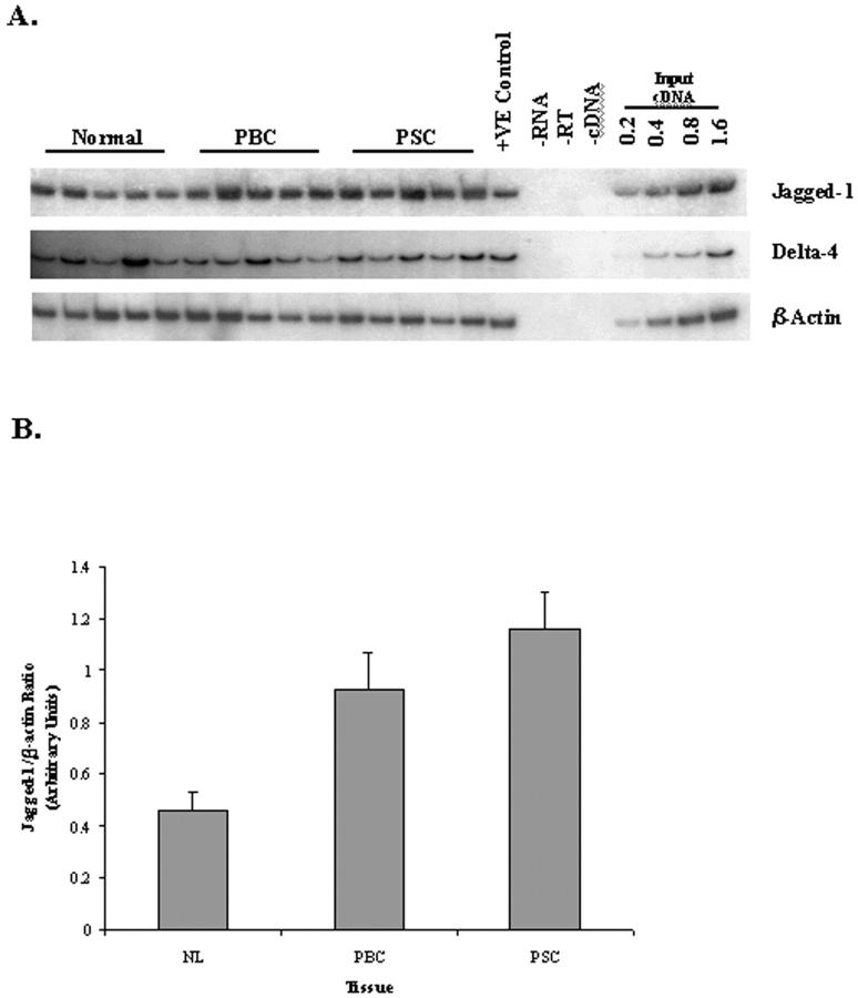
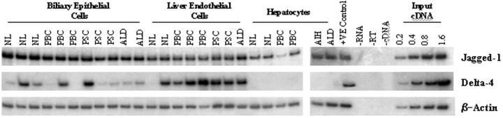
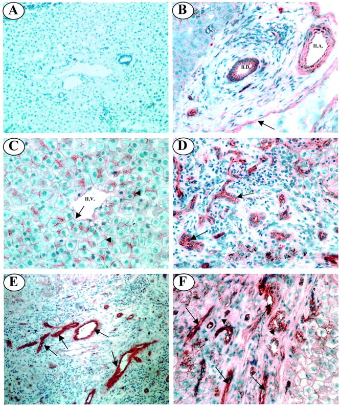
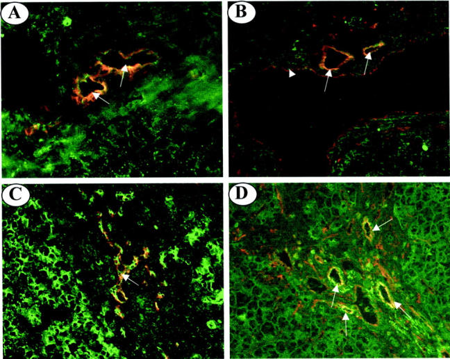
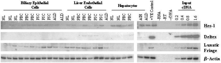
The Jagged and Delta family of transmembrane proteins are ligands for Notch receptors, which control the proliferation and/or differentiation of many cell lineages. Expression and localization of these ligands in the adult human liver has not been fully elucidated, nor whether dysregulation of these proteins contributes to liver disease processes. We have examined expression of the five known Notch ligands in human liver. Expression of Jagged-1 and Delta-4 mRNA was seen in normal and diseased liver tissue, whereas Jagged-2, Delta-1, and Delta-3 mRNA was undetectable. In primary liver cell isolates, Jagged-1 expression was found in all cell types, whereas Delta-4 was present in biliary epithelial and liver endothelial cells, but absent in hepatocytes. Interestingly, Jagged-1 mRNA expression was significantly up-regulated in diseased liver tissue. By immunohistochemistry, Jagged-1 expression was present on most structures in normal tissue. However in disease, strikingly strong Jagged-1 immunoreactivity was observed on many small neovessels and bile ductules. The expression of downstream modulators and effectors of Notch signaling was also detectable in purified cell isolates. This, together with aberrant Jagged-1 expression suggests that the Notch signaling pathway may play a role in the neovascularization and biliary defects observed in the liver during the development of cirrhosis.


Figures

Figure 1.
Figure 1.
A: RT-PCR analysis of Jagged-1 and Delta-4 gene expression in normal and diseased liver tissue. First-strand cDNA prepared from total RNA extracted from NIH 3T3 cells expressing the human Jagged-1 gene was used as a positive and linearity control. For Delta-4 PCR, cDNA prepared from human umbilical vein endothelial cells was used as a positive and linearity control. B: Densitometric analysis of Jagged-1 gene expression in normal and diseased liver tissue. The graph shows values for Jagged-1 mRNA levels obtained by densitometry normalized with respect to β-actin signals. Data represents the mean densitometric value for Jagged-1 ±SEM (n = 5). A paired Student’s t-test shows that the data for PBC (P < 0.05) and PSC (P < 0.01) is significantly different.
Figure 2.
Figure 2.
RT-PCR analysis of Jagged-1 and Delta-4 mRNA expression in cell isolates. RT-PCR analysis was performed on RNA extracted from primary cell cultures. NL, normal liver; AIH, autoimmune hepatitis.
Figure 3.
Figure 3.
Immunolocalization of Jagged-1 expression in normal and diseased adult liver tissue. A: No primary antibody control. B: In the portal tract of normal liver tissue Jagged-1 staining is present on the endothelium of the portal vein (black arrow), together with the walls and endothelium of the hepatic artery. Jagged-1 expression is also evident on the epithelium of bile ducts. C: In the parenchyma of normal liver tissue, Jagged-1 is localized to the cell membrane of hepatocytes (arrowheads) and the endothelium of the hepatic vein (arrow). D: Strong Jagged-1 immunoreactivity is associated with bile ductules in PBC liver tissue (black arrows). E: In PSC liver strong Jagged-1 staining is seen on numerous small vessels in fibrous septa (black arrows). F: In alcoholic liver disease liver, strong Jagged-1 expression was evident on bile ductules (arrowheads) and numerous small vessels (black arrows) within the portal tract. B. D., bile duct; H. A., hepatic artery. Original magnifications: ×100 (A and E); ×200 (B, D, and F); ×400 (C).
Figure 4.
Figure 4.
Dual-immunofluorescence staining of frozen liver sections from normal and PBC livers. A: In normal liver, double-immunofluorescent staining with CK19 (red fluorescence) and Jagged-1 (green fluorescence) reveals co-localization on bile ducts (yellow fluorescence, arrows). B: In normal liver, double-immunofluorescent staining with CD31 (red fluorescence) and Jagged-1 (green fluorescence) reveals co-localization (yellow fluorescence) to the endothelium of the hepatic arteries (arrows) and the endothelium of the portal vein (arrowhead). C: CK19 (red fluorescence) and Jagged-1 (green fluorescence) staining co-localizes to bile ductules (yellow fluorescence, arrow) in PBC liver tissue. D: CD31 (red fluorescence) and Jagged-1 (green fluorescence) co-localize (yellow fluorescence, arrows) to the endothelium of neovessels in the portal tract of PBC liver tissue (arrows). Original magnifications, ×200.
Figure 5.
Figure 5.
The expression of downstream modulators of Notch signaling in cell isolates. RT-PCR analysis was performed on RNA extracted from primary cell cultures. First-strand cDNA prepared from total RNA extracted from Jurkat cells was used as a positive and linearity control for Deltex and Hes-1 PCR. For Lunatic Fringe PCR, cDNA prepared from human umbilical vein endothelial cells was used as a positive and linearity control. NL, normal liver; AIH, autoimmune hepatitis.

Similar articles

Cited by

References

    1. Artavanis-Tsakonas S, Rand MD, Lake RJ: Notch signaling: cell fate control and signal integration in development. Science 1999, 284:770-776 - PubMed
    1. Mumm JS, Kopan R: Notch signaling: from the outside in. Dev Biol 2000, 228:151-165 - PubMed
    1. Ellisen LW, Bird J, West DC, Soreng AL, Reynolds TC, Smith SD, Sklar J: TAN-1, the human homologue of the Drosophila gene, is broken by chromosomal translocations in T lymphoblastic neoplasms. Cell 1991, 66:649-661 - PubMed
    1. Stifani S, Blaumueller CM, Redhead NJ, Hill RE, Artavanis-Tsakonas S: Human homologs of a Drosophila enhancer of split gene product define a novel family of nuclear proteins. Nat Genet 1992, 2:119-127 - PubMed
    1. Joutel A, Corpechot C, Ducros A, Vahedi K, Chabriat H, Mouton P, Alamowitch S, Domenga V, Cecillion M, Marechal E, Maciazek J, Vayssiere C, Cruaud C, Cabanis E-A, Ruchoux MM, Weissenbach J, Bach JF, Bousser MG, Tournier-Lasserve E: Notch3 mutations in CADASIL, an hereditary adult onset condition causing stroke and dementia. Nature 1996, 383:707-710 - PubMed

Publication types

MeSH terms