. 2002 May;160(5):1767-78.
doi: 10.1016/S0002-9440(10)61123-6.

Carp, a cardiac ankyrin-repeated protein, and its new homologue, Arpp, are differentially expressed in heart, skeletal muscle, and rhabdomyosarcomas

Affiliations

Carp, a cardiac ankyrin-repeated protein, and its new homologue, Arpp, are differentially expressed in heart, skeletal muscle, and rhabdomyosarcomas

Naoko Ishiguro et al. Am J Pathol. 2002 May.

Abstract

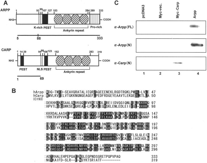
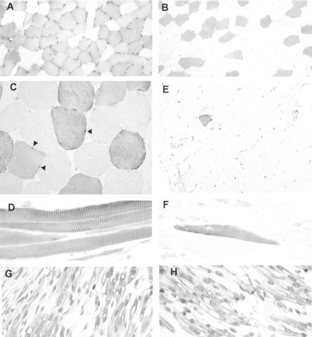
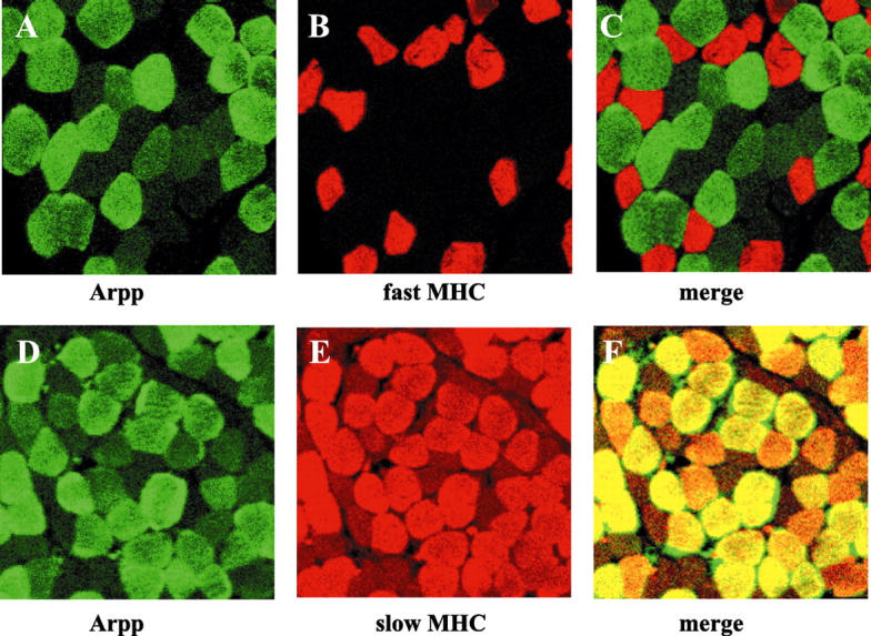
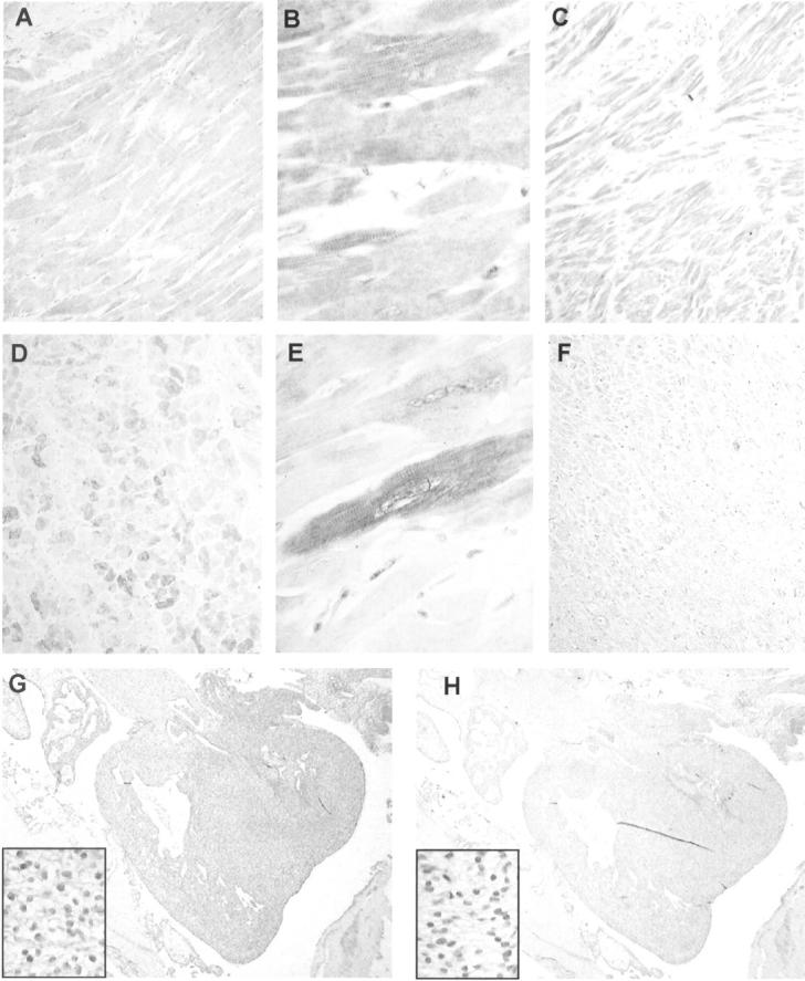
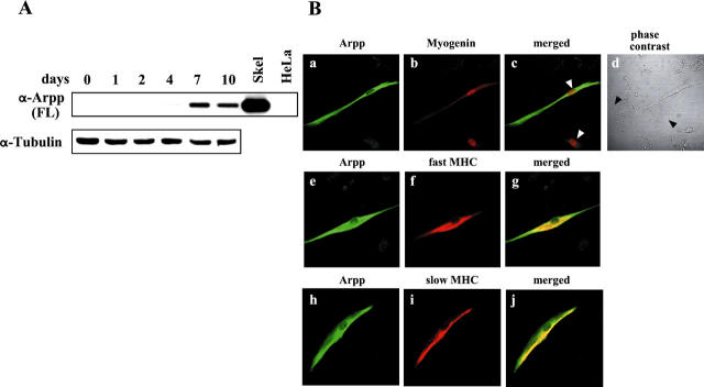
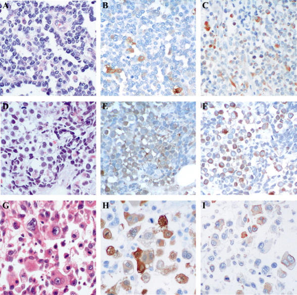
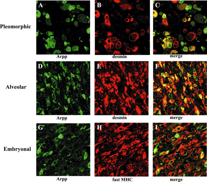
Arpp, a protein containing an ankyrin repeat domain, PEST sequence, and proline-rich region, is a novel ankyrin-repeated protein highly homologous to Carp, which is proposed to be the putative genetic marker for cardiac hypertrophy. In this study, we comparatively analyzed expression of Arpp and Carp protein in skeletal and cardiac muscles and rhabdomyosarcomas (RMSs). In adult skeletal muscle, Arpp was preferentially expressed in the nucleus and cytoplasm of type I fibers, whereas Carp was barely detectable in skeletal muscle. On the other hand, in adult cardiac muscle, interestingly, Arpp was expressed in ventricles mostly, whereas Carp was expressed throughout the atrium and ventricle. Furthermore, although Carp was identified in fetal heart at 11 developmental weeks, Arpp was very low or undetectable in these fetal hearts. These results suggest that Arpp and Carp are differentially expressed and function in both skeletal and cardiac muscle of fetus and adult. We found that Arpp expression was induced during the differentiation of C2C12 cells in vitro, suggesting that Arpp-expression may be associated with the differentiation stage during myogenesis. Both Arpp and Carp were found to be expressed in all of the RMS cases studied. Because the expression patterns of Arpp in RMS were different from those of muscle actin or desmin, Arpp may be detectable in RMS cases that do not express other existing RMS markers.


Figures

Figure 1.
Figure 1.
Identification of Carp and its homologue, Arpp, by specific Abs. A: Schematic representation of the domain structures of Arpp and Carp proteins. The amino acid sequences used for immunization [Arpp(5-333) for α-Arpp(FL) Ab, Arpp(5-88) for α-Arpp(N) Ab, and Carp(1-69) for α-Carp(N) Ab] are indicated by bold bars. B: Comparison of the amino acid sequence of Arpp with that of human Carp (C-193). Shaded boxes show identical residues. C: Western blotting of Arpp- and Carp-transfected HeLa cells. HeLa cells were transfected with pcDNA3-vector (lane 1), myc-vector (lane 2), myc-Carp (lane 3), and pcDNA3-Arpp (lane 4). The cell lysates were subsequently subjected to Western blotting with α-Arpp(FL) Ab (top), α-Arpp(N) Ab (middle), and α-Carp(N) Ab (bottom).
Figure 2.
Figure 2.
Expression of Arpp and Carp in human skeletal muscle tissues demonstrated immunohistochemically. A: Cross-section of biceps femoris muscle immunostained with α-Arpp(FL) Ab. Arpp-positive myofibers are scattered randomly in a checkerboard-like pattern. B: Cross-section of quadriceps femoris muscle immunostained with α-Arpp(FL) Ab. C: Cross-section of skeletal muscle immunostained with α-Arpp(FL) Ab. Both nuclei (arrowheads) and cytoplasm of myofibers are positively stained. D: Longitudinal section of skeletal muscle immunostained with α-Arpp(FL) Ab. Positive immunoreactions coincide with muscle striation in Arpp-positive muscle fibers. E: Cross-section of skeletal muscle immunostained with α-Carp(N) Ab. Carp is undetectable in almost all myofibers. Very small numbers of Carp-positive myofibers were detected. F: Longitudinal section of skeletal muscle immunostained with α-Carp(N) Ab. Positive immunoreaction coincides with muscle striation in Carp-positive muscle fibers. G and H: Immunohistochemistry of skeletal muscle of fetus at 11 developmental weeks with α-Carp(N) Ab (G) and α-Arpp(FL) Ab (H). Both nuclei and cytoplasms are positively stained with α-Arpp(FL) Ab and α-Carp(N) Ab. Myocytes positively immunostained and those stained at only a trace level are observed (G, H). Original magnifications: ×100 (A, B, E); ×400 (C, D, F–H).
Figure 3.
Figure 3.
Muscle-type-specific expression of Arpp protein. Paraffin-embedded human skeletal muscle tissue sections were analyzed by double-immunostaining analysis using confocal microscopy. Skeletal muscle tissue sections were incubated with α-Arpp(FL) Ab and α-MHC(fast) Ab (A–C), or with α-Arpp(FL) Ab and α-MHC(slow) Ab (D–F). Subsequently, these first Abs were detected by Alexa Fluor 488-conjugated goat anti-rabbit secondary Ab (green) or Alexa Fluor 546-conjugated goat anti-mouse secondary Ab (red). Skeletal muscle fibers expressing Arpp (A and D, green), fast MHC (B, red) and slow MHC (E, red) were detected. When the signals reflecting the expression of Arpp and fast MHC or Arpp and slow MHC were merged (C and F), the resulting yellow signal indicated co-expression of Arpp and fast MHC or Arpp and slow MHC.
Figure 4.
Figure 4.
Arpp and Carp expression in human heart. Paraffin-embedded human heart tissue sections were analyzed by immunohistochemistry with α-Carp(N) Ab (A, B, C, G) and α-Arpp(FL) Ab (D, E, F, H). Carp is expressed in both ventricle (A) and atrium (C). Ventricular cardiomyocytes diffusely express Carp protein (B). Carp is also expressed in fetal heart at 11 developmental weeks (G). Arpp protein is strongly expressed in ventricular cardiomyocytes (D) but rarely expressed in the atria (F). Ventricular cardiomyocytes expressing Arpp at high, low, or undetectable levels were admixed (E). Arpp expression is barely detectable in fetal heart at 11 developmental weeks (H). Original magnifications: ×100 (A, C, D, F); ×400 (B, insets in G and H, E); ×20 (G, H).
Figure 5.
Figure 5.
Arpp is induced during differentiation of C2C12 cells. A: Western blot analysis of differentiating C2C12 cells for Arpp expression. After culture of C2C12 cells in DM for 0, 1, 2, 4, 7, or 10 days, the cells were collected and their lysates subjected to Western blotting using α-Arpp(FL) Ab. Mouse skeletal muscle (Skel) and HeLa cells (HeLa) were subjected to the same analysis as a positive and negative control, respectively. The same amount of the lysate was subjected to Western blotting using α-tubulin Ab as a loading control. B: Arpp is expressed in differentiated C2C12 cells. After culture for 7 days in DM, C2C12 cells were fixed and incubated with a mixture of α-Arpp(FL) Ab and α-myogenin Ab (a–c), α-MHC(fast) Ab (e–g), or α-MHC(slow) Ab (h–j). These first Abs were detected by Alexa Fluor 488-conjugated goat anti-rabbit secondary Ab (green) or Alexa Fluor 546-conjugated goat anti-mouse secondary Ab (red). Arpp was detected as green signals (a, e, h). Myogenin (b), fast MHC (f), and slow MHC (i) were detected as red signals. When both signals were merged, the resulting yellow signals reflect co-expression of Arpp and myogenin (c), Arpp and fast MHC (g), or Arpp and slow MHC (j). Myogenin was detected in the differentiated myocytes (red in b). Among the myogenin-expressing myotube-like cells, Arpp-positive myotube-like cells and Arpp undetectable myotube-like cells (white arrowheads in c) were admixed. After culture of C2C12 cells for 7 days in DM, among undifferentiated myoblast-like cells (black arrowheads in d), differentiated myotube-like cells were distributed.
Figure 6.
Figure 6.
Arpp and Carp are strongly expressed in RMS cells. RMS tissues were analyzed by H&E staining (A, D, G) and immunohistochemistry with α-Arpp(FL) Ab (B, E, H) and α-Carp(N) Ab (C, F, I). In a case of alveolar-type RMS (A–C), Arpp-positive and -negative RMS cells (B) and Carp-positive and -negative RMS cells (C) are admixed. In a case of embryonal-type RMS (D–F), strongly immunoreactive RMS cells are scattered among those weakly stained (E). Population of Carp-positive RMS cells is larger than that of Arpp (F). Both nuclei and cytoplasm are positively stained (F). In a case of pleomorphic-type RMS (G–I), Arpp-positive and -negative RMS cells are scattered. Both cytoplasms and nuclei are positively immunostained (H). Carp-positive and -negative RMS cells are diffusely distributed (I). The population of Carp-positive RMS cells is lower than that of Arpp (I). Original magnifications, ×200.
Figure 7.
Figure 7.
Double immunohistochemistry of Arpp and other existing muscle-specific markers for RMS. Paraffin-embedded RMS tissue sections were analyzed by double-immunostaining analysis using α-Arpp(FL) Ab together with α-desmin Ab or α-MHC(fast) Ab as the first Ab, followed by incubation with a mixture of Alexa Fluor 488-conjugated goat anti-rabbit secondary Ab (green), and Alexa Fluor 546-conjugated goat anti-mouse secondary Ab (red). In a case of pleomorphic type RMS (A–C) and a case of alveolar type RMS (D–F), RMS cells expressing Arpp (A and D, green) and desmin (B and E, red) were detected. When both the green signals and the red signals were merged, the detected yellow signals reflected co-expressions of Arpp and desmin (C and F, yellow). In a case of embryonal type RMS (G–I), RMS cells expressing Arpp (G, green) and fast myosin (H, red) were detected. When merged, the detected yellow signals reflected co-expression of Arpp and fast myosin (I, yellow).

References

    1. Moriyama M, Tsukamoto Y, Fujiwara M, Kondo G, Nakada C, Baba T, Ishiguro N, Miyazaki A, Nakamura K, Hori N, Sato K, Shomori K, Takeuchi K, Satoh H, Mori S, Ito H: Identification of a novel human ankyrin-repeated protein homologous to CARP. Biochem Biophys Res Comm 2001, 285:715-723 - PubMed
    1. Kemp TJ, Sadusky TJ, Saltisi F, Carey N, Moss J, Yang SY, Sassoon DA, Goldspink G, Coulton GR: Identification of Ankrd2, a novel skeletal muscle gene coding for a stretch-responsive ankyrin-repeat protein. Genomics 2000, 66:229-241 - PubMed
    1. Zou Y, Evans S, Chen J, Kuo H-C, Harvey RP, Chien KR: CARP, a cardiac ankyrin repeat protein, is downstream in the Nkx2-5 homeobox gene pathway. Development 1997, 124:793-804 - PubMed
    1. Kuo H, Chen J, Ruiz-Lozano P, Zou Y, Nemer M, Chien KR: Control of segmental expression of the cardiac-restricted ankyrin repeat protein gene by distinct regulatory pathways in murine cardiogenesis. Development 1999, 126:4223-4234 - PubMed
    1. Jeyaseelan R, Poizat C, Baker RK, Abdishoo S, Isterabadi LB, Lyons GE, Kedes L: A novel cardiac-restricted target for doxorubicin. CARP, a nuclear modulator of gene expression in cardiac progenitor cells and cardiomyocytes. J Biol Chem 1997, 272:22800-22808 - PubMed

MeSH terms