. 2002 May;160(5):1787-98.
doi: 10.1016/s0002-9440(10)61125-x.

Expression of transforming growth factor-beta 1 by pancreatic stellate cells and its implications for matrix secretion and turnover in chronic pancreatitis

Affiliations

Expression of transforming growth factor-beta 1 by pancreatic stellate cells and its implications for matrix secretion and turnover in chronic pancreatitis

Fanny Wai-Tsing Shek et al. Am J Pathol. 2002 May.

Expression of concern in

  • Note of Concern.
    [No authors listed] [No authors listed] Am J Pathol. 2018 Jan;188(1):265. doi: 10.1016/j.ajpath.2017.10.002. Am J Pathol. 2018. PMID: 29249254 Free PMC article. No abstract available.

Abstract

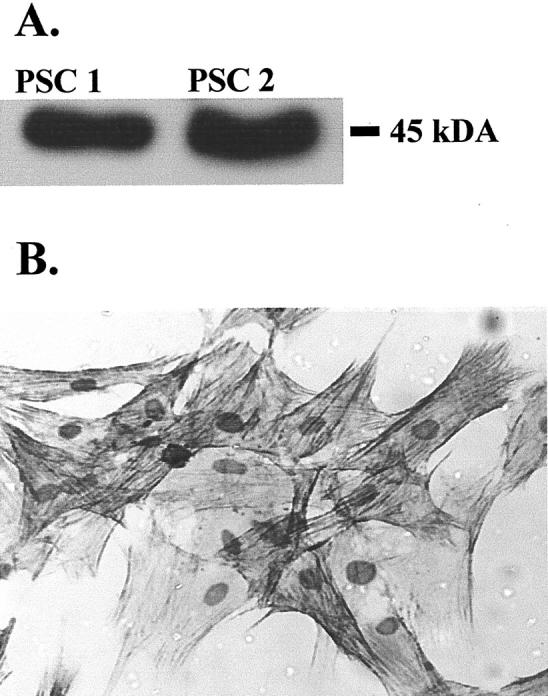
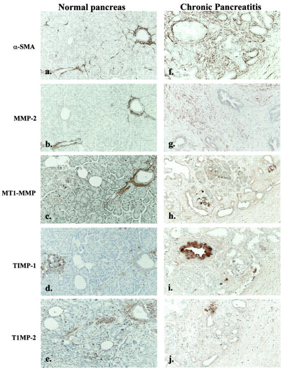
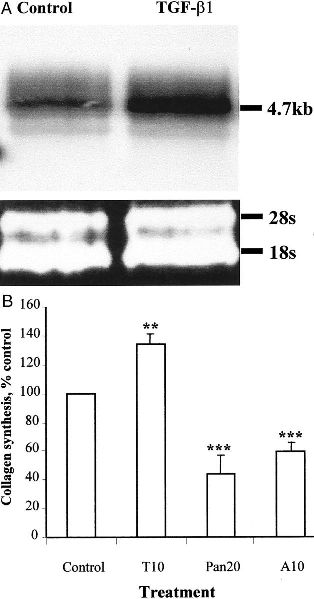
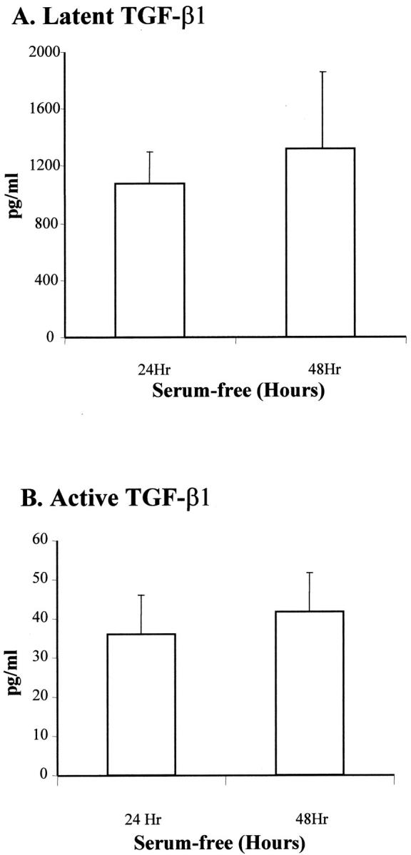
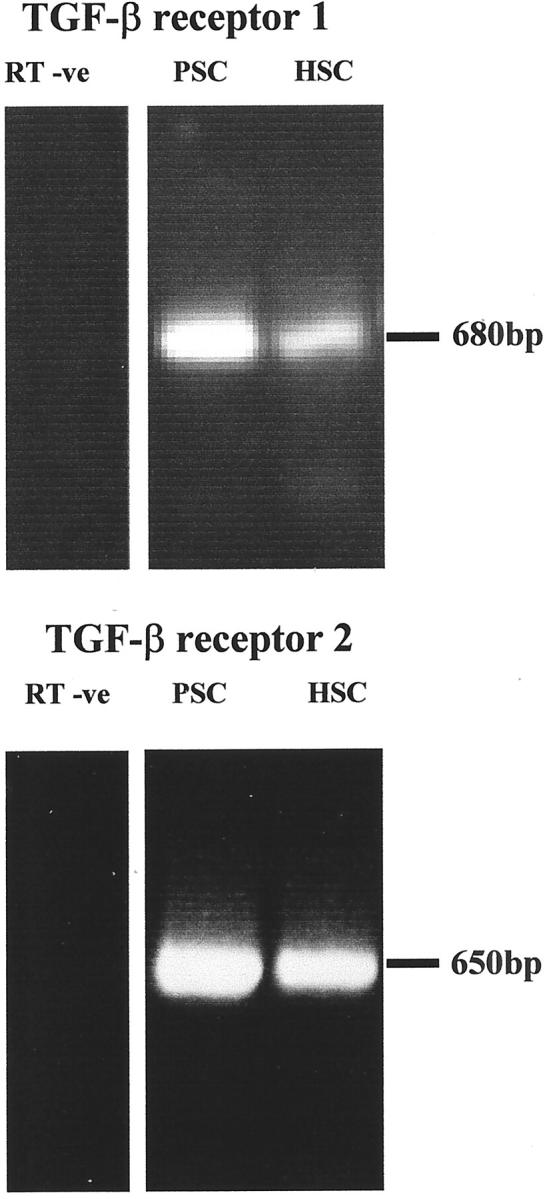
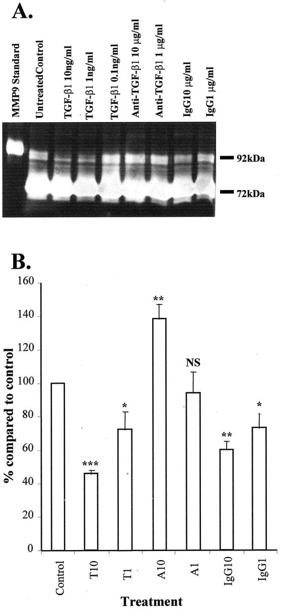
Pancreatic stellate cells mediate fibrosis in chronic pancreatitis. Matrix metalloproteinases (MMPs) and tissue inhibitors of metalloproteinases (TIMPs)-1 and -2 are crucial modulators of fibrosis. Transforming growth factor-beta (TGF-beta) is a key regulator of extracellular matrix production and myofibroblast proliferation. We have examined MMP and TIMP synthesis by transformed cultured pancreatic stellate cells and their regulation by TGF-beta 1. By Northern analysis they expressed mRNAs for procollagen 1, TIMP-1, TIMP-2, and MMP-2. Expression of membrane type-1 MMP was confirmed by Western blotting. By immunohistochemistry these enzymes localized to fibrotic areas in human chronic pancreatitis. Active TGF-beta 1 constitutes 2 to 5% of total TGF-beta 1 secreted by pancreatic stellate cells; they express TGF-beta receptors I and II. Exogenous TGF-beta 1 (10 ng/ml) significantly increased procollagen-1 mRNA by 69% and collagen protein synthesis by 34%. Similarly TGF-beta 1 at 0.1, 1, and 10 ng/ml significantly reduced cellular proliferation rate by 37%, 44%, and 44%, respectively, whereas pan-TGF-beta-neutralizing antibody increased proliferation by 40%. TGF-beta1 (10 ng/ml) down-regulated MMP-9 by 54% and MMP-3 by 34% whereas TGF-beta 1-neutralizing antibody increased MMP-9 expression by 39%. Pancreatic stellate cells express both mediators of matrix remodeling and the regulatory cytokine TGF-beta 1 that, by autocrine inhibition of MMP-3 and MMP-9, may enhance fibrogenesis by reducing collagen degradation.


Figures

Figure 1.
Figure 1.
A: α-SMA was detected by Western analysis of 2 μg of protein extracts of two separate preparations (PSC1, PSC2) of activated PSCs. After autoradiography, a single signal of appropriate molecular weight (45 kd) was observed. Data are representative of three further PSC preparations. B: Activated PSCs (passage 2) express α-SMA. Cell cultures were fixed and immunostained for α-SMA using a monoclonal antibody. Data are representative of three separate preparations.
Figure 2.
Figure 2.
Total cellular RNA was examined by Northern blotting for the expression of α-SMA, collagen-I, MMP 2, TIMP-1, and TIMP-2. A single signal of appropriate size was observed for each of the mRNAs for α-SMA, procollagen-1, MMP 2, and TIMP-1 whereas TIMP-2 mRNA was expressed as two splice variants of 1.0 and 3.4 kb from activated PSCs passaged two to four times. Similar results were observed in four separate preparations of PSCs.
Figure 3.
Figure 3.
Protein extracts (15 μg) from two separate preparations of passages 2 to 4 rat PSCs (PSC1, PSC2) and one preparation of passage 3 human PSCs (PSC3) were subjected to Western analysis for MT1-MMP. After autoradiography a single signal consistent with presence of active MT1-MMP of 45 kd was observed.
Figure 4.
Figure 4.
Representative sections of normal pancreas from one patient were immunostained for α-SMA, MMP-2, MT1-MMP, TIMP-1, and TIMP-2 as described in Materials and Methods. Positive staining for α-SMA (a), MMP-2 (b), and MT1-MMP (c) was observed in the vascular and perivascular stroma. In addition, islets also stained strongly for TIMP-1 (d) and TIMP-2 (e). Representative sections of resected samples from a chronic pancreatitis patient were immunostained for α-SMA (f), MMP-2 (g), MT1-MMP (h), TIMP-1 (i), and TIMP-2 (j). The vascular and perivascular stroma was positive for all of the proteins. In addition, each of the fibrosis mediators displayed a similar distribution pattern to α-SMA in the fibrosis area. (f–j). Original magnifications, ×25.
Figure 5.
Figure 5.
Effect of TGF-β1 on the PSC collagen synthesis. A: Northern analysis compared procollagen type 1 mRNA in cells treated for 24 hours with TGF-β1 (10 ng/ml) and untreated control. The ribosomal bands confirmed equal loading of total mRNA in the experiments. Results were representative of three independent experiments. B: Effects of TGF-β1 10 ng/ml (T10), neutralizing antibody to pan-TGF-β 20 μg/ml (Pan20), and neutralizing antibody to TGF-β1 10 μg/ml (A10) on collagen protein synthesis. Results are expressed as a percentage (mean ± SE) of control values pooled from six separate PSC preparations. **, P < 0.05; ***, P < 0.005.
Figure 6.
Figure 6.
Effect of TGF-β1 at concentrations of 0.1 (T0.1), 1 (T1), and 10 (T10) ng/ml and neutralizing antibody to pan-TGF-β at 20 μg/ml (Pan20) and nonimmune IgG at 20 μg/ml (IgG) on proliferation rate of activated PSCs passaged two to four times. Results were expressed as a percentage of control values and were representative of 10 separate PSC preparations. ***, P < 0.005.
Figure 7.
Figure 7.
A: Quantification of latent TGF-β1 in supernatant of PSCs cultured for 24 hours and 48 hours in serum-free conditioned media. Using commercial ELISA, latent TGF-β1 was determined after hydrochloric acid activation. Data were representative of six separate PSC preparations. B: Quantification of active TGF-β1 in supernatant of PSCs cultured for 24 and 48 hours in serum-free conditioned media. Active TGF-β1 was measured directly by ELISA. Data were representative of six separate PSC preparations. The amount of active TGF-β1 ranged from 2 to 5% of the total TGF-β1 expressed by PSCs.
Figure 8.
Figure 8.
Expression of TGF-β receptors I and II by PSCs. RT-PCR showed that mRNA of TGF-β receptors I and II were detectable in activated PSCs and mRNA from activated hepatic stellate cells were used as a positive control. Data were representative of four separate PSC preparations.
Figure 9.
Figure 9.
A: A representative Western blot demonstrating the regulation of MMP3 by TGF-β1 at 10 ng/ml and 1 ng/ml. Equal quantity of serum-free conditioned media protein were used in all of the experiments. Similar results were observed in three separate PSC preparations. B: Densitometric analysis of the Western blots demonstrating the effect of TGF-β1 at concentrations of 1 ng/ml (T1) and 10 ng/ml (T10) on the regulation of MMP3. The results were expressed as a percentage (mean ± SE) of the control values with data pooled from three separate PSC preparations. Results were corrected for the small variation in DNA in the culture of each condition. **, P < 0.05.
Figure 10.
Figure 10.
A: A representative gelatin zymography demonstrating the expression of MMP9 and MMP2 at 92 kd and 72 kd, respectively, by PSCs. This also illustrates the effect of TGF-β1 in the regulation of MMP9 at the concentration shown. Eight μl was used in each lane. Similar results were observed in three separate PSC preparations. B: Densitometric analysis of the gelatin zymography demonstrating the effects of TGF-β1 at 10 ng/ml (T10) and 1 ng/ml (T1) and neutralizing antibody to TGF-β1 at 10 μg/ml (A10) and 1 μg/ml (A1) on the regulation of MMP9. A nonimmune IgG at 10 μg/ml (IgG10) and 1 μg/ml (IgG1) were included as a control. The results were expressed as percentage (mean ± SE) of the control (nontreatment) values. Results were corrected for the small variations in DNA in the culture of each condition. Data were pooled from three separate PSC preparations. ***, P < 0.005; **, P < 0.05; *, P < 0.1).
Figure 11.
Figure 11.
A schematic diagram proposing how TGF-β1 supports a net accumulation of collagen in pancreatic fibrosis through inhibition of MMPs and stimulation of collagen synthesis.

Similar articles

Cited by

References

    1. Apte MV, Haber PS, Darby SJ, Rodgers SC, McCaughan GW, Korsten MA, Pirola RC, Wilson JS: Pancreatic stellate cells are activated by proinflammatory cytokines: implications for pancreatic fibrogenesis. Gut 1999, 44:534-541 - PMC - PubMed
    1. Apte MV, Haber PS, Applegate TL, Norton ID, McCaughan GW, Korsten MA, Pirola RC, Wilson JS: Periacinar stellate shaped cells in rat pancreas: identification, isolation, and culture. Gut 1998, 43:128-133 - PMC - PubMed
    1. Bachem MG, Schneider E, Gross H, Weidenbach H, Schmid RM, Menke A, Siech M, Beger H, Grunert A, Adler G: Identification, culture, and characterization of pancreatic stellate cells in rats and humans. Gastroenterology 1998, 115:421-432 - PubMed
    1. Friedman SL: Seminars in medicine of the Beth Israel Hospital, Boston. The cellular basis of hepatic fibrosis. Mechanisms and treatment strategies. N Engl J Med 1993, 328:1828-1835 - PubMed
    1. Arthur MJ: Collagenases and liver fibrosis. J Hepatol 1995, 22:43-48 - PubMed

Publication types

MeSH terms

Substances