. 2002 May 1;16(9):1150-62.
doi: 10.1101/gad.219302.

Hox gene control of segment-specific bristle patterns in Drosophila

Affiliations

Hox gene control of segment-specific bristle patterns in Drosophila

Marion Rozowski et al. Genes Dev. .

Abstract

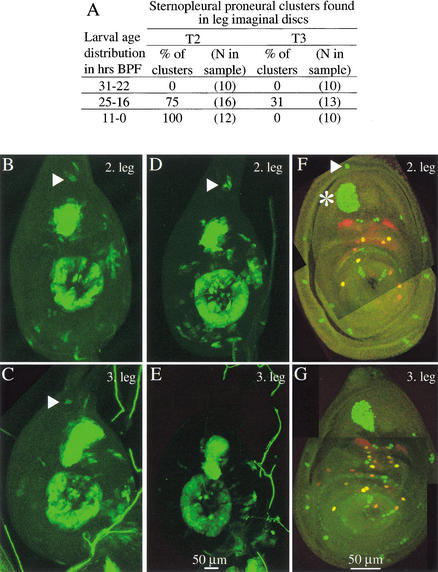
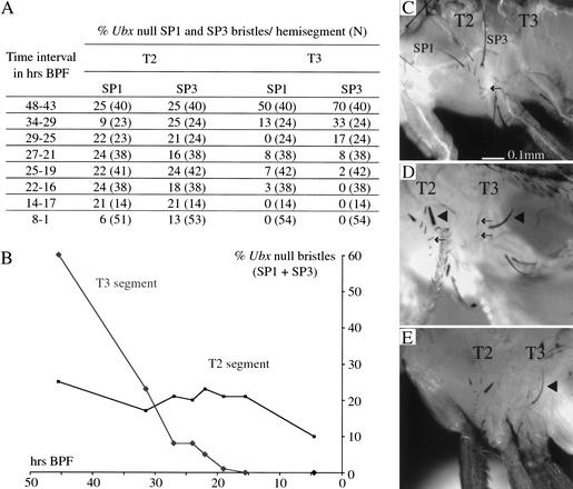
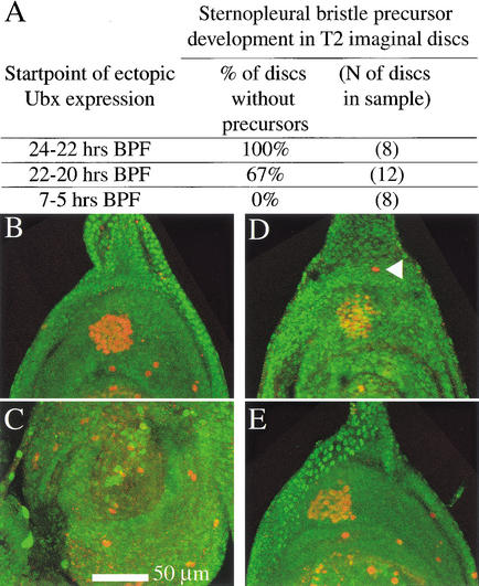
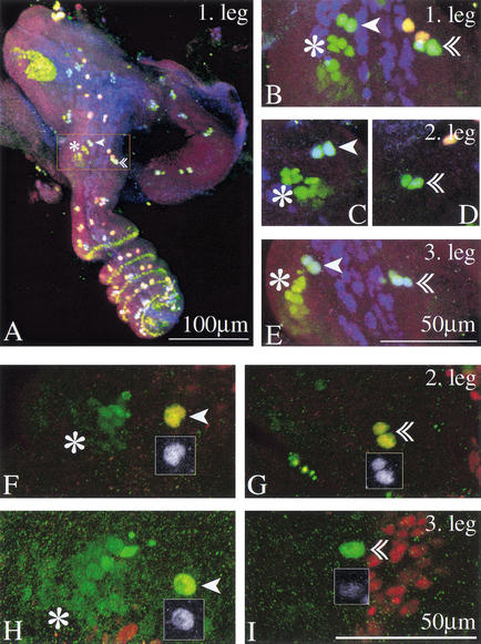
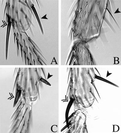
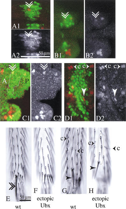
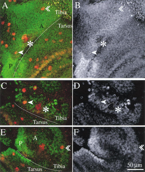
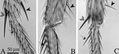
Hox genes specify the different morphologies of segments along the anteroposterior axis of animals. How they control complex segment morphologies is not well understood. We have studied how the Hox gene Ultrabithorax (Ubx) controls specific differences between the bristle patterns of the second and third thoracic segments (T2 and T3) of Drosophila melanogaster. We find that Ubx blocks the development of two particular bristles on T3 at different points in sensory organ development. For the apical bristle, a precursor is singled out and undergoes a first division in both the second and third legs, but in the third leg further differentiation of the second-order precursors is blocked. For the posterior sternopleural bristle, development on T3 ceases after proneural cluster initiation. Analysis of the temporal requirement for Ubx shows that in both cases Ubx function is required shortly before bristle development is blocked. We suggest that interactions between Ubx and the bristle patterning hierarchy have evolved independently on many occasions, affecting different molecular steps. The effects of Ubx on bristle development are highly dependent on the context of other patterning information. Suppression of bristle development or changes in bristle morphology in response to endogenous and ectopic Ubx expression are limited to bristles at specific locations.


Figures

Figure 1
Figure 1
Development of the posterior sternopleural bristle in the third leg ceases after proneural cluster initiation—no precursor segregation takes place. (A) Summary table. Sternopleural proneural clusters were identified as cells staining for GFP expressed with the sca-Gal4 line. (BE) Leg imaginal discs of two individuals from the time interval 25–16 h BPF during which the cluster (triangle) emerges in T2 and T3 and also disappears in T3. One individual shows a few GFP positive cells in the dorsal outer ring in the second (B) and the third leg (C). The other individual shows several GFP positive cells in the dorsal outer ring only in the second leg (D), not in the third leg (E). (F,G) Sensory organ precursors at the white prepupa stage. A sternopleural bristle precursor is found in the second leg (triangle) but not the third leg. Discs of the neuralized-lacZ line were stained with anti-β-galactosidase (green) and with anti-Pox-neuro (red). In the second leg disc (F), the sternopleural bristle precursor (triangle) is located in the outer ring of the disc dorsal to the large femoral chordotonal organ (asterisk).
Figure 2
Figure 2
Formation of anterior and posterior sternopleural macrochaetes (SP1 and SP3) in T3 following timed induction of Ubx null clones and Ubx null clone induction targeted to the sternopleural proneural cluster. (A) Summary table. (B) Graph showing the frequency of T2 and T3 anterior and posterior sternopleural bristles as a function of the time of Ubx null clone induction. (C) Lateral thorax of a wild-type fly. Sternopleural bristles are found exclusively on T2 (SP1 anterior and SP3 posterior sternopleural macrochaete; the arrow points to the row of microchaetes). (D) Fly with Sb+-marked Ubx null clones induced with a heatshock 28 h BPF. Ubx null clones induced at this time give rise to sternopleural macrochaetes (triangle) and microchaetes (arrows) on T3; corresponding bristles on T2 are marked in the same way. (E) Fly with Sb+-marked Ubx null clones induced with the scabrous-Gal4 line. sca-Gal4 is transiently expressed in the T3 sternopleural proneural cluster when the cluster is first formed on T2 and T3. Ubx null clones generated at this time give rise to sternopleural macrochaetes on T3 (triangle in E).
Figure 3
Figure 3
Ectopic Ubx expression from 24–22 h BPF can suppress sternopleural bristle precursor development on T2, but ectopic expression from shortly before puparium formation cannot. Larvae were subjected to heatshock regimes covering different spans of third larval instar development. Larvae were examined for sensory organ precursors at a stage corresponding to the white prepupa. Precursors were visualized by staining with anti-β-galactosidase in the neuralized-lacZ background (red). Ubx levels were assayed by costaining for Ubx protein (green). (A) Summary table. (BC) Individual for which the heatshock-Ubx regime started between 24 and 22 h BPF. (B) Second leg disc of this individual. No sternopleural bristle precursor is found in the dorsal leg periphery. (C) Third leg disc of this individual. (DE) Individual for which the hs-Ubx regime started 7 h BPF. (D) Second leg discs of this animal. The sternopleural bristle precursor is present in the dorsal leg periphery (arrowhead). (E) Third leg of this animal. (C,E) In both treatments, ectopic Ubx levels at the time of collection have not sunk below endogenous third leg levels.
Figure 4
Figure 4
Second-order precursors for both the apical and the preapical bristle form on all three legs. The apical bristle precursors on T3 lose their neural markers but survive. (AE) Leg imaginal discs of the neu-lacZ line 4–4.5 h APF stained with anti-β-galactosidase (green), with anti-Pox-neuro (red) and with anti-Cut (blue). Mechanosensory bristles appear blue-green. (A) First leg. The inset frames the distal tibia and is magnified in B. (BE) Close-up of the distal tibia in the three legs. Second-order precursors for the preapical (arrowhead) and the apical bristle (double arrowhead) are found in all three legs. Preapical bristle precursors are in close proximity to the tibial chordotonal organ precursors (asterisk). (FI) Leg discs from a white prepupa of the neu-lacZ line, 4.5 h APF stained with anti-β-galactosidase (green) and anti-Cut (red). Sensory organs are marked as above. Insets show Cut expression of the precursors. (F) Preapical and (G) apical bristle precursors of the second leg; (H) preapical and (I) apical bristle precursors of the third leg. Cut expression has faded from the apical bristle precursors in the third leg, while anti-β-galactosidase expression still marks the cells (I).
Figure 5
Figure 5
Removal of Ubx from the second-order precursors results in shaft and socket development of the apical bristle on T3 and converts preapical bristle morphology on T3 towards T2. (AD) Double arrowhead marks the apical bristle, arrowhead marks the preapical bristle. (A) Wild-type second leg and (B) wild-type third leg. (C) Second leg of experimental animal expressing the Sb mutation in apical and preapical bristle. (D) Third leg in which mitotic recombination driven in the sensory organ lineage resulted in Sb+ macrochaetes.
Figure 6
Figure 6
In a regime of prolonged ectopic Ubx expression, neural markers disappear from the apical bristle precursors on T2. The adult flies show selective loss of the apical bristle. (AD) Confocal images of the apical bristle precursors in prepupal legs with ectopic Ubx expression (Ubx isoform Ia) driven by dpp-Gal4 in the neu-lacZ background. Precursors are marked by anti-β-galactosidase staining (red), and Ubx expression is shown (green). Double arrowhead marks the apical bristle precursors, arrowhead marks the preapical bristle precursors. (A) Second leg about 3 h APF. Second-order precursors for the apical bristle are present within the domain of ectopic Ubx expression. (A2) shows the Ubx channel. (B) Second leg 5–6 h APF. Very low levels of anti-β-galactosidase staining remain in the apical bristle precursors (see red channel in B2). Anti-β-galactosidase staining is also reduced in the apical bristle precursors of the third leg of this individual (C). The stripe of ectopic Ubx expression in the third leg (white outlines in C1) compares to Ubx levels in the posterior third leg (marked P), exceeding levels in the anterior third leg (marked A). (D) In the dorsal second leg, the stripe of ectopic Ubx expression occupies most of the width between the chemosensory precursors (small arrowheads accompanied by the letter c). (E) Ventral aspect of the distal tibia of an adult wild-type fly and (F) of a fly from the above regime of ectopic Ubx expression—the apical bristle is missing from the ventral side of the tibia. From the dorsal domain of ectopic Ubx expression (D) the preapical bristle (arrowhead in H) and mechanosensory microchaetes on the dorsal leg (circle in H) arise normally. (G) Dorsal aspect of the distal tibia of a wild-type fly.
Figure 7
Figure 7
Ubx protein levels are modulated in the third leg epithelium, but the levels in apical and preapical bristle precursors are similar. Confocal images of distal tibia and basitarsus of third leg of neu-lacZ flies 2.5 h APF (A,B) and 4.5 h APF (CF). In green and right-hand panels anti-Ubx staining; in red anti-β-galactosidase staining. (A,B) Third leg 2.5 h APF. In the area reproduced here, Ubx protein levels are modulated: Expression is higher in posterior (P) than anterior leg (A); levels are higher in anterior distal tibia than anterior basitarsus; levels are reduced in anterior distal tibia in a dorsal strip containing sensory organ precursors and in the invaginating cells of the tibial chordotonal organ (see outline marked by asterisk in B). The first-order precursors of apical (arrowhead) and preapical bristle (double arrowhead) show similar levels of Ubx expression (see outlines marked with arrowheads in B). (C,D) Dorsal and (E,F) ventral aspects of distal tibia and basitarsus of a third leg at 4.5 h APF. By this time, the second-order precursors of apical and preapical bristle have formed. Ubx levels in these precursors are still comparable; Ubx levels in the tibial-chordotonal organ remain reduced (see outlines marked with arrowheads respectively asterisk in D and F).
Figure 8
Figure 8
Ectopic Ubx expression in the sensory organ lineage has different effects on bristle morphology depending on bristle identity or bristle location. (A) Wild-type second leg and (B) wild-type third leg. (C) Second leg in which Ubx has been expressed in the sensory organ lineage with the 109-68 Gal4 driver at 18°C. In the ectopic Ubx expression regime (C), the stout preapical bristle on T2 is transformed toward the morphology of the thin preapical bristle normally found on T3 (arrowhead), whereas short bristles on the ventral basitarsus of T2 can be transformed toward the long stout morphology of those on the T3 leg (arrows). The morphology of the T2 apical bristle remains largely unchanged in this ectopic Ubx expression regime (double arrowhead).

Similar articles

Cited by

References

    1. Abu-Shaar M, Mann RS. Generation of multiple antagonistic domains along the proximodistal axis during Drosophila leg development. Development. 1998;125:3821–3830. - PubMed
    1. Akam M. The molecular basis for metameric pattern in the Drosophila embryo. Development. 1987;101:1–22. - PubMed
    1. Auerbach C. The development of the legs, wings, and halteres in wild type and some mutant strains of Drosophila melanogaster. Proc R Soc Edinb B. 1936;58:787–815.
    1. Baonza A, Roch F, Martin-Blanco E. DER signaling restricts the boundaries of the wing field during Drosophila development. Proc Natl Acad Sci. 2000;97:7331–7335. - PMC - PubMed
    1. Blochlinger K, Bodmer R, Jan LY, Jan YN. Patterns of expression of Cut, a protein required for external sensory organ development in wild-type and cut mutant Drosophila embryos. Genes & Dev. 1990;4:1322–1331. - PubMed

Publication types

LinkOut - more resources