

doi: 10.1093/nar/30.10.e47.
Telomere measurement by quantitative PCR
Affiliations
- PMID: 12000852
- PMCID: PMC115301
- DOI: 10.1093/nar/30.10.e47
Item in Clipboard
Telomere measurement by quantitative PCR
Nucleic Acids Res.
.
Abstract
It has long been presumed impossible to measure telomeres in vertebrate DNA by PCR amplification with oligonucleotide primers designed to hybridize to the TTAGGG and CCCTAA repeats, because only primer dimer-derived products are expected. Here we present a primer pair that eliminates this problem, allowing simple and rapid measurement of telomeres in a closed tube, fluorescence-based assay. This assay will facilitate investigations of the biology of telomeres and the roles they play in the molecular pathophysiology of diseases and aging.
Figures
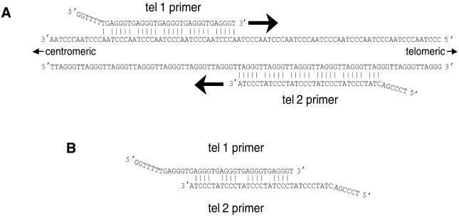
Annealing of primers tel 1 and tel 2 to genomic DNA and to each other in the first round of PCR. (A) Annealing of primers to genomic DNA. The tel 1 primer can hybridize to any available partially complementary 31 bp stretch along the strand of telomeric DNA oriented 5′→3′ toward the centromere. The tel 2 primer can hybridize to any partially complementary 33 bp stretch along the strand oriented 5′→3′ toward the end of the chromosome. In both of these primer–template hybridizations, every sixth base is mismatched, however, the last five bases at the 3′-end of the primers are perfectly matched to complementary bases in the template. Addition of bases by DNA polymerase begins at the 3′-ends of the annealed primers and proceeds in the direction of the large arrows. (B) Annealing of primers to each other. The strongest possible hybridizations of the primers to each other involve a repeated pattern of six bases containing four consecutive paired bases followed by two mismatched bases, an example of which is shown here. Note that the 3′-terminal base of each primer cannot form a stable base pair with the base opposite it, thereby blocking addition of bases by DNA polymerase.
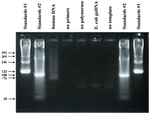
Agarose gel electrophoresis following PCR with the tel 1 and tel 2 primers. PCR was as described in Materials and Methods, except that 22 cycles of PCR were performed instead of 18 cycles. Eight microliters of reaction product were then loaded into each lane of a 4% (Nusieve 3:1) agarose gel and electrophoresis carried out at ∼2.5 V/cm in 45 mM Tris–borate, 1 mM EDTA with ethidium bromide present at a concentration of 0.5 µg/ml. The gel was then transilluminated with UV light and digital photography was performed with the Stratagene EagleEye System. Lanes 1, 2, 8 and 9, size standards. For lane 3 the template for PCR was 35 ng of total human genomic DNA. The remaining samples differed from the lane 3 sample as follows: lane 4, no primers; lane 5, no polymerase; lane 6, E.coli genomic DNA as template instead of human genomic DNA; lane 7, no template. The bottom of the smear in lane 3 was estimated to contain a 76 bp product, based on a comparison of its mobility with the mobilities of the size standards in a plot of log(number of base pairs) versus distance migrated from the origin.
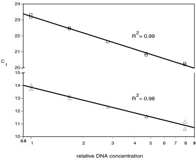
Standard curves used to measure the relative T/S ratio. Five DNA concentrations over an 8-fold range were generated by serial dilution (dilution factor ∼1.68) and aliquoted to microtiter plate wells; the final amounts per well ranged from 12.64 to 100 ng, with the middle quantity approximately matching that of the samples being assayed. The Ct of a DNA sample is the fractional number of PCR cycles to which the sample must be subjected in order to accumulate enough product to cross a set threshold of magnitude of fluorescent signal. Any individual or pooled human DNA sample may be used to create the standard curves, as long as the Ct of each assayed sample falls within the range of Ct values of the standard curves. Circles, single copy gene 36B4; triangles, telomere.
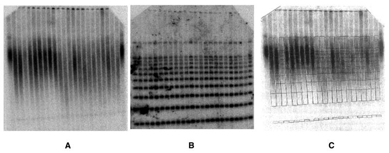
Determination of mean terminal restriction fragment lengths in human DNA samples spiked with DNA size standards. The size standards, a 1 kb ladder ranging from 1 to 10 kb and HindIII-digested phage λ DNA, were added to each individual’s DNA sample after complete restriction enzyme digestion of the human genomic DNA and heat inactivation of the restriction enzyme. (A) Southern blot hybridized with a 32P-end-labeled (TTAGGG)7 oligonucleotide probe. (B) The same blot after stripping the telomere repeat probe and rehybridizing with a mixture of two radiolabeled oligonucleotides, one that binds all components of the 1 kb ladder, the other specific for the 23.13 kb λ HindIII fragment. (C) A grid image of the standard ladders of (B), generated using ImageQuaNT software tools (Molecular Dynamics), was superimposed on the image of the telomere smears of (A) to locate the positions of the size intervals within the smears.
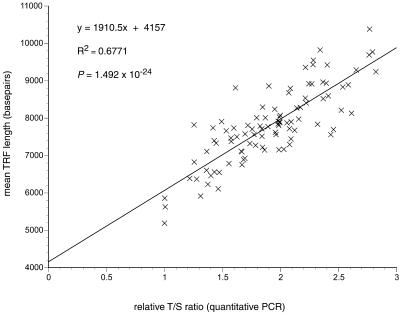
Correlation of relative T/S ratios determined by quantitative PCR and mean TRF lengths determined by Southern blot analysis in DNA samples prepared directly from blood draws on 95 individuals. All relative T/S ratios plotted here have values ≥1.0, because the initial T/S ratios determined using the standard curves have all been normalized to the lowest T/S ratio (0.69) observed among the samples. The equation for the linear regression line best fitting the data is shown.
Similar articles
-
Real-time PCR assay for measurement of mouse telomeres.Comp Med. 2006 Feb;56(1):17-22. Comp Med. 2006. PMID: 16521855
-
Analysis of telomeres and telomerase.Curr Protoc Cell Biol. 2003 Nov;Chapter 18:Unit 18.6. doi: 10.1002/0471143030.cb1806s20. Curr Protoc Cell Biol. 2003. PMID: 18228424
-
Estimation of the amount of telomere molecules in different human age groups and the telomere increasing effect of acupuncture and shiatsu on St.36, using synthesized basic units of the human telomere molecules as reference control substances for the bi-digital O-ring test resonance phenomenon.Acupunct Electrother Res. 1998;23(3-4):185-206. doi: 10.3727/036012998816356472. Acupunct Electrother Res. 1998. PMID: 10193703
-
New developments in telomere length analysis.Exp Gerontol. 2005 May;40(5):363-8. doi: 10.1016/j.exger.2005.02.008. Epub 2005 Mar 29. Exp Gerontol. 2005. PMID: 15919587 Review.
-
The polymerase chain reaction: new variations on an old theme.Curr Opin Biotechnol. 1995 Feb;6(1):24-9. doi: 10.1016/0958-1669(95)80005-0. Curr Opin Biotechnol. 1995. PMID: 7894078 Review.
Cited by
-
DNA-methylation-based telomere length estimator: comparisons with measurements from flow FISH and qPCR.Aging (Albany NY). 2021 Jun 3;13(11):14675-14686. doi: 10.18632/aging.203126. Epub 2021 Jun 3. Aging (Albany NY). 2021. PMID: 34083495 Free PMC article.
-
The association between leukocyte telomere lengths and sleep instability based on cardiopulmonary coupling analysis.Sleep Breath. 2015 Sep;19(3):963-8. doi: 10.1007/s11325-014-1110-x. Epub 2015 Jan 28. Sleep Breath. 2015. PMID: 25628010
-
Eroded human telomeres are more prone to remain uncapped and to trigger a G2 checkpoint response.Nucleic Acids Res. 2013 Jan;41(2):900-11. doi: 10.1093/nar/gks1121. Epub 2012 Nov 27. Nucleic Acids Res. 2013. PMID: 23193277 Free PMC article.
-
Parental responsiveness moderates the association between early-life stress and reduced telomere length.Dev Psychopathol. 2013 Aug;25(3):577-585. doi: 10.1017/S0954579413000011. Epub 2013 Mar 26. Dev Psychopathol. 2013. PMID: 23527512 Free PMC article.
-
Early exclusive breastfeeding is associated with longer telomeres in Latino preschool children.Am J Clin Nutr. 2016 Aug;104(2):397-405. doi: 10.3945/ajcn.115.115428. Epub 2016 Jul 20. Am J Clin Nutr. 2016. PMID: 27440083 Free PMC article.
References
-
- Lansdorp P.M., Verwoerd,N.P., van de Rijke,F.M., Dragowska,V., Little,M.T., Dirks,R.W., Raap,A.K. and Tanke,H.J. (1996) Heterogeneity in telomere length of human chromosomes. Hum. Mol. Genet., 5, 685–691. - PubMed
-
- Bryant J.E., Hutchings,K.G., Moyzis,R.K. and Griffith,J.K. (1997) Measurement of telomeric DNA content in human tissues. Biotechniques, 23, 476–478, 480, 482, passim. - PubMed
-
- Norwood D. and Dimitrov,D.S. (1998) Sensitive method for measuring telomere lengths by quantifying telomeric DNA content of whole cells. Biotechniques, 25, 1040–1045. - PubMed
-
- Nakamura Y., Hirose,M., Matsuo,H., Tsuyama,N., Kamisango,K. and Ide,T. (1999) Simple, rapid, quantitative and sensitive detection of telomere repeats in cell lysate by a hybridization protection assay. Clin. Chem., 45, 1718–1724. - PubMed
Publication types
MeSH terms
Substances
Grants and funding
LinkOut - more resources
Full Text Sources
Other Literature Sources
