. 2002 Apr 25;2(1):2.
doi: 10.1186/1472-6823-2-2.

Islet expression of the DNA repair enzyme 8-oxoguanosine DNA glycosylase (Ogg1) in human type 2 diabetes

Affiliations

Islet expression of the DNA repair enzyme 8-oxoguanosine DNA glycosylase (Ogg1) in human type 2 diabetes

Björn Tyrberg et al. BMC Endocr Disord. .

Abstract

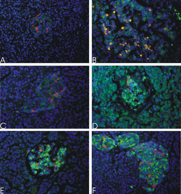
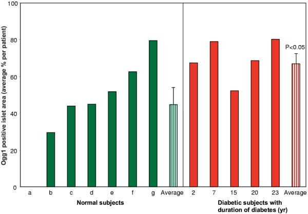
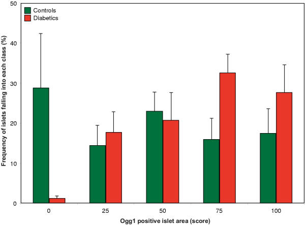
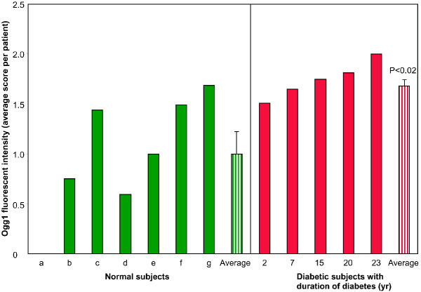
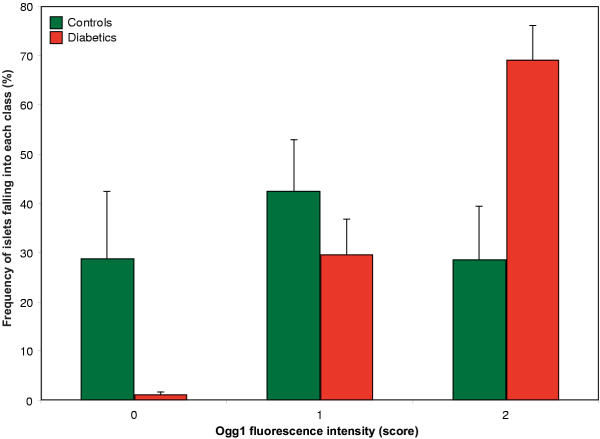
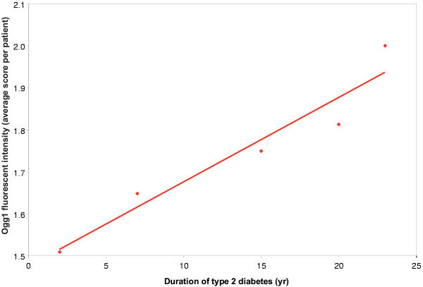
BACKGROUND: It has become increasingly clear that beta-cell failure plays a critical role in the pathogenesis of type 2 diabetes. Free-radical mediated beta-cell damage has been intensively studied in type 1 diabetes, but not in human type 2 diabetes. Therefore, we studied the protein expression of the DNA repair enzyme Ogg1 in pancreases from type 2 diabetics. Ogg1 was studied because it is the major enzyme involved in repairing 7,8-dihydro-8-oxoguanosine DNA adducts, a lesion previously observed in a rat model of type 2 diabetes. Moreover, in a gene expression screen, Ogg1 was over-expressed in islets from a human type 2 diabetic. METHODS: Immunofluorescent staining of Ogg1 was performed on pancreatic specimens from healthy controls and patients with diabetes for 2-23 years. The intensity and islet area stained for Ogg1 was evaluated by semi-quantitative scoring. RESULTS: Both the intensity and the area of islet Ogg1 staining were significantly increased in islets from the type 2 diabetic subjects compared to the healthy controls. A correlation between increased Ogg1 fluorescent staining intensity and duration of diabetes was also found. Most of the staining observed was cytoplasmic, suggesting that mitochondrial Ogg1 accounts primarily for the increased Ogg1 expression. CONCLUSION: We conclude that oxidative stress related DNA damage may be a novel important factor in the pathogenesis of human type 2 diabetes. An increase of Ogg1 in islet cell mitochondria is consistent with a model in which hyperglycemia and consequent increased beta-cell oxidative metabolism lead to DNA damage and the induction of Ogg1 expression.


Figures

Figure 1
Figure 1
Ogg1 immunofluorescent staining in human pancreata. Ogg1 staining (green) and somatostatin (red), as a marker for islets of Langerhans, in normal human pancreata (A, C, E); and pancreata from type 2 diabetic subjects (B, D, F). Micrograph A is representative of an islet with no or very little Ogg1 staining; B of an islet with moderate staining intensity of 25% of the area; C, strong staining of 50% of the area; D, strong staining of 100% of the area; E, strong staining of 75% of the area; and F, strong staining of 100% of the area. Notably, the surrounding exocrine tissue is positive for Ogg1 at varying degrees (B and D vs. the rest). The micrographs were captured with identical camera settings. Original optical magnification 400×.
Figure 2
Figure 2
Islet area stained for Ogg1. Islets were semi-quantitatively scored according to Methods into five different groups depending on the percentage of the area stained positive for Ogg1. Islets from type 2 diabetic subjects (2, 7, 15, 20, 23 years of diabetes duration, red) have significantly larger areas stained for Ogg1 (P < 0.05) than islets from healthy controls (a-g, green).
Figure 3
Figure 3
Islet area stained for Ogg1. Islets were semi-quantitatively scored according to Methods into five different groups depending on the percentage of the area stained positive for Ogg1. The frequency distribution of the scoring in normal and diabetic subjects shows that it is shifted to the left in islets from type 2 diabetic subjects.
Figure 4
Figure 4
Ogg1 fluorescent staining intensity. Islets were semi-quantitatively scored according to Methods into three different groups depending on the islet Ogg1 fluorescent intensity. Islets from type 2 diabetic subjects (2, 7, 15, 20, 23 years of diabetes duration, red) have significantly higher islet Ogg1 fluorescent intensity (P < 0.02) than islets from healthy controls (a-g, green).
Figure 5
Figure 5
Ogg1 fluorescent staining intensity. Islets were semi-quantitatively scored according to Materials and Methods into three different groups depending on the islet Ogg1 fluorescent intensity. The frequency distribution of the scoring in normal and diabetic subjects shows that it is shifted to the left in islets from type 2 diabetic subjects. Fluorescent intensity is expressed as arbitrary units.
Figure 6
Figure 6
Diabetes duration affects the Ogg1 expression. Increased type 2 diabetes duration increases the Ogg1 fluorescent intensity and expression. The slope of the linear regression significantly differs from zero (P < 0.01) and the regression coefficient (R2) is 0.92.

Similar articles

Cited by

References

    1. Tyrberg B, Levine F. Current and future treatment strategies for type 2 diabetes: the β-cell as a therapeutic target. Curr Opin Investig Drugs. 2001;2:1568–1574. - PubMed
    1. Finegood DT, Scaglia L, Bonner-Weir S. Dynamics of beta-cell mass in the growing rat pancreas. Estimation with a simple mathematical model. Diabetes. 1995;44:249–256. - PubMed
    1. Sorenson RL, Brelje TC. Adaptation of islets of Langerhans to pregnancy: beta-cell growth, enhanced insulin secretion and the role of lactogenic hormones. Horm Metab Res. 1997;29:301–307. - PubMed
    1. Kahn BB. Type 2 diabetes: when insulin secretion fails to compensate for insulin resistance. Cell. 1998;92:593–6. doi: 10.1016/S0092-8674(00)81125-3. - DOI - PubMed
    1. Farnier M, Picard S. Diabetes: Statins, Fibrates, or Both? Curr Atheroscler Rep. 2001;3:19–28. - PubMed