. 2002 Jun;184(11):2906-13.
doi: 10.1128/JB.184.11.2906-2913.2002.

Plasmid-encoded asp operon confers a proton motive metabolic cycle catalyzed by an aspartate-alanine exchange reaction

Affiliations

Plasmid-encoded asp operon confers a proton motive metabolic cycle catalyzed by an aspartate-alanine exchange reaction

Keietsu Abe et al. J Bacteriol. 2002 Jun.

Abstract

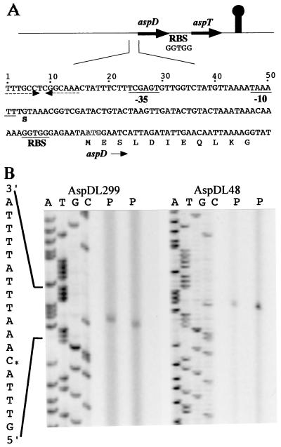
Tetragenococcus halophila D10 catalyzes the decarboxylation of L-aspartate with nearly stoichiometric release of L-alanine and CO(2). This trait is encoded on a 25-kb plasmid, pD1. We found in this plasmid a putative asp operon consisting of two genes, which we designated aspD and aspT, encoding an L-aspartate-beta-decarboxylase (AspD) and an aspartate-alanine antiporter (AspT), respectively, and determined the nucleotide sequences. The sequence analysis revealed that the genes of the asp operon in pD1 were in the following order: promoter --> aspD --> aspT. The deduced amino acid sequence of AspD showed similarity to the sequences of two known L-aspartate-beta-decarboxylases from Pseudomonas dacunhae and Alcaligenes faecalis. Hydropathy analyses suggested that the aspT gene product encodes a hydrophobic protein with multiple membrane-spanning regions. The operon was subcloned into the Escherichia coli expression vector pTrc99A, and the two genes were cotranscribed in the resulting plasmid, pTrcAsp. Expression of the asp operon in E. coli coincided with appearance of the capacity to catalyze the decarboxylation of aspartate to alanine. Histidine-tagged AspD (AspDHis) was also expressed in E. coli and purified from cell extracts. The purified AspDHis clearly exhibited activity of L-aspartate-beta-decarboxylase. Recombinant AspT was solubilized from E. coli membranes and reconstituted in proteoliposomes. The reconstituted AspT catalyzed self-exchange of aspartate and electrogenic heterologous exchange of aspartate with alanine. Thus, the asp operon confers a proton motive metabolic cycle consisting of the electrogenic aspartate-alanine antiporter and the aspartate decarboxylase, which keeps intracellular levels of alanine, the countersubstrate for aspartate, high.


Figures

FIG. 1.
FIG. 1.
Determination of the aspDT mRNA 5′ end and gene organization of the asp operon of T. halophila and its putative 5′ flanking region. (A) At the top, the genes are indicated by arrows (not to scale). The directions of transcription are also indicated by the arrows. The noncoding region following the aspT gene contained a sequence with a potential structure in the mRNA that was similar to the rho-independent transcription termination signal of E. coli (indicated by a mushroom-like symbol). The two open reading frames each contained an ATG codon and were preceded by possible RBSs at distances of 7 to 9 nucleotides. The assigned initiation codons are putative. The termination codons were TAA for both aspD and aspT. The intergenic noncoding region in the asp cluster was 25 bp between aspD and aspT. At the bottom, the nucleotide sequence of the 5′ flanking region for aspD is shown. ATG in white letters indicates the location of the start codon for aspD. The dashed arrows indicate a palindrome. The transcription initiation site is labeled S, and putative −35 and −10 regions deduced from the transcript analysis described below are underlined. An RBS is also underlined. (B) Primer extension analysis was carried out with oligonucleotides AspDL229 and AspDL48 and RNA from aspartate-grown cells of T. halophila D10. Lanes P, primer extension aliquot (duplicate); lanes C, G, T, and A, sequence ladder generated with AspDL229 or AspDL48. The +1 site is marked with an asterisk.
FIG. 2.
FIG. 2.
Similarity of the amino acid sequence of T. halophila AspD (AspD/Th) (DDBJ/EMBL/GenBank accession no. AB072729) to the amino acid sequences of P. dacunhae aspartate-β-decarboxylase (AspD/Pd) (22) and A. faecalis aspartate-β-decarboxylase (AsdA/Af) (DDBJ/EMBL/GenBank accession no. AF168368). Asterisks indicate identical amino acid residues. The box and triangles indicate predicted pyridoxal phosphate attachment sites and substrate binding sites, respectively. The numbers for each line are the numbers of amino acid residues starting from the amino terminus. Dashes indicate gaps.
FIG. 3.
FIG. 3.
Aspartate decarboxylation by E. coli cells harboring pTrcAsp (asp operon). The asp operon in pTrcAsp was expressed by induction with 200 μM IPTG for 12 h at 30°C. Cells were harvested, resuspended in 50 mM potassium MES buffer (pH 6) containing 10 mM aspartate, and incubated for 20 min at 30°C. The concentrations of aspartate (open symbols) and alanine (solid symbols) in the reaction mixtures were monitored (□ and ♦, pTrcAsp; ○ and ▴, pTrc99A). The vector without inserts was pTrc99A. The error bars indicate standard errors.
FIG. 4.
FIG. 4.
Purification of AspDHis. (A) SDS-PAGE profiles obtained during purification of AspDHis. Lanes 2 and 3, 114.0 μg of protein of the crude extract and the immediate flowthrough from an Ni2+-NTA column, respectively; lane 4, wash fluid taken just prior to elution of AspDHis by 50 mM sodium phosphate buffer (pH 8) containing 300 mM NaCl and 250 mM imidazole; lane 5, 2.4 μg of purified AspDHis. An arrow indicates the position of AspDHis. (B) Western blot of the profiles described for panel A. The contents of a duplicate SDS-PAGE gel were transferred to a polyvinylidene difluoride membrane and probed with antiserum against the histidine tag.
FIG. 5.
FIG. 5.
AspT expressed in E. coli catalyzes electroneutral aspartate self-exchange and electrogenic exchange of aspartate with alanine. (A) A detergent extract of IPTG-induced cells carrying pTrcAsp or pTrc99A was used to prepare proteoliposomes (or liposomes). Proteoliposomes loaded with 100 mM potassium aspartate and 100 mM potassium phosphate (pH 7) were placed in an NMG-based medium (0.1 M NMG sulfate, 0.1 M NMG phosphate; pH 7) at a concentration of 10 to 25 μg of protein/ml along with 100 μM [3H]aspartate and either 1 μM valinomycin (open symbols) or ethanol (solid symbols). After 8 min (arrow), aliquots from each tube received 10 mM NMG-aspartate (▵ and ▴, pTrcAsp; ⋄, pTrc99A), 10 mM NMG-alanine (○ and •, pTrcAsp; □, pTrc99A), or the equivalent volume of assay buffer (▹). Samples were taken and used for filtration and washing at the times indicated. Control liposomes (▿) were also examined without the addition of unlabeled substrates. Error bars are omitted in the case of pTrc99A (⋄ and □) and control liposomes (▿) for clarity. (B) Proteoliposomes (or liposomes) were loaded with 200 mM alanine plus 100 mM phosphate (pH 7) as either the NMG salt (•, □, and ▪, pTrcAsp; ▵, pTrc99A) or the potassium salt (○, pTrcAsp) and diluted 100-fold into assay media containing 200 μM [3H]aspartate along with 100 mM sulfate plus 100 mM phosphate as the NMG salt (○, □, and ▪, pTrcAsp) or the potassium salt (•, pTrcAsp); with one exception (□) (−Val), 1 μM valinomycin (+Val) was also present. Samples were taken and used for filtration and washing at the times indicated. The presence of external potassium (Kout) or internal potassium (Kin) is indicated. The control proteoliposomes (No K[+Val] or No K[−Val]), whose behavior was largely unaffected by valinomycin, showed aspartate transport virtually identical to that found for the potassium-loaded proteoliposomes not exposed to valinomycin. Liposomes (⊕) without protein loaded with the NMG-based buffer containing 200 mM alanine were also diluted into the potassium-based buffer with 200 μM [3H]aspartate plus 1 μM valinomycin. Error bars are omitted for Kin[+Val] (○), pTrc99A (▵), and control liposomes (⊕) for clarity.

References

    1. Abe, K., H. Hayashi, and P. C. Maloney. 1996. Exchange of aspartate and alanine. J. Biol. Chem. 271:3079-3084. - PubMed
    1. Abe, K., Z. S. Ruan, and P. C. Maloney. 1996. Cloning, sequencing, and expression in Escherichia coli of OxlT, the oxalate:formate exchange protein of Oxalobacter formigenes. J. Biol. Chem. 271:6789-6793. - PubMed
    1. Ambudkar, S. V., and P. C. Maloney. 1986. Bacterial anion exchange, use of osmolyte during solubilization and reconstitution of phosphate-linked antiport from Streptococcus lactis. J. Biol. Chem. 261:10079-10086. - PubMed
    1. Anantharam, V., M. J. Allison, and P. C. Maloney. 1989. Oxalate:formate exchange. The basis for energy coupling in Oxalobacter. J. Biol. Chem. 264:7244-7250. - PubMed
    1. Anderson, D. G., and L. L. McKay. 1983. Simple and rapid method for isolating large plasmid DNA from lactic streptococci. Appl. Environ. Micorobiol. 46:549-552. - PMC - PubMed

MeSH terms

Associated data

LinkOut - more resources