. 2002 Jun;184(11):3008-16.
doi: 10.1128/JB.184.11.3008-3016.2002.

Characterization of interactions between the transcriptional repressor PhlF and its binding site at the phlA promoter in Pseudomonas fluorescens F113

Affiliations

Characterization of interactions between the transcriptional repressor PhlF and its binding site at the phlA promoter in Pseudomonas fluorescens F113

Abdelhamid Abbas et al. J Bacteriol. 2002 Jun.

Abstract

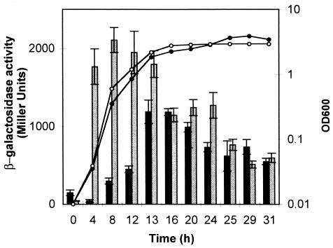
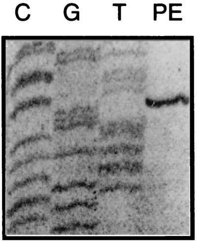
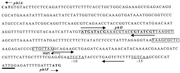
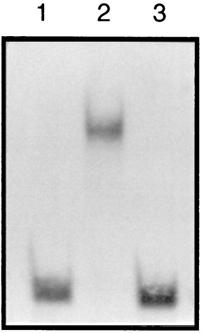
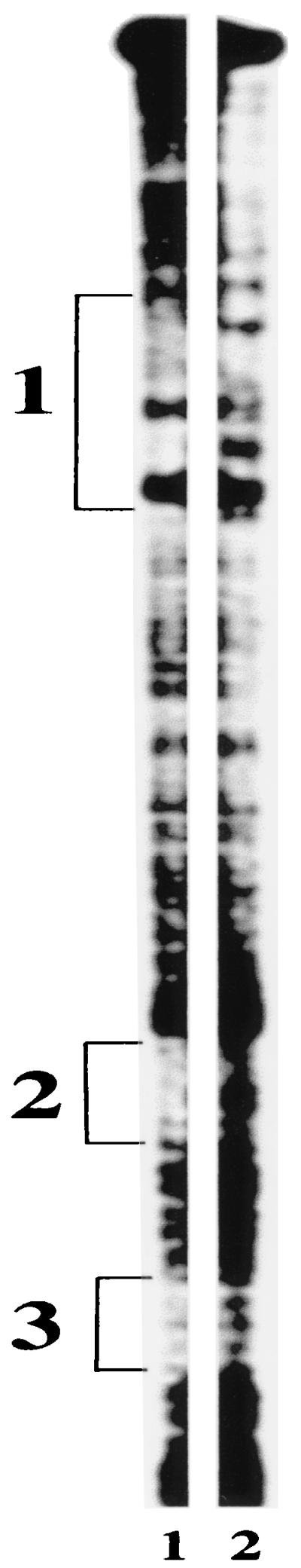
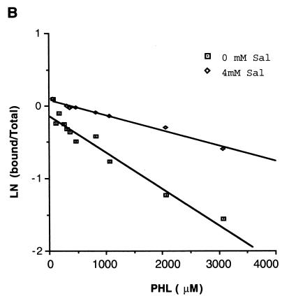
The phlACBD genes responsible for the biosynthesis of the antifungal metabolite 2,4-diacetylphloroglucinol (PHL) by the biocontrol strain Pseudomonas fluorescens F113 are regulated at the transcriptional level by the pathway-specific repressor PhlF. Strong evidence suggests that this regulation occurs mainly in the early logarithmic phase of growth. First, the expression of the phlF gene is relatively high between 3 and 13 h of growth and relatively low thereafter, with the phlACBD operon following an opposite expression profile. Second, the kinetics of PHL biosynthesis are specifically altered in the logarithmic phase in a P. fluorescens F113 phlF mutant. The phlA-phlF intergenic region presents a complex organization in that phlACBD is transcribed from a sigma(70) RNA polymerase-dependent promoter that is likely to overlap the promoter of the divergently transcribed phlF gene. The repression by PhlF is due to its interaction with an inverted repeated sequence, phO, located downstream of the phlA transcriptional start site. Cross-linking experiments indicate that PhlF can dimerize in solution, and thus PhlF may bind phO as a dimer or higher-order complex. Furthermore, it is now demonstrated that certain regulators of PHL synthesis act by modulating PhlF binding to phO. PHL, which has previously been shown to be an autoinducer of PHL biosynthesis, interacts with PhlF to destabilize the PhlF-phO complex. Conversely, the PhlF-phO complex is stabilized by the presence of salicylate, which has been shown to be an inhibitor of phlA expression.


Figures

FIG. 1.
FIG. 1.
Time course of expression of the phl operon. The transcriptional fusions phlACBD-lacZ (pCU107) and phlF-lacZ (pCU109) were introduced into P. fluorescens F113. Cultures were grown on minimal medium, and absorbance was measured at 600 nm (solid circles, phlF-lacZ; open circles, phlACBD-lacZ). Expression of the fusions was assessed by measuring levels of β-galactosidase. Grey shading represents phlF-lacZ expression, and dark shading represents phlACBD-lacZ expression. Triplicate cultures were assayed. The standard deviations are represented with error bars.
FIG. 2.
FIG. 2.
Primer extension analysis mapping the phlA transcriptional start site. Primer extension (PE) on total RNA from P. fluorescens F113 was performed using primer PE-A, which maps inside the phlA coding region. Plasmid DNA was sequenced using the same primer. The gel shows the only primer extension stop detected by this analysis.
FIG. 3.
FIG. 3.
Sequence organization of the phlA-phlF intergenic region. phlA and phlF indicate the start of the phlA and phlF open reading frames, respectively. Start indicates the transcriptional start of the phlACBD operon. The putative σ70 −10 and −35 elements for phlA are underlined. The PhlF binding site, phO, is indicated, with its two inverted repeated sequences shown by arrows. The three regions protected by the PhlF footprint are boxed. From 5′ to 3′, these are box 1, box 2, and box 3, as referred to in the text. The CCAAT (ATTGG) motif of low-temperature-induced genes is underlined. The inverted repeated sequences flanking the −10 element are indicated with arrows. The accession number for this sequence is AF497760.
FIG. 4.
FIG. 4.
Map of probes used in the mobility shift assays in this study. The names of probes are presented on the left of the figure. Binding of PhlF protein to the probes is indicated as + or −. IGR, intergenic region. The 5′ ends of the phlA and phlF ORFs are indicated
FIG. 5.
FIG. 5.
Identification of the PhlF binding site. Probe 7EH (2 ng) was analyzed for reduced mobility in a native gel in the presence of 3 μg of purified PhlF. Lane 1 contains labeled 7EH probe; lane 2 contains labeled 7EH probe and 3 μg of PhlF-6xHis; lane 3 contains labeled 7EH probe, 3 μg of PhlF-6xHis, and an excess of unlabeled 7EH probe.
FIG. 6.
FIG. 6.
Footprint of PhlF. DNA probe 7EH that was bound by PhlF (lane 1) or unbound (lane 2) was treated with a chemical footprinting agent to locate the PhlF footprint. Three protected regions of the probe were identified and labeled 1, 2, and 3. The positions of these protected regions are shown in Fig. 3.
FIG. 7.
FIG. 7.
PhlF forms a dimer in vitro. SDS-PAGE analysis of purified PhlF-6xHis samples (3 μg) without (lane 1) and with (lane 2) treatment with 10 mM glutaraldehyde. Midrange molecular size standards were loaded the left lane; sizes are shown in kilodaltons.
FIG. 8.
FIG. 8.
(A) Impact of PHL on the electrophoretic mobility of the PhlF-7EH probe complex. Increasing amounts of PHL were added to the binding mixture containing 3 μg of PhlF-6xHis and 2 ng of radiolabeled 7EH probe. Pro, free 7EH probe. Samples 1 to 11 contain 0 mM, 0.05 mM, 0.1 mM, 0.2 mM, 0.25 mM, 0.3 mM, 0.4 mM, 0.75 mM, 1 mM, 2 mM, and 3 mM PHL, respectively. (B) Mobility shift assay using an unrelated system composed of repressor protein cI2009 of phage Tuc2009 and a DNA fragment (2880 to 3030) from the genetic switch of Tuc2009. Lanes: Pro, probe without repressor; 0, probe with repressor cI2009; 3 mM, probe with repressor cI2009 and PHL (3 mM).
FIG. 9.
FIG. 9.
Salicylate enhances PhlF binding to phO. Binding reaction mixtures containing 2 ng of radiolabeled 7EH probe, 3 μg of PhlF-6xHis, and increasing amounts of PHL were prepared with or without 4 mM salicylate. Binding was measured by densitometry, and the relative PhlF binding activity in the presence and absence of salicylate was calculated for each concentration of PHL. This is presented as the natural log of the intensity of bound probe divided by the total intensity of the probe. Sal, salicylate.

References

    1. Aarons, S., A. Abbas, C. Adams, A. Fenton, and F. O'Gara. 2000. A regulatory RNA (prrB RNA) modulates expression of secondary metabolite genes in Pseudomonas fluorescens F113. J. Bacteriol. 182:3913-3919. - PMC - PubMed
    1. Backes, H., C. Berens, V. Helbl, S. Walter, F. X. Schmid, and W. Hillen. 1997. Combinations of the alpha-helix-turn-alpha-helix motif of TetR with respective residues from LacI or 434Cro: DNA recognition, inducer binding, and urea-dependent denaturation. Biochemistry 36:5311-5322. - PubMed
    1. Bangera, M. G., and L. S. Thomashow. 1996. Characterization of a genomic locus required for synthesis of the antibiotic 2,4-diacetylphloroglucinol by the biological control agent Pseudomonas fluorescens Q2-87. Mol. Plant-Microbe Interact. 9:83-90. - PubMed
    1. Bangera, M. G., and L. S. Thomashow. 1999. Identification and characterization of a gene cluster for synthesis of the polyketide antibiotic 2,4-diacetylphloroglucinol from Pseudomonas fluorescens Q2-87. J. Bacteriol. 181:3155-3163. - PMC - PubMed
    1. Barthelmebs, L., B. Lecomte, C. Divies, and J.-F. Cavin. 2000. Inducible metabolism of phenolic acids in Pediococcus pentosaceus is encoded by an autoregulated operon which involves a new class of negative transcriptional regulator. J. Bacteriol. 182:6724-6731. - PMC - PubMed

Publication types

MeSH terms

LinkOut - more resources