. 2002 May;23(5):872-81.

MR imaging assessment of myelination in the very preterm brain

Affiliations

MR imaging assessment of myelination in the very preterm brain

Serena J Counsell et al. AJNR Am J Neuroradiol. 2002 May.

Abstract

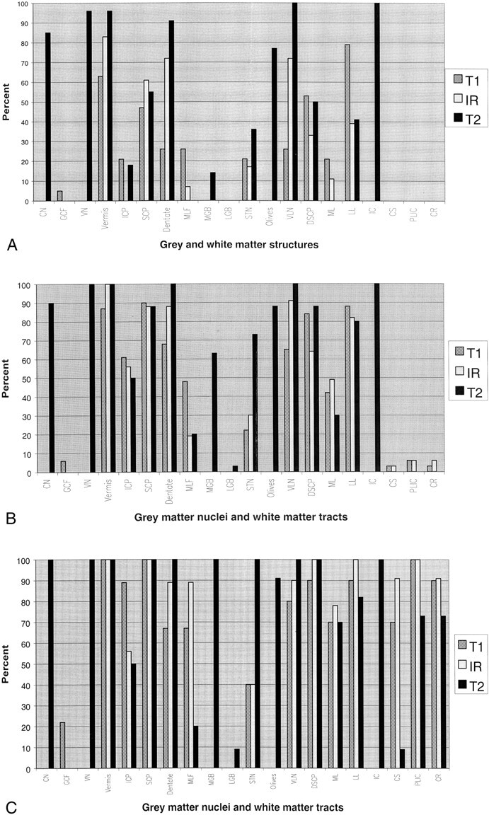
Background and purpose: MR imaging was performed in very preterm infants by using an MR imager in the neonatal intensive care unit. The aims of this study were to assess the development of myelination in the preterm brain based on MR imaging findings and to compare the ability of T1-weighted conventional spin-echo, inversion recovery fast spin-echo, and T2-weighted fast spin-echo MR imaging to show myelination in these infants.

Methods: MR imaging was performed for 26 preterm infants with a median gestational age of 28 weeks who had normal neurodevelopmental outcomes at 2 years corrected age.

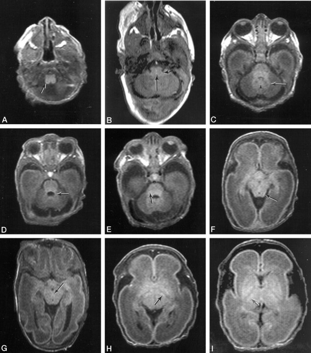
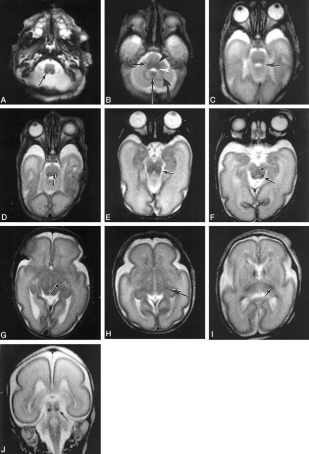
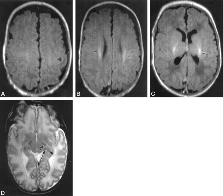
Results: Myelin was evident in the gracile and cuneate nuclei and fasciculi, vestibular nuclei, cerebellar vermis, inferior and superior cerebellar peduncles, dentate nucleus, medial longitudinal fasciculus, medial geniculate bodies, subthalamic nuclei, inferior olivary nuclei, ventrolateral nuclei of the thalamus, decussation of the superior cerebellar peduncles, medial lemnisci, lateral lemnisci, and inferior colliculi at < or = 28 weeks gestational age. From this gestational age, myelination was not visualized at any new site until 36 weeks gestational age, when myelin was visualized in the corona radiata, posterior limb of the internal capsule, corticospinal tracts of the precentral and postcentral gyri, and lateral geniculate bodies. T2-weighted fast spin-echo MR imaging showed myelin in gray matter nuclei at an earlier gestational age than did T1-weighted conventional spin-echo or inversion recovery fast spin-echo MR imaging. T1-weighted conventional spin-echo MR imaging showed myelin earlier in some white matter tracts in the preterm brain.

Conclusion: Myelination was evident in numerous gray and white matter structures in the very preterm brain. A knowledge of myelination milestones will allow delays to be detected at an early stage.


Figures

F<sc>ig</sc> 1.
Fig 1.
Percentages of different types of images showing myelination in the gray matter nuclei and white matter tracts identified in this study at <30 weeks gestational age, between 30 and 36 weeks gestational age, and between 37 and 42 weeks gestational age. CN, region of gracile and cuneate nuclei; GCF, gracile and cuneate fasciculi; VN, vestibular nuclei; Vermis, cerebellar vermis; ICP, inferior cerebellar peduncles; SCP, superior cerebellar peduncles; Dentate, dentate nucleus of the cerebellum; MLF, medial longitudinal fasciculus; MGB, medial geniculate bodies; LGB, lateral geniculate bodies; STN, subthalamic nuclei; Olives, inferior olivary nuclei; VLN, ventrolateral nuclei of the thalamus; DSCP, decussation of the superior cerebellar peduncles; ML, medial lemnisci; LL, lateral lemnisci; IC, inferior colliculi; CS, corticospinal tracts of the precentral and postcentral gyri; PLIC, posterior limb of the internal capsule; CR, corona radiata; T1, T1-weighted conventional spin-echo MR images; IR, inversion recovery fast spin-echo MR images; T2, T2-weighted fast spin-echo MR images. A, Graph illustrates the percentages of T1-weighted conventional spin-echo, inversion recovery fast spin-echo, and T2-weighted fast spin-echo MR images showing myelination at <30 weeks gestational age. B, Graph illustrates the percentages of T1-weighted conventional spin-echo, inversion recovery fast spin-echo, and T2-weighted fast spin-echo MR images showing myelination between 30 and 36 weeks gestational age. C, Graph illustrates the percentages of T1-weighted conventional spin-echo, inversion recovery fast spin-echo, and T2-weighted fast spin-echo MR images showing myelination between 37 and 42 weeks gestational age.
F<sc>ig</sc> 2.
Fig 2.
Myelin is shown in numerous gray and white matter structures in the preterm brain on T1-weighted conventional spin-echo images. A, Transverse T1-weighted conventional spin-echo image of an infant at 28 weeks gestational age shows myelin in the gracile and cuneate fasciculi (arrow) as high signal intensity. B, Transverse T1-weighted conventional spin-echo image of an infant at 28 weeks gestational age shows myelin in the medial longitudinal fasciculus (long arrow) and in the inferior cerebellar peduncles (short arrow) as high signal intensity. C, Transverse T1-weighted conventional spin-echo image of an infant at 28 weeks gestational age shows myelin in the cerebellar vermis (arrowhead) and in the dentate nucleus of the cerebellum (arrow) as high signal intensity. D, Transverse T1-weighted conventional spin-echo image of an infant at 30 weeks gestational age shows myelin in the superior cerebellar peduncles (arrow) as high signal intensity. E, Transverse T1-weighted conventional spin-echo image of an infant at 28 weeks gestational age shows myelin in the medial lemnisci as high signal intensity (arrow). F, Transverse T1-weighted conventional spin-echo image of an infant at 28 weeks gestational age, obtained at the level of the mesencephalon, shows myelin in the lateral lemnisci (arrow) as high signal intensity. G, Transverse T1-weighted conventional spin-echo image of an infant at 28 weeks gestational age shows myelin in the decussation of the superior cerebellar peduncles (arrow) as high signal intensity. H, Transverse T1-weighted conventional spin-echo image of an infant at 28 weeks gestational age shows myelin in the subthalamic nuclei (arrow) as high signal intensity. I, Transverse T1-weighted conventional spin-echo image of an infant at 28 weeks gestational age, obtained at the level of the basal ganglia, shows myelin in the ventrolateral nuclei of the thalmus (arrow) as high signal intensity.
F<sc>ig</sc> 3.
Fig 3.
Myelin is shown in numerous gray and white matter structures in the preterm brain on T2-weighted fast spin-echo MR images. A, Transverse T2-weighted fast spin-echo MR image of an infant at 25 weeks gestational age shows myelin in the region of the gracile and cuneate nuclei as low signal intensity (arrow). B, Transverse T2-weighted fast spin-echo MR image of an infant at 28 weeks gestational age shows myelin in the cerebellar vermis (long arrow), dentate nucleus of the cerebellum (curved arrow), vestibular nuclei (white arrowhead), inferior olivary nuclei (black arrowhead), and inferior cerebellar peduncle (short arrow) as low signal intensity. C, Transverse T2-weighted fast spin-echo MR image of an infant at 29 weeks gestational age shows myelin in the superior cerebellar peduncles (arrow) as low signal intensity. D, Transverse T2-weighted fast spin-echo MR image of an infant at 30 weeks gestational age shows myelin in the medial lemnisci (arrow) as low signal intensity. E, Transverse T2-weighted fast spin-echo MR image of an infant at 29 weeks gestational age shows myelin in the lateral lemnisci (arrow) as low signal intensity. F, Transverse T2-weighted fast spin-echo MR image of an infant at 30 weeks gestational age shows myelin in the decussation of the superior cerebellar peduncles (arrowhead) and inferior colliculi (arrow) as low signal intensity. G, Transverse T2-weighted fast spin-echo MR image of an infant at 29 weeks gestational age shows myelin in the subthalamic nuclei as low signal intensity (arrow). H, Transverse T2-weighted fast spin-echo MR image of an infant at 29 weeks gestational age shows myelin in the medial geniculate bodies (arrow) as low signal intensity. I, Transverse T2-weighted fast spin-echo MR image of an infant at 27 weeks gestational age shows myelin in the ventrolateral nuclei of the thalamus as low signal intensity (arrow). J, Coronal T2-weighted fast spin-echo MR image of an infant at 27 weeks gestational age shows myelin in the inferior colliculi (arrow) as low signal intensity.
F<sc>ig</sc> 4.
Fig 4.
Myelination is shown in the sites that myelinate at term-equivalent age. A, Transverse inversion recovery fast spin-echo image of an infant at 41 weeks gestational age, who was born at 28 weeks gestational age, shows myelin in the corticospinal tracts of the precentral and postcentral gyri as high signal intensity (arrowhead). B, Transverse inversion recovery fast spin-echo image of an infant at 41 weeks gestational age, who was born at 28 weeks gestational age, shows myelin in the corona radiata as high signal intensity (arrow). C, Transverse inversion recovery fast spin-echo image at the level of the basal ganglia of an infant at 42 weeks gestational age, who was born at 28 weeks gestational age, shows myelin in the posterior limb of the internal capsule as high signal intensity (arrow). D, Transverse T2-weighted fast spin-echo MR image at the level of the mesencephalon of an infant at 36 weeks gestational age, who was born at 28 weeks gestational age, shows myelin in the medial geniculate body (long arrow), the lateral geniculate body (arrowhead), and the subthalamic nuceli (short arrow) as low signal intensity.

References

    1. Johnson MA, Pennock JM, Bydder GM, et al. Clinical NMR imaging of the brain in children: normal and neurologic disease. AJR Am J Roentgenol 1983;141:1005–1018 - PubMed
    1. Holland BA, Haas DK, Norman D, Brant-Zawadzki M, Newton TH. MRI of normal brain maturation. AJNR Am J Neuroradiol 1986;7:201–208 - PMC - PubMed
    1. Barkovich AJ, Kjos BO, Jackson DE, Norman D. Normal maturation of the neonatal and infant brain: MR imaging at 1.5 T. Radiology 1988;166:173–180 - PubMed
    1. Dietrich RB, Bradley WG, Zagaroza EJ VI, et al. MR evaluation of early myelination patterns in normal and developmentally delayed infants. AJR Am J Roentgenol 1988;150:889–896 - PubMed
    1. Bird CR, Hedberg M, Drayer BP, Keller PJ, Flom RA, Hodak JA. MR assessment of myelination in infants and children: usefulness of marker sites. AJNR Am J Neuroradiol 1989;10:731–740 - PMC - PubMed

Publication types