. 2002 May 15;21(10):2332-42.
doi: 10.1093/emboj/21.10.2332.

Synaptic multiprotein complexes associated with 5-HT(2C) receptors: a proteomic approach

Affiliations

Synaptic multiprotein complexes associated with 5-HT(2C) receptors: a proteomic approach

Carine Bécamel et al. EMBO J. .

Abstract

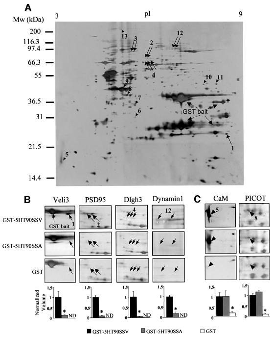
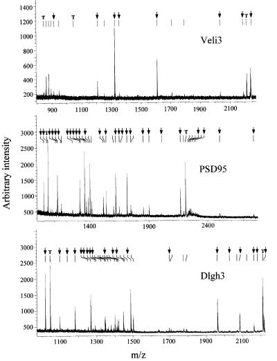
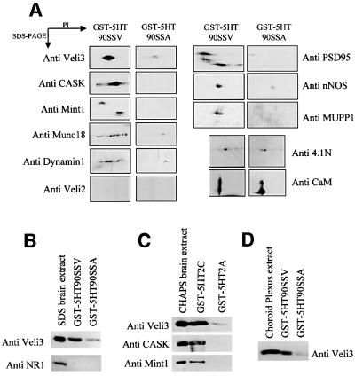
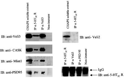
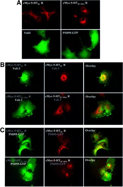
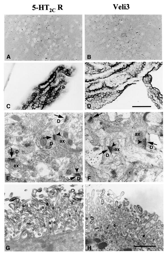
Membrane-bound receptors such as tyrosine kinases and ionotropic receptors are associated with large protein networks structured by protein-protein interactions involving multidomain proteins. Although these networks have emerged as a general mechanism of cellular signalling, much less is known about the protein complexes associated with G-protein-coupled receptors (GPCRs). Using a proteomic approach based on peptide affinity chromatography followed by mass spectrometry and immunoblotting, we have identified 15 proteins that interact with the C- terminal tail of the 5-hydroxytryptamine 2C (5-HT(2C)) receptor, a GPCR. These proteins include several synaptic multidomain proteins containing one or several PDZ domains (PSD95 and the proteins of the tripartite complex Veli3-CASK-Mint1), proteins of the actin/spectrin cytoskeleton and signalling proteins. Coimmunoprecipitation experiments showed that 5-HT(2C) receptors interact with PSD95 and the Veli3-CASK-Mint1 complex in vivo. Electron microscopy also indicated a synaptic enrichment of Veli3 and 5-HT(2C) receptors and their colocalization in microvilli of choroidal cells. These results indicate that the 5-HT(2C) receptor is associated with protein networks that are important for its synaptic localization and its coupling to the signalling machinery.


Figures

None
Fig. 1. Two-dimensional analysis of the 5-HT2C receptor protein complex. (A) Proteins that bind to the C-terminal tail of the 5-HT2C receptor were purified by affinity chromatography using the 90-amino-acid C-terminal sequence of the receptor fused to GST (GST–5HT90SSV), separated by 2D electrophoresis and silver stained. A typical 2D gel is illustrated. Proteins that interact specifically with the PDZ ligand of the receptor (arrows) were detected by comparing protein patterns obtained with GST–5HT90SSV and a mutant bait in which the last residue was replaced by alanine (GST–5HT90SSA). Arrowheads indicate proteins that interact equally with both wild-type and mutated baits but that were less represented in gels from experiments using GST alone. (B and C) Areas of interest of gels obtained in experiments performed with GST–5HT90SSV, GST–5HT90SSA and GST alone. The quantification of proteins (spot volume relative to the volume of all spots) was performed with Image Master. Data (means ± SEM of values from four gels) were normalized for each spot to the value measured in experiments using the wild-type bait (ND, not detectable). *P < 0.05 versus GST–5HT90SSV (ANOVA followed by Student–Newman–Keul’s test).
None
Fig. 2. MALDI-TOF peptide mass maps obtained from spots 1, 2 and 4. Ion signals with measured masses (Table II) that matched calculated masses of protonated tryptic peptides of mouse Veli3, PSD95 and Dlgh3 are indicated by arrows. T indicates the ion signals corresponding to the autolysis products of trypsin that were used for internal calibration of spectra (mol. wts 842.51, 1045.56 and 2211.10, respectively).
None
Fig. 3. Detection of proteins interacting with the 5-HT2C receptor by western blotting. (A) CHAPS-solubilized proteins from whole brain, retained by the GST–5HT90SSV and GST–5HT90SSA baits, were resolved on 2D gels and transferred electrophoretically on to nitrocellulose membranes. (B) Proteins were solubilized with 1% SDS instead of CHAPS and incubated with the GST–5HT90SSV and GST–5HT90SSA baits. (C) CHAPS-solubilized proteins from whole brain were passed over affinity columns containing the C-terminal tails of 5-HT2C and 5-HT2A receptors fused to GST. (D) CHAPS-solubilized proteins from choroid plexus were incubated with the GST–5HT90SSV and GST–5HT90SSA baits. Immunoblotting was performed with antibodies raised against the indicated proteins. For each protein, the immunoreactive signals were found at the expected isoelectric points (A) and molecular weights. The data illustrated are representative of three experiments.
None
Fig. 4. Association of PSD95 and the Veli3–CASK–Mint1 complex with 5-HT2C receptors of mice brain. Solubilized membranes of mice brain were immunoprecipitated with either the anti-5-HT2C, the anti-Veli3 or the anti-PSD95 antibody. Immunoprecipitated proteins were analysed by western blotting using antibodies to Veli3, CASK, Mint1, PSD95, Veli2 and the 5-HT2C receptor. Input (CHAPS-soluble extract) represents 10% of the total protein used for the immunoprecipitation.
None
Fig. 5. Interaction of 5-HT2C receptors with Veli3 and PSD95 via PDZ-based scaffolds in transiently transfected COS-7 cells. (A) Cells expressing the c-Myc-tagged 5-HT2C receptor or the c-Myc-tagged 5-HT2C-SSA receptor were immunostained using the anti-c-Myc antibody. Cells expressing Veli3 were immunostained using the anti-Veli3 antibody. PSD95 was revealed by its GFP tag. (B) COS-7 cells were cotransfected with Veli3 and either the c-Myc-tagged 5-HT2C receptor (top panels) or the c-Myc-tagged 5-HT2C-SSA receptor (bottom panels). The left panels illustrate Veli3 staining, medium c-Myc staining. Both proteins were colocalized in cells coexpressing the c-Myc-tagged 5-HT2C receptor and Veli3, in contrast with cells coexpressing the 5-HT2C-SSA receptor mutant and Veli3. (C) COS-7 cells were cotransfected with PSD95–GFP and either the c-Myc-tagged 5-HT2C receptor (top panels) or the c-Myc-tagged 5-HT2C-SSA receptor (bottom panels). The left panels illustrate GFP fluorescence, medium c-Myc staining. Both proteins were colocalized in clusters in cells coexpressing the c-Myc-tagged 5-HT2C receptor and PSD95, whereas PSD95 failed to form clusters with the 5-HT2C-SSA receptor.
None
Fig. 6. Immunolocalization of the 5-HT2C receptor and Veli3 in the anterior olfactory nucleus and the choroid plexus. Light microscope observations (AD) indicated similar distribution patterns for both 5-HT2C receptor and Veli3 immunostainings in the anterior olfactory nucleus (A and B) and the choroid plexus (C and D). Electron microscope observations (EH) demonstrated that electron-dense precipitates corresponding to immunostainings for both antibodies were associated with the same subcellular structures, i.e. (i) post-synaptic (arrows) and, to a lesser extent, presynaptic (arrowheads) thickenings of axo-dendritic synapses in the olfactory nucleus (E and F); and (ii) the membrane of distal portions of microvilli in choroidal cells (G and H). D, dendritic profile; ax, axonal profile. Scale bars (A–D) = 50 µm, (E–H) = 1 µm.

References

    1. Abramowski D., Rigo,M., Duc,D., Hoyer,D. and Staufenbiel,M. (1995) Localization of the 5-hydroxytryptamine2C receptor protein in human and rat brain using specific antisera. Neuropharmacology, 34, 1635–1645. - PubMed
    1. Bécamel C., Figge,A., Poliak,S., Dumuis,A., Peles,E., Bockaert,J., Lubbert,H. and Ullmer,C. (2001) Interaction of serotonin 5-hydroxytryptamine type 2C receptors with PDZ10 of the multi-PDZ domain protein MUPP1. J. Biol. Chem., 276, 12974–12982. - PubMed
    1. Biederer T. and Südhof,T.C. (2000) Mints as adaptors. Direct binding to neurexins and recruitment of Munc18. J. Biol. Chem., 275, 39803–39806. - PubMed
    1. Biederer T. and Südhof,T.C. (2001) Cask and protein 4.1 support F-actin nucleation on neurexins. J. Biol. Chem., 276, 47869–47876. - PubMed
    1. Borg J.P., Straight,S.W., Kaech,S.M., de Taddeo-Borg,M., Kroon,D.E., Karnak,D., Turner,R.S., Kim,S.K. and Margolis,B. (1998) Identification of an evolutionarily conserved heterotrimeric protein complex involved in protein targeting. J. Biol. Chem., 273, 31633–31636. - PubMed

Publication types

MeSH terms