. 2002 May 15;21(10):2343-53.
doi: 10.1093/emboj/21.10.2343.

Essential role of calcineurin in response to endoplasmic reticulum stress

Affiliations

Essential role of calcineurin in response to endoplasmic reticulum stress

Myriam Bonilla et al. EMBO J. .

Abstract

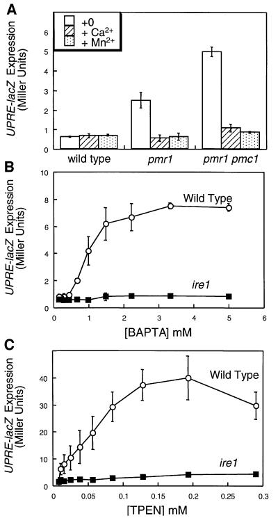
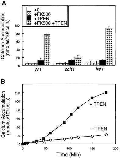
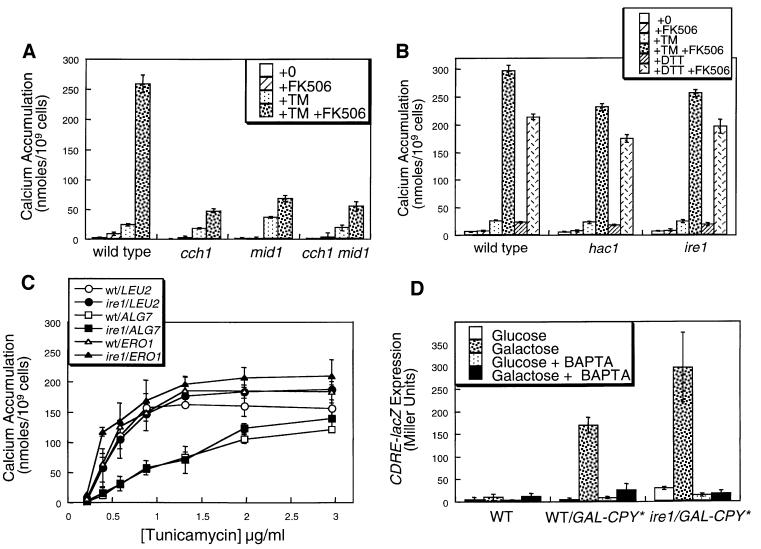
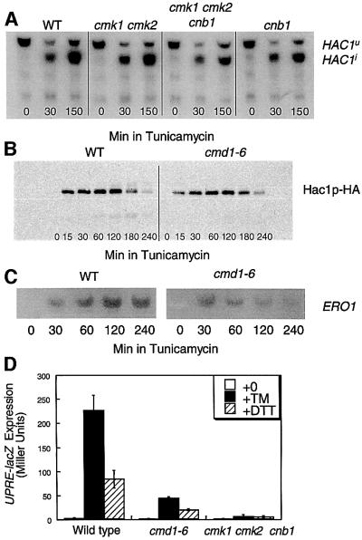
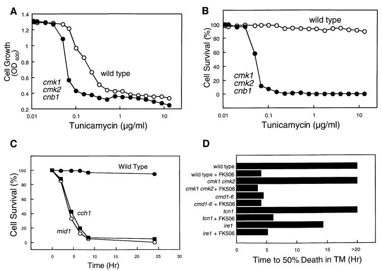
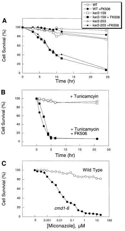
Depletion of calcium ions (Ca2+) from the endoplasmic reticulum (ER) of yeast cells resulted in the activation of the unfolded protein response (UPR) signaling pathway involving Ire1p and Hac1p. The depleted ER also stimulated Ca2+ influx at the plasma membrane through the Cch1p-Mid1p Ca2+ channel and another system. Surprisingly, both Ca2+ influx systems were stimulated upon accumulation of misfolded proteins in the ER even in the presence of Ca2+. The ability of misfolded ER proteins to stimulate Ca2+ influx at the plasma membrane did not require Ire1p or Hac1p, and Ca2+ influx and signaling factors were not required for initial UPR signaling. However, activation of the Ca2+ channel, calmodulin, calcineurin and other factors was necessary for long-term survival of cells undergoing ER stress. A similar calcium cell survival (CCS) pathway operates in the pathogenic fungi and promotes resistance to azole antifungal drugs. These findings reveal an unanticipated new regulatory mechanism that couples ER stress to Ca2+ influx and signaling pathways, which help to prevent cell death and promote resistance to an important class of fungistatic drugs.


Figures

None
Fig. 1. Ca2+ starvation and chelation can stimulate the UPR signaling pathway. (A) Wild-type, pmr1 mutant and pmr1 pmc1 double mutant cells (strains K601, K609 and K613, respectively) bearing the UPRE-lacZ reporter gene (plasmid pCZY1) were grown to log phase in SC medium lacking uracil, Ca2+ and Mn2+ with or without a supplement of 10 mM CaCl2 or 100 µM MnCl2. After 4 h, the cells were harvested and assayed for β-galactosidase activity. Bars represent the mean of three independent transformants (± SD). The Pmr1p Ca2+ pump prevented activation of the UPR signaling pathway in medium lacking Ca2+. (B and C) Wild-type and ire1 mutant cells (strains K1257 and K1259, respectively) bearing the UPRE-lacZ reporter gene were grown to log phase in SC medium lacking uracil and containing 50 mM K-MES buffer pH 6.5, and then treated with the indicated concentrations of either the high-affinity membrane-impermeant Ca2+ chelator BAPTA or the low-affinity membrane-permeant Ca2+ chelator TPEN. Cells were incubated and processed for β-galactosidase activity as in (A).
None
Fig. 2. TPEN stimulates Ca2+ influx via the Cch1p–Mid1p Ca2+ channel independently of Ire1p. (A45Ca2+ accumulation in wild-type cells, cch1 mutants and ire1 mutants (strains K1257, RG05040 and K1259 respectively) was measured after incubation for 4 h in SC medium supplemented with trace amounts of 45CaCl2 plus 2.0 µg/ml FK506 and 129 µM TPEN as indicated. Bars represent the mean of three independent experiments (± SD). (B45Ca2+ accumulation in wild-type cells (strain K601) was measured at various time-points after treatment with either FK506 (open symbols) or TPEN plus FK506 (filled symbols) as in (A).
None
Fig. 3. Accumulation of misfolded proteins in the ER stimulates Ca2+ influx and signaling independently of Ire1p. (A45Ca2+ accumulation in wild type, cch1 and mid1 mutants was measured as in Figure 2A except that TM (2.5 µg/ml) was used instead of TPEN. (B45Ca2+ accumulation in wild type, ire1 and hac1 mutants (strains K1257, K1259 and RG05650) was measured as in (A) except that DTT (2.5 mM) was included as indicated. (C45Ca2+ accumulation in wild-type (open symbols) and ire1 mutant cells (closed symbols) that were overexpressing Alg7p, Ero1p or no additional proteins in the ER (plasmids pMK13XRV, pKT001 or vector control) was measured after treatment with varying amounts of TM in the presence of 2 µg/ml FK506. Complementary results were obtained using DTT instead of TM (data not shown). (D) Expression of the CDRE-lacZ reporter gene (plasmid pAMS366) was measured in wild-type cells and ire1 mutants expressing a folding-incompetent mutant of carboxypeptidase Y (denoted CPY*) under control of the galactose promoter (strains DNY1047 and DNY1048) after growth for 4 h in SC medium lacking uracil but supplemented with either 2% glucose or 2% galactose as indicated. Induction of CPY* in galactose medium stimulated CDRE-lacZ expression through a process that was sensitive to both 4 mM BAPTA and FK506 (data not shown). Wild-type cells lacking CPY* (strain K601) were included as a control. Bars represent the mean of three independent transformants (± SD).
None
Fig. 4. Calcium signaling factors are not required for efficient UPR signaling. (A) Alternative splicing of HAC1 transcripts was stimulated by tunicamycin independently of calcium signaling factors. HAC1 splicing was monitored by northern blot analysis of total RNA isolated from wild-type (K601), cmk1 cmk2 (K481), cmk1 cmk2 cnb1 (K488) and cnb1 (K603) strains after 0, 30 or 150 min in 2.5 µg/ml TM. (B) Accumulation of Hac1p was stimulated by tunicamycin treatment independently of calcium signaling factors. Hac1p accumulation was monitored by western blotting of total cell protein extracted from wild-type (K601) cells and cmd1-6 mutants (JGY148) carrying epitope-tagged Hac1p–HA (plasmid pMB004) that had been treated with 2.5 µg/ml TM for the indicated periods of time. (C) Northern blot analysis of ERO1 gene expression was performed as in (B) on wild-type and cmd1-6 mutant cells at various times after treatment with TM. Mutants defective in calmodulin signaling reproducibly induced ERO1 transcripts much more transiently than wild-type cells. (DUPRE-lacZ expression was measured in wild type (strain K601), cmd1-6 mutants (strain JGY148) and cmk1 cmk2 cnb1 triple mutants (strain K488) after 4 h growth in SC medium supplemented with either 2.5 mg/ml TM or 2.5 mM DTT as indicated.
None
Fig. 5. Tunicamycin treatment arrests growth of wild-type cells and causes death of cells deficient in Ca2+ influx and signaling factors. After treatment of log-phase yeast cultures with TM, dead cells were stained with methylene blue and counted. Cell survival reflects the percentage live cells of total cells in each culture. Wild type and cmk1 cmk2 cnb1 triple mutants (strains K601 and K488) were treated with varying amounts of TM for 6 h and then assayed for cell growth (A) or cell survival (B) as described in Materials and methods. (C) Survival of wild type, cch1 and mid1 mutants (strains K601, ELY117, ELY138) was measured at various times after addition of TM (2.5 µg/ml). (D) The time required for various yeast strains to undergo 50% cell death was determined in time course experiments similar to those in (C).
None
Fig. 6. Calcineurin signaling is required for survival of kar2-ts mutants and for survival of Saccharomyces and Candida species in response to azoles or other ER stress agents. (A) FK506 prevents survival of kar2-159 (strain MS137) and kar2-203 (strain MS1028) at a semi-permissive temperature (30°C). Cell survival assays were performed as described in Figure 5. (B) FK506 (filled symbols) prevents survival of C.albicans (circles) and C.glabrata (squares) in medium containing TM. (C) Calmodulin signaling was required for survival in medium containing miconazole. Survival of wild type and cmd1-6 mutants was measured after treatment with varying amounts of miconazole for 6 h in SC medium.

References

    1. Antebi A. and Fink,G.R. (1992) The yeast Ca2+-ATPase homologue, PMR1, is required for normal Golgi function and localizes in a novel Golgi-like distribution. Mol. Biol. Cell, 3, 633–654. - PMC - PubMed
    1. Brachmann C.B., Davies,A., Cost,G.J., Caputo,E., Li,J., Hieter,P. and Boeke,J.D. (1998) Designer deletion strains derived from Saccharomyces cerevisiae S288C: a useful set of strains and plasmids for PCR-mediated gene disruption and other applications. Yeast, 14, 115–132. - PubMed
    1. Burk S.E., Lytton,J., MacLennan,D.H. and Shull,G.E. (1989) cDNA cloning, functional expression and mRNA tissue distribution of a third organellar Ca2+ pump. J. Biol. Chem., 264, 18561–18568. - PubMed
    1. Carnero E., Ribas,J.C., Garcia,B., Duran,A. and Sanchez,Y. (2000) Schizosaccharomyces pombe ehs1p is involved in maintaining cell wall integrity and in calcium uptake. Mol. Gen. Genet., 264, 173–183. - PubMed
    1. Cormack B.P. and Falkow,S. (1999) Efficient homologous and illegitimate recombination in the opportunistic yeast pathogen Candida glabrata. Genetics, 151, 979–987. - PMC - PubMed

Publication types

MeSH terms