. 2002 May 15;21(10):2364-72.
doi: 10.1093/emboj/21.10.2364.

Polyploid measles virus with hexameric genome length

Affiliations

Polyploid measles virus with hexameric genome length

Monika Rager et al. EMBO J. .

Abstract

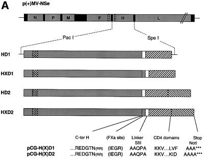
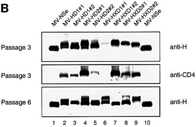
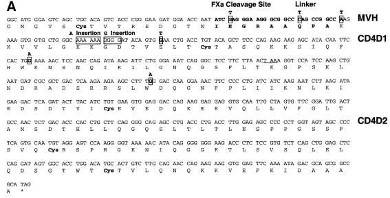
Particles of most virus species accurately package a single genome, but there are indications that the pleomorphic particles of parainfluenza viruses incorporate multiple genomes. We characterized a stable measles virus mutant that efficiently packages at least two genomes. The first genome is recombinant and codes for a defective attachment protein with an appended domain interfering with fusion-support function. The second has one adenosine insertion in a purine run that interrupts translation of the appended domain and restores function. In that genome, a one base deletion in a different purine run abolishes polymerase synthesis, but restores hexameric genome length, thus ensuring accurate RNA encapsidation, which is necessary for efficient replication. Thus, the two genomes are complementary. The infection kinetics of this mutant indicate that packaging of multiple genomes does not negatively affect growth. We also show that polyploid particles are produced in standard infections at no expense to infectivity. Our results illustrate how the particles of parainfluenza viruses efficiently accommodate cargoes of different volume, and suggest a mechanism by which segmented genomes may have evolved.


Figures

None
Fig. 1. Genomic structure and protein expression of recombinant MV. (A) Plasmid p(+)MV-NSe encoding the MV anti-genome (top), PacI–SpeI fragments used for subcloning (center), and amino acid sequences (one-letter code) of the junctions between the H protein ectodomain and the CD4 domains (bottom). Coding regions of the six MV cistrons are represented by gray boxes, intergenic regions by solid black boxes, transmembrane domains by checkered boxes, the FXa cleavage site by a dotted box, the flexible linker by a white box and the CD4 domains by hatched boxes. Arginine residues in parentheses were deleted to avoid the possibility of introducing an undesired furin cleavage site (Schneider et al., 2000). (B) Western blot analysis of H/CD4 hybrid protein expression. Vero cells infected with the recombinant viruses were lysed at passage 3 (top and center) or passage 6 (bottom). The proteins were seperated by SDS–PAGE and probed with either an H-specific antiserum (top and bottom) or with a CD4-specific antiserum (center).
None
Fig. 2. Stop mutations in the recombinant viral genomes and propagation of the insertion mutant MV-HXD2#2. (A) The genomes of recovered recombinant viruses were analyzed by RT–PCR of infected Vero cell RNA. For simplicity, all the mutations found in the different clones of MV-HD1, MV-HD2, MV-HXD1 and MV-HXD2 are represented on the sequence of MV-HXD2. The last 15 codons of the H ORF are shown, connected to CD4 domains 1 and 2 by a four-amino-acid flexible linker and the five-amino-acid factor Xa cleavage site (bold). CD4D1, sequence of domain 1 of CD4; CD4D2, sequence of domain 2 of CD4. The premature stop codon resulting from the single nucleotide insertion is underlined. (B) Western blot analysis of MV-HXD2#2 clones. Six distinct syncytia from a MV-HXD2#2 infection were picked and grown on fresh Vero cells. Cell lysates were fractionated by SDS–PAGE and proteins were detected with an anti-H cytoplasmic tail antiserum. (C) Genomic sequence of the A insertion site (I–IV) and the P-editing site (V) in MV-HXD2#2 at different passages as determined by RT–PCR of RNA extracted from infected cells (I–III) and from purified viral particles (IV and V).
None
Fig. 3. RT–PCR analysis of MV-HXD2#2. (A) The MV-HXD2#2 genome is depicted as the putative concatemer. The 1932 bp fragment that would have resulted from RT–PCR on purified particles of a virus carrying concatemeric genomes, and the 1532 bp fragment amplified to investigate linkage between the A insertion in the CD4 domain and the A deletion in the L reading frame, are indicated. Numbers near the arrows correspond to the positions in the 16 398-nucleotide MV-HXD2 genome. (B) Sequence of the polypurine stretches at the P editing site, the insertion site and the deletion site. The mutated sequence is shown below the parental sequence.
None
Fig. 4. Growth kinetics of MV-HXD2#2. (A) Dilution assay with MV-NSe (squares), MV-eGFP (diamonds) and MV-HXD2#2 (triangles). Vero cells in six-well plates were infected with 1:2 serial dilutions of the respective viruses starting at a concentration of 200 p.f.u./ml. Syncytia were counted 5 days post-infection. The number of syncytia in the well containing undiluted virus was set to 100% and relative percentages were calculated. (B) Time course of cell-associated (closed symbols, left panel) and released (open symbols, right panel) virus production in Vero cells infected with MV-HXD2#2 (triangles), or the control viruses MV-NSe (squares) and MV-eGFP (diamonds). Cells were infected at a m.o.i. of 0.03 and incubated at 37°C for the times indicated. Viral titers were determined by 50% end-point dilution.
None
Fig. 5. Co-infection assay with MV-eGFP and MV-DsRed1. Vero cells were simultaneously infected with MV-eGFP and MV-DsRed1, and syncytia expressing both eGFP and DsRed1 were passaged. The percentage of syncytia expressing only eGFP (gray), only DsRed1 (black), and both eGFP and DsRed1 (white) was determined. The total number of syncytia counted for each passage is given in parentheses. In the aggregation control experiment, MV-eGFP and MV-DsRed1 were mixed and incubated on ice for 1 h prior to infection.

References

    1. Baczko K. et al. (1993) Clonal expansion of hypermutated measles virus in a SSPE brain. Virology, 197, 188–195. - PubMed
    1. Bellini W.J., Englund,G., Rozenblatt,S., Arnheiter,H. and Richardson,C.D. (1985) Measles virus P gene codes for two proteins. J. Virol., 53, 908–919. - PMC - PubMed
    1. Bluming A.Z. and Ziegler,J.L. (1971) Regression of Burkitt’s lymphoma in association with measles infection. Lancet, 2, 105–106. - PubMed
    1. Calain P. and Roux,L. (1993) The rule of six, a basic feature for efficient replication of Sendai virus defective interfering RNA. J. Virol., 67, 4822–4830. - PMC - PubMed
    1. Cathomen T., Buchholz,C.J., Spielhofer,P. and Cattaneo,R. (1995) Preferential initiation at the second AUG of the measles virus F mRNA: a role for the long untranslated region. Virology, 214, 628–632. - PubMed

Publication types

Substances