. 2002 Jul 26;277(30):26796-803.
doi: 10.1074/jbc.M200754200. Epub 2002 May 10.

Ceramide channels increase the permeability of the mitochondrial outer membrane to small proteins

Affiliations

Ceramide channels increase the permeability of the mitochondrial outer membrane to small proteins

Leah J Siskind et al. J Biol Chem. .

Abstract

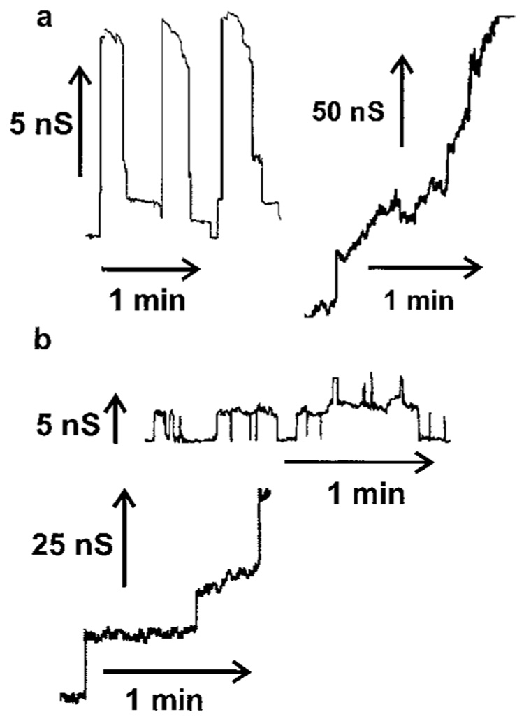
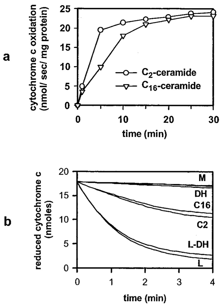
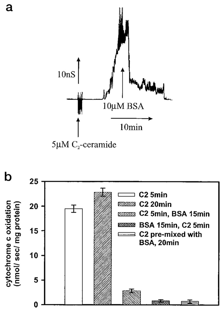
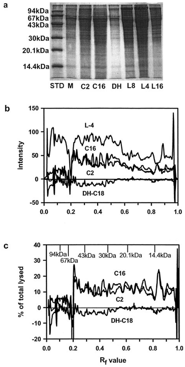
Ceramides are known to play a major regulatory role in apoptosis by inducing cytochrome c release from mitochondria. We have previously reported that C(2)- and C(16)-ceramide, but not dihydroceramide, form large channels in planar membranes (Siskind, L. J., and Colombini, M. (2001) J. Biol. Chem. 275, 38640-38644). Here we show that ceramides do not trigger a cytochrome c secretion or release mechanism, but simply raise the permeability of the mitochondrial outer membrane, via ceramide channel formation, to include small proteins. Exogenously added reduced cytochrome c was able to freely permeate the mitochondrial outer membrane with entry to and exit from the intermembrane space facilitated by ceramides in a dose- and time-dependent manner. The permeability pathways were eliminated upon removal of C(2)-ceramide by bovine serum albumin, thus ruling out a detergent-like effect of C(2)-ceramide on membranes. Ceramide channels were not specific to cytochrome c, as ceramides induced release of adenylate kinase, but not fumerase from isolated mitochondria, showing some specificity of these channels for the outer mitochondrial membrane. SDS-PAGE results show that ceramides allow release of intermembrane space proteins with a molecular weight cut-off of about 60,000. These results indicate that the ceramide-induced membrane permeability increases in isolated mitochondria are via ceramide channel formation and not a release mechanism, as the channels that allow cytochrome c to freely permeate are reversible, and are not specific to cytochrome c.


Figures

FIG. 1
FIG. 1. Sample traces of C2- and C16-ceramide channels in planar membranes
Continuous current recordings induced by the addition of ceramide to the aqueous solution as described under “Experimental Procedures.” The applied voltage was clamped at 10 mV. a, 1 µm C16-ceramide was added only to one side (the cis side) of the membrane.b, 5 µm C2-ceramide was added to both sides of the membrane.
FIG. 2
FIG. 2. C2- and C16-ceramide increase the permeability of the mitochondrial outer membrane to cytochrome c in a dose- and time-dependent manner
Mitochondria were incubated for the indicated time periods and with the indicated concentrations of C2- (circles) or C16-ceramide (triangles) as described under “Experimental Procedures.” Reduced cytochrome c was then added and the absorbance was monitored at 550 nm. The initial rates plotted were in nanomoles of cytochrome c oxidized per s/mg of mitochondrial protein. Results are representative experiment of at least 3 performed on separate mitochondrial preparations. a mitochondria incubated for the indicated time periods with 20 µm ceramides. b, mitochondria incubated with the following treatments: mitochondrial controls (M; untreated mitochondria, vehicle controls, 83 µm BSA for 15 min, with overlapping lines); 20 µm C2-dihydroceramide for 10 min (DH); 20 µm C2-ceramide for 10 min (C2); 20 µm C16-ceramide for 10 min (C16); lysed mitochondria were incubated for 10 min with 20 µm C2-dihydroceramide (L-DH); lysed mitochondrial controls (L; untreated lysed mitochondria, lysed mitochondria incubated for 10 min with 20 µm C2-ceramide, C16-ceramide, or vehicles or 15 min with 83 µm BSA, with overlapping lines).
FIG. 3
FIG. 3. The permeability increase induced by C2-ceramide can be reversed with BSA
a, BSA was added (10 µm final) to the aqueous phase on both sides of a planar phospholipid membrane containing C2-ceramide channels (5 µm C2-ceramide on both sides). The applied voltage was clamped at 10 mV. b, mitochondria were incubated with 20 µm C2-ceramide for the indicated time periods and where indicated with 83 µm BSA for the indicated time periods as described under “Experimental Procedures.” Reduced cytochrome c was then added and the absorbance was monitored at 550 nm. The initial rates were plotted as nanomoles of cytochrome c oxidized per s/mg of mitochondrial protein. Error bars represent standard deviations. Results are a representative experiment of at least 3 performed on separate mitochondrial preparations.
FIG. 4
FIG. 4. C2-C16-ceramide increase the permeability of the mitochondrial outer membrane to intermembrane space proteins with a cut-off of about 60 kDa
Mitochondria were incubated with or without ceramides as described under “Experimental Procedures” and the released proteins were run on a 15% acrylamide SDS-PAGE. The ceramide level was 18 nmol of ceramide/mg of mitochondrial protein. Results are a representative experiment of 3 performed on separate mitochondrial preparations. The following abbreviations are used: STD, standard proteins; M, mitochondrial control; C2, C2-ceramide; C16, C16-ceramide; DH or DH-C18, C18-dihydroceramide; L8, lysed mitochondria diluted 8-fold; L4, lysed mitochondria diluted 4-fold; L16, lysed mitochondria diluted 16-fold. a, SDS-PAGE was stained with GelCode Blue stain (Pierce). b, densitometry was performed on the gel from a with individual backgrounds for each lane and the mitochondrial control was subtracted out. Results were expressed as intensity of staining versus the RF value. c, results were expressed as the % of lysed (undiluted) versus the RF value.

References

    1. Radin NS. Eur. J. Biochem. 2001;268:193–204. - PubMed
    1. Kolesnick RN, Krönke M. Annu. Rev. Physiol. 1998;60:643–665. - PubMed
    1. Ariga T, Jarvis WD, Yu RK. J. Lipid Res. 1998;39:1–16. - PubMed
    1. Hannun YA. Science. 1996;274:1855–1859. - PubMed
    1. Kolesnick RN, Goñi FM, Alonoso A. J. Cell. Physiol. 2000;184:285–300. - PubMed

Publication types