. 2002 May;13(5):1536-49.
doi: 10.1091/mbc.02-02-0010.

Targeted destruction of DNA replication protein Cdc6 by cell death pathways in mammals and yeast

Affiliations
Free PMC article

Targeted destruction of DNA replication protein Cdc6 by cell death pathways in mammals and yeast

Frederic Blanchard et al. Mol Biol Cell. 2002 May.
Free PMC article

Abstract

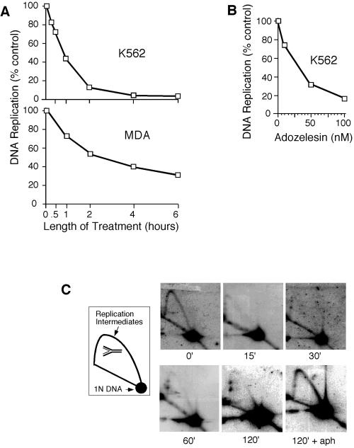
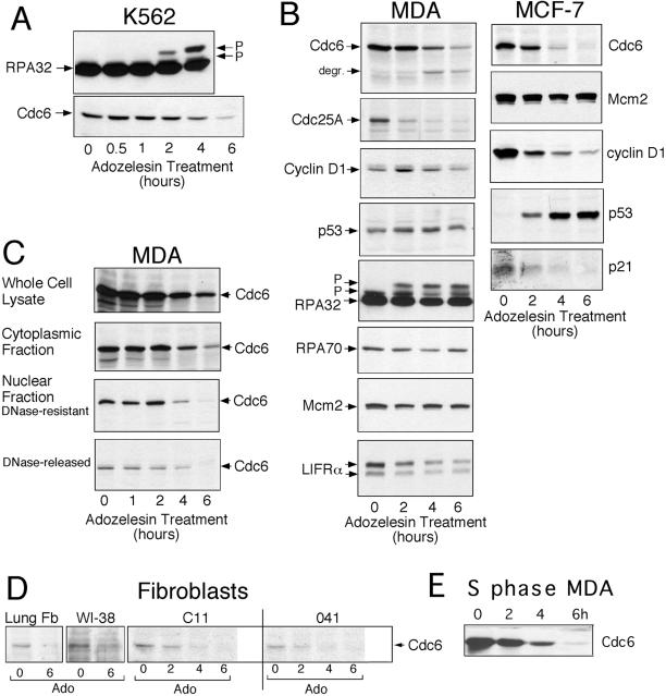
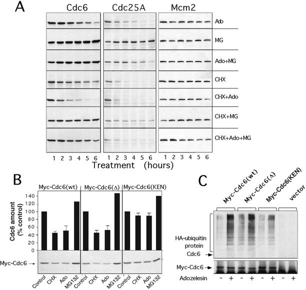
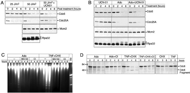
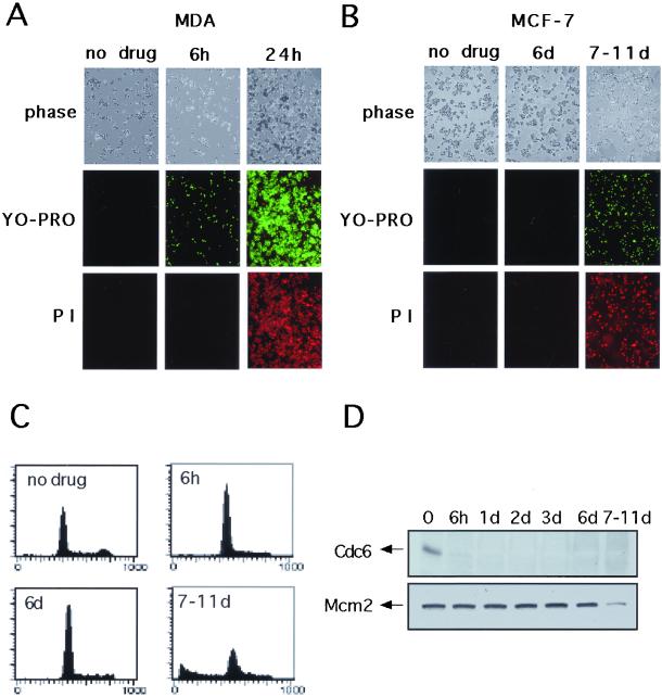
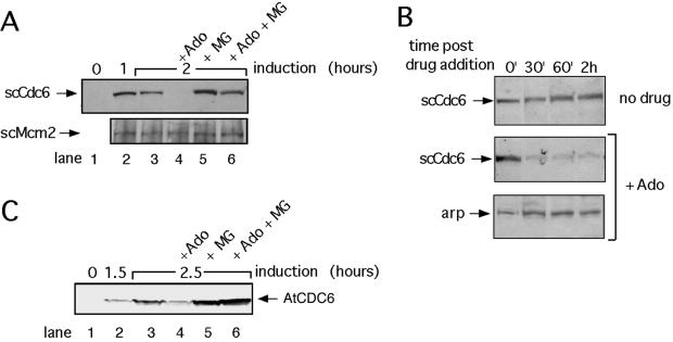
The highly conserved Cdc6 protein is required for initiation of eukaryotic DNA replication and, in yeast and Xenopus, for the coupling of DNA replication to mitosis. Herein, we show that human Cdc6 is rapidly destroyed by a p53-independent, proteasome-, and ubiquitin-dependent pathway during early stages of programmed cell death induced by the DNA-damaging drug adozelesin, or by a separate caspase-dependent pathway in cells undergoing apoptosis through an extrinsic pathway induced by tumor necrosis factor-alpha and cycloheximide. The proteasome-dependent pathway induced by adozelesin is conserved in the budding yeast Saccharomyces cerevisiae. The destruction of Cdc6 may be a primordial programmed death response that uncouples DNA replication from the cell division cycle, which is reinforced in metazoans by the evolution of caspases and p53.


Figures

Figure 1
Figure 1
Adozelesin inhibits initiation of mammalian chromosomal DNA replication. (A) K562 and MDA cells were treated with 40 nM adozelesin for indicated times and DNA synthesis was assessed by measuring tritiated thymidine incorporation into acid-precipitable DNA. (B) K562 cells were treated with indicated concentrations of adozelesin for 2 h and DNA synthesis was assessed as in A. (C) Replicating and nonreplicating DNA isolated from K562 cells treated with adozelesin or adozelesin + aphidicolin (aph) for the indicated times were separated from one another by neutral-neutral two-dimensional gel electrophoresis, and blots of gels were probed with a fragment of DNA from the tandemly repeated human rDNA locus. Schematic shows migration of single branched replication intermediates and nonreplicating DNA molecules (1N DNA).
Figure 2
Figure 2
Levels of Cdc6 and other proteins are reduced by adozelesin treatment. (A–D) Cells were treated for the indicated times with 40 nM adozelesin and cellular proteins were extracted, separated on SDS-PAGE gels, and immunoblotted to detect the indicated proteins. (C) Nuclei were separated from cytoplasm of drug-treated cells to obtain a “cytoplasmic fraction” and a “nuclear fraction” and isolated nuclei were treated with DNase I to obtain the “DNase I-resistant” and “DNase I-released” fractions. (D) Cultures of normal lung fibroblasts (Lung Fb) and established fibroblast cell lines were treated for 16 h with 5 μg/ml aphidicolin to accumulate large numbers of cells in S phase. Then 40 nM adozelesin (Ado) was added for the additional times indicated before proteins were extracted and analyzed by immunoblotting. (E) MDA cells were treated with aphidicolin for 16 h to synchronize them in S phase and then treated with 40 nM adozelesin and aphidicolin for an additional 2–6 h.
Figure 3
Figure 3
Adozelesin induces ubiquitination and proteasome-dependent degradation of Cdc6. (A) MDA cells were treated with 40 nM adozelesin (Ado) for the indicated times in presence or absence of 10 μM MG132 (MG), 10 μg/ml cycloheximide (CHX), or combinations as indicated. Cell extracts were analyzed for Cdc6, Cdc25A, or MCM2 levels by immunoblotting. (B) MCF-7 cells were transfected with vectors expressing Myc-Cdc6 wild-type [Myc-Cdc6 (wt)] or mutated destruction box [Myc-Cdc6(Δ)] or KEN motif [Myc-Cdc6(KEN)] proteins. Cells were treated 6 h with adozelesin (Ado), cycloheximide (CHX), or MG beginning 24 h after transfection and analyzed for the level of transfected Cdc6 by immunoblotting with anti-Myc antibodies. Signals from immunoblots in three independent experiments were quantitated by densitometry and the averages and SEs presented above each lane. (C) MCF-7 cells were cotransfected with expression vectors for HA-tagged ubiquitin and the Myc-tagged wild-type and mutant Cdc6 described above. Lysates were made from cells after adozelesin treatment for 6 h beginning 24 h after transfection, and Myc-tagged Cdc6 proteins were immunoprecipitated with Myc antibodies. Immunoprecipitated proteins were analyzed for HA-ubiquitin and then for Myc-tagged Cdc6 by immunoblotting with anti-HA (top) and anti-Myc (bottom) antibodies. hIg is the IgG light chain that reacts with the secondary antibody.
Figure 4
Figure 4
Cdc6 destruction is induced by UV radiation and TNF-α and coincides with a caspase-dependent apoptotic response. (A) MDA cells were irradiated (25 or 50 J/m2 UV radiation) and returned to normal culture conditions in the presence or absence of 1 μM of the kinase inhibitor UCN-01 for 0–6 h. Proteins extracted at indicated times after irradiation were analyzed by immunoblotting. (B) MDA cells were treated with 1 μM UCN01, 40 nM adozelesin (Ado), or both (Ado+UCN01) for indicated times and extracted proteins analyzed by immunoblotting. (C) DNA was isolated from MDA cells treated with various combinations of Ado, 100 ng/ml TNF-α (TNF), and 10 μg/ml cycloheximide (TNF-CHX) with or without 10 μM MG132 (MG) or 10 μM caspase inhibitor I (CI) for indicated times and then size-fractionated on ethidium bromide-containing agarose gels. The standard (std) is a 100-base pair ladder. (D) Cells were treated with the indicated compounds for 0–6 h and protein extracts analyzed by immunoblotting.
Figure 5
Figure 5
Proteasome-dependent destruction of Cdc6 occurs during early apoptosis. MDA (A) or MCF-7 (B) cells were treated or not treated with adozelesin for 6 h and adozelesin-containing medium was replaced with fresh drug-free medium for an additional 24 h. Apoptosis was assessed at the indicated times after addition of drug by phase contrast microscopy, YO-PRO staining, and staining with propidium iodide. (C) FACS analysis of MCF-7 cells after adozelesin treatment. (D) Western blot analysis of Cdc6 and Mcm2 in MCF-7 cells after adozelesin treatment.
Figure 6
Figure 6
Adozelesin induces the proteasome-dependent destruction of Cdc6 in budding yeast. (A and C) S. cerevisiae cells harboring plasmids expressing the S. cerevisiae Cdc6 (scCdc6) or Arabidopsis CDC6 (AtCDC6) genes regulated by a galactose-inducible promoter were changed from raffinose- to galactose-containing medium to induce the ectopic expression of scCdc6 or AtCDC6. They were treated with 4 μM adozelesin and/or 250 μM MG132 beginning 1 h (scCdc6 experiments) or 1.5 h (AtCDC6 experiments) after galactose induction. Total protein lysates made at the indicated times after induction were analyzed by immunoblotting for S. cerevisiae Cdc6 (scCdc6) or arabidopsis Cdc6 (AtCDC6) proteins, or for scMcm2 protein as a control. (B) cdc4 cells were synchronized in early S phase by release from an α factor G1 block into HU and then cultured in the continued presence of HU at the nonpermissive temperature (37°C) for 2 h to destroy the activity of Cdc4p. Cycloheximide and adozelesin were then added for the times indicated before cells were lysed for Western blot analysis. Arp, actin-related protein.

References

    1. Agami R, Bernards R. Distinct initiation and maintenance mechanisms cooperate to induce G1 cell cycle arrest in response to DNA damage. Cell. 2000;102:55–66. - PubMed
    1. Anderson GR, Stoler DL, Brenner BM. Cancer: the evolved consequence of a destabilized genome. Bioessays. 2001;23:1037–1046. - PubMed
    1. Bhuyan BK, Smith KS, Adams EG, Petzold GL, McGovren JP. Lethality, DNA alkylation, and cell cycle effects of adozelesin (U-73975) on rodent and human cells. Cancer Res. 1992a;52:5687–5692. - PubMed
    1. Bhuyan BK, Smith KS, Adams EG, Wallace TL, Von Hoff DD, Li LH. Adozelesin, a potent new alkylating agent: cell-killing kinetics and cell-cycle effects. Cancer Chemother Pharmacol. 1992b;30:348–354. - PubMed
    1. Brewer BJ, Fangman WL. The localization of replication origins on ARS plasmids in S. cerevisiae. Cell. 1987;51:463–471. - PubMed

Publication types

MeSH terms

LinkOut - more resources