. 2002 May;13(5):1550-65.
doi: 10.1091/mbc.02-02-0015.

Paxillin-dependent paxillin kinase linker and p21-activated kinase localization to focal adhesions involves a multistep activation pathway

Affiliations
Free PMC article

Paxillin-dependent paxillin kinase linker and p21-activated kinase localization to focal adhesions involves a multistep activation pathway

Michael C Brown et al. Mol Biol Cell. 2002 May.
Free PMC article

Abstract

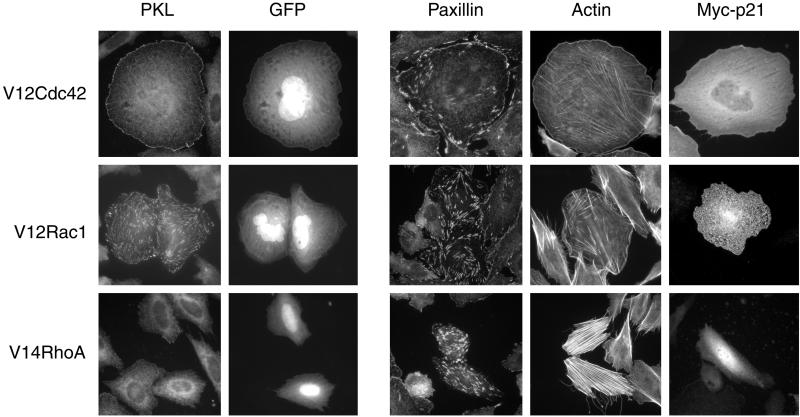
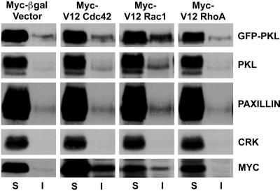
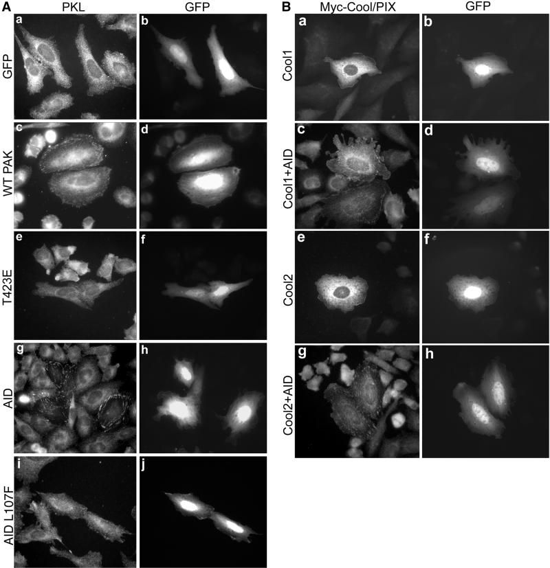
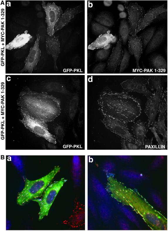
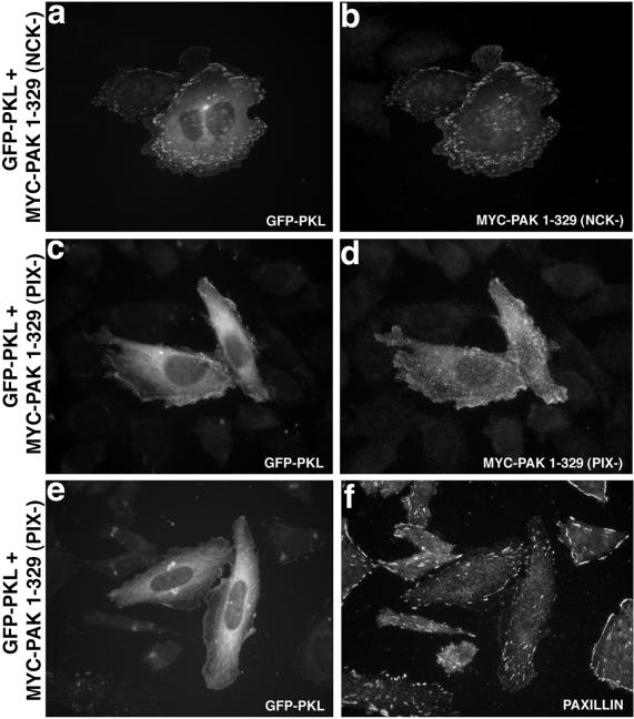
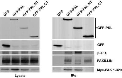
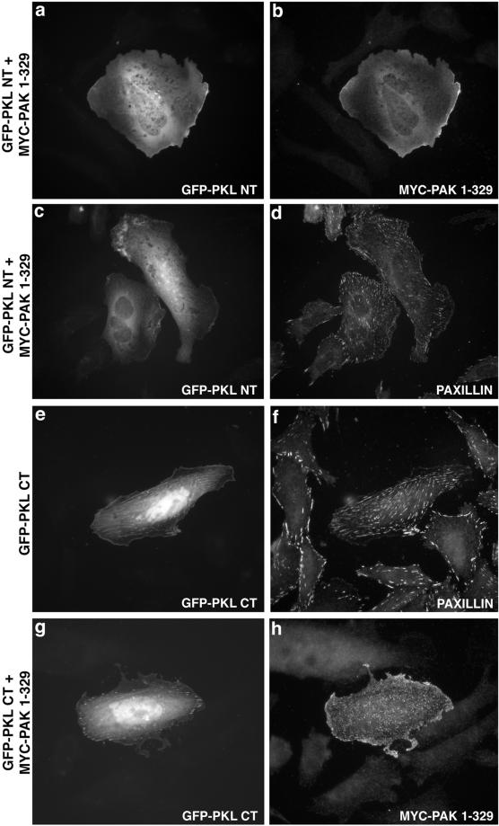
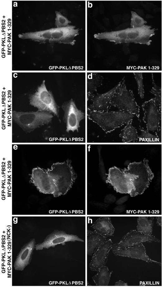
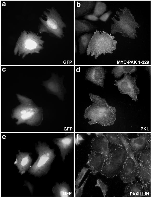
The precise temporal-spatial regulation of the p21-activated serine-threonine kinase PAK at the plasma membrane is required for proper cytoskeletal reorganization and cell motility. However, the mechanism by which PAK localizes to focal adhesions has not yet been elucidated. Indirect binding of PAK to the focal adhesion protein paxillin via the Arf-GAP protein paxillin kinase linker (PKL) and PIX/Cool suggested a mechanism. In this report, we demonstrate an essential role for a paxillin-PKL interaction in the recruitment of activated PAK to focal adhesions. Similar to PAK, expression of activated Cdc42 and Rac1, but not RhoA, stimulated the translocation of PKL from a generally diffuse localization to focal adhesions. Expression of the PAK regulatory domain (PAK1-329) or the autoinhibitory domain (AID 83-149) induced PKL, PIX, and PAK localization to focal adhesions, indicating a role for PAK scaffold activation. We show PIX, but not NCK, binding to PAK is necessary for efficient focal adhesion localization of PAK and PKL, consistent with a PAK-PIX-PKL linkage. Although PAK activation is required, it is not sufficient for localization. The PKL amino terminus, containing the PIX-binding site, but lacking paxillin-binding subdomain 2 (PBS2), was unable to localize to focal adhesions and also abrogated PAK localization. An identical result was obtained after PKLDeltaPBS2 expression. Finally, neither PAK nor PKL was capable of localizing to focal adhesions in cells overexpressing paxillinDeltaLD4, confirming a requirement for this motif in recruitment of the PAK-PIX-PKL complex to focal adhesions. These results suggest a GTP-Cdc42/GTP-Rac triggered multistep activation cascade leading to the stimulation of the adaptor function of PAK, which through interaction with PIX provokes a functional PKL PBS2-paxillin LD4 association and consequent recruitment to focal adhesions. This mechanism is probably critical for the correct subcellular positioning of PAK, thereby influencing the ability of PAK to coordinate cytoskeletal reorganization associated with changes in cell shape and motility.


Figures

Figure 1
Figure 1
Induction of PKL focal adhesion localization by activated Cdc42 and Rac1, but not RhoA. CHO.K1 cells were cotransfected with myc-tagged V12Cdc42 (top), V12Rac1 (middle), or V14RhoA (bottom) and GFP. Immunofluorescence analysis was performed to examine the effects of active p21 expression on endogenous PKL subcellular localization. GFP was cotransfected to identify transfectants. In addition, paxillin localization, actin organization, and ectopic myc-p21 expression was examined in GFP and p21 cotransfectants; 75 ± 8% (n = 3) of active Cdc42 and 90 ± 6% (n = 3) of active Rac1 transfected cells revealed PKL in focal adhesions, whereas PKL within active RhoA transfected cells was primarily diffuse. Transfectants were determined by GFP expression; colabeling is only shown for PKL immunostaining.
Figure 2
Figure 2
Cdc42 and Rac1 stimulate PKL transition to a Triton X-100–insoluble fraction. The subcellular distribution of PKL was examined in cells expressing active Cdc42, Rac1, or RhoA. Overexpression of Cdc42 and Rac1 but not RhoA stimulated an increase in both endogenous PKL and ectopic GFP-PKL detected within Triton X-100 detergent-insoluble (I) cell fractions consistent with a translocation of PKL to focal adhesions in these cells (Figure 1). Similarly, a shift in paxillin, Cdc42, and Rac1, but not RhoA or the SH2-SH3 adaptor protein Crk, to the detergent insoluble fraction was detected.
Figure 3
Figure 3
Role for PAK scaffold function in the stimulation of PKL focal adhesion localization. (A) CHO.K1 cells were transfected with GFP only (a and b) or WT PAK1 and GFP (to identify cotransfectants, c and d) followed by examination of endogenous PKL (a and c) by immunofluorescence microscopy; 2 ± 1% (n = 3) of WT PAK1 (c) and 3.75 ± 2.5% (n = 3) of WT PAK3 (our unpublished data) transfectants showed PKL in focal adhesions vs. none with GFP alone (a). Constitutively active PAK1 T423E expression did not stimulate PKL focal adhesion localization (e). However, expression of GFP-AID (g and h) but not inactive GFP-AID L107F (i and j) resulted in the induction of endogenous PKL localization to focal adhesions (g) in 90 ± 6.5% (n = 5) of transfectants. (B) Cool1–2/βα-PIX localize to focal adhesions upon GFP-AID expression. CHO.K1 cells were cotransfected with myc-Cool-1/β-PIX and GFP (a and b), myc-Cool-2/α-PIX and GFP (e and f), myc-Cool-1/β-PIX and GFP-AID (c and d), or myc-Cool-2/α-PIX and GFP-AID (h and i) followed by immunofluorescence examination of myc-Cool localization.
Figure 4
Figure 4
Scaffold domain of PAK triggers PKL and PAK focal adhesion localization. (A) GFP-WT PKL (a) and myc-PAK1–329 (b) were cotransfected into CHO.K1 cells and focal adhesion localization observed by immunofluorescence microscopy; 86 ± 9% (n = 5) of transfectants showed PAK and PKL in focal adhesions. GFP-WT PKL (c) colocalizes with paxillin within focal adhesions (d) when coexpressed with PAK1–329. (B) GFP-WT PKL and myc-PAK1–329 colocalize (white) with paxillin within focal adhesions in CHO.K1 (a and b). The cell at the bottom right with red focal adhesion staining (a) is expressing only WT avian paxillin.
Figure 5
Figure 5
PIX- but not NCK-binding is required for PAK1–329 or PKL localization to focal adhesions. CHO.K1 cells were cotransfected with GFP-WT PKL (a) and myc-PAK1–329/P13A (NCK-binding defective, NCK−) (b), followed by immunofluorescence microscopy that confirmed focal adhesion localization (84 ± 6.9%, n = 4). However, expression of myc-PAK1–329 P191G/R192A (PIX binding defective, PIX−) with GFP-WT PKL resulted in an attenuation in the capacity of PKL (c) and PAK (d) to localize efficiently to focal adhesions, 28% (myc-PAK1–329 P191G/R192A) vs. 86% (myc-PAK1–329, n = 5. e shows the weak capacity of GFP-PKL to localize to focal adhesions when cotransfected with myc-PAK1–329 (PIX−). The presence of paxillin-containing focal adhesions in these cotransfectants was confirmed (f).
Figure 6
Figure 6
In vivo formation of a PAK–PIX–PKL–paxillin complex. CHO.K1 cells were cotransfected with GFP, GFP-PKL, GFP-PKL NT, or GFP-PKL CT and myc-PAK1–329 followed by immunoprecipitation with anti-GFP and blotting for GFP, β-PIX, paxillin, and myc. Full-length PKL associates with β-PIX, paxillin, and PAK, whereas the amino terminus binds efficiently to β-PIX and PAK, and the carboxyl terminus binds to paxillin but not β-PIX or PAK.
Figure 7
Figure 7
PKL amino terminus cannot support PKL or PAK localization to focal adhesions, whereas the PKL carboxyl terminus is unmasked and is constitutively in focal adhesions. CHO.K1 cells were cotransfected with GFP-PKL NT and myc-PAK1–329 followed by immunofluorescence analysis of GFP-PKL (a) and myc-PAK1–329 (b). Both PKL and PAK were diffusely distributed. Paxillin localization (d) to focal adhesions in cotransfected cells (GFP-PKL NT, c) was confirmed. PKL CT is constitutively localized (e) to paxillin-containing (f) focal adhesions; further, coexpression (g) prevents PAK1–329 localization to focal adhesions (h).
Figure 8
Figure 8
Deletion of PKL PBS2 abrogates localization of PKL and PAK to focal adhesions. CHO. K1 cells cotransfected with GFP-PKLΔPBS2 (a) and myc-PAK1–329 (b) were examined by immunofluorescence, demonstrating a diffuse as well as discrete dorsal peripheral membrane localization. Paxillin localization (d) to focal adhesions in cotransfected cells (GFP-PKLΔPBS2, c) was confirmed. A striking dorsal peripheral membrane localization of GFP-PKL ΔPBS2 (e) and myc-PAK1–329 (f) was observed in 25% of cotransfected cells. Mutation of the NCK-binding site (NCK−) on PAK (myc-PAK1–329 P13A) abrogated this membranous localization of GFP-PKL ΔPBS2 (g). Normal paxillin staining was observed as is shown in h.
Figure 9
Figure 9
PKL and PAK cannot localize to focal adhesions in cells expressing paxillin lacking the LD4 motif confirming paxillin-dependent recruitment of the complex to focal adhesions. CHO.K1 paxillinΔLD4 cells were cotransfected with GFP (to identify transfectants; a, c, and e) and myc-PAK1–329 followed by immunofluorescence microscopy to characterize endogenous PKL (d) and myc-PAK1–329 (b) localization. Both PKL and PAK demonstrated a diffuse as well as peripheral membrane compartmentalization distinct from the focal adhesion localization of paxillin (f).

References

    1. Al-Awar O, Radhakrishna H, Powell NN, Donaldson JG. Separation of membrane trafficking and actin remodeling functions of ARF6 with an effector domain mutant. Mol Cell Biol. 2000;20:5998–6007. - PMC - PubMed
    1. Aplin AE, Howe AK, Juliano RL. Cell adhesion molecules, signal transduction and cell growth [see comments] Curr Opin Cell Biol. 1999;11:737–744. - PubMed
    1. Bagrodia S, Cerione RA. PAK to the future. Trends Cell Biol. 1999;9:350–355. - PubMed
    1. Bagrodia S, Taylor SJ, Jordon KA, Van Aelst L, Cerione RA. A novel regulator of p21-activated kinases. J Biol Chem. 1998;273:23633–23636. - PubMed
    1. Bishop AL, Hall A. Rho GTPases, and their effector proteins. Biochem J. 2000;348:241–255. - PMC - PubMed

Publication types

MeSH terms