. 2002 May;13(5):1750-64.
doi: 10.1091/mbc.01-12-0592.

Inhibitors of COP-mediated transport and cholera toxin action inhibit simian virus 40 infection

Affiliations
Free PMC article

Inhibitors of COP-mediated transport and cholera toxin action inhibit simian virus 40 infection

Ayanthi A Richards et al. Mol Biol Cell. 2002 May.
Free PMC article

Abstract

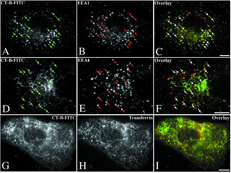
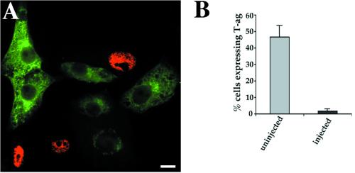
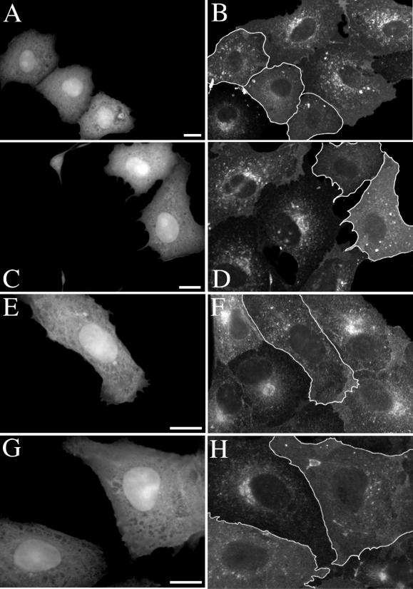
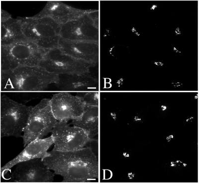
Simian virus 40 (SV40) is a nonenveloped virus that has been shown to pass from surface caveolae to the endoplasmic reticulum in an apparently novel infectious entry pathway. We now show that the initial entry step is blocked by brefeldin A and by incubation at 20 degrees C. Subsequent to the entry step, the virus reaches a domain of the rough endoplasmic reticulum by an unknown pathway. This intracellular trafficking pathway is also brefeldin A sensitive. Infection is strongly inhibited by expression of GTP-restricted ADP-ribosylation factor 1 (Arf1) and Sar1 mutants and by microinjection of antibodies to betaCOP. In addition, we demonstrate a potent inhibition of SV40 infection by the dipeptide N-benzoyl-oxycarbonyl-Gly-Phe-amide, which also inhibits late events in cholera toxin action. Our results identify novel inhibitors of SV40 infection and show that SV40 requires COPI- and COPII-dependent transport steps for successful infection.


Figures

Figure 1
Figure 1
Electron microscopic localization of SV40 in PDI-positive subdomain of ER. Vero cells were incubated with SV40 for 18 to 21 h at 37°C before fixation and processing for either epon embedding (A and B) or frozen sectioning (C). (A and B) SV40 accumulates in a domain of the rough ER. Virus particles (large arrowheads) are in smooth-membraned areas of the rough ER; ribosomes (small arrowheads in B) are evident on cisternae connected to the SV40-containing structures. (C) Frozen section labeled for SV40 (large gold, large arrowheads) and for PDI (small gold, small arrowheads) showing colocalization of the virus with the ER marker. N, nucleus. Bars, 200 nm.
Figure 2
Figure 2
BFA sensitivity of early events in SV40 infection. Treatments of 0.1 μg/ml BFA for 3 h (B) were found to disrupt the Golgi complex of Vero cells (cf. untreated cells in A) as revealed by immunolabeling for the Golgi marker giantin. In Vero cells treated with 0.1 μg/ml BFA for 3 h and subsequently infected with SV40 for 21 h in the continued presence of the drug, a potent inhibition of viral infection was observed (E; +BFA control). Immunolabeling for SV40 (green) and nuclear staining with DAPI (blue) revealed an accumulation of the virus at the periphery of cells treated with BFA (D), a pattern clearly different from that observed in untreated cells (C). (E) When the 3-h BFA treatment was applied at various time points after the commencement of infection, a sensitivity to the drug of early but not later events in SV40 infection was evident. A representative experiment is shown. (F) BFA inhibits SV40 internalization. Vero cells cultured in 0.5 μg/ml BFA for 1 h were allowed to internalize SV40 for 2 h at 37°C in the presence or absence of the drug. Subsequent exposure of the cells to an SV40 neutralizing antiserum allowed inactivation of all surface-exposed virus before a further 46 h of infection in the absence of the drug. Fixation and immunolabeling for T-ag revealed a substantially greater sensitivity of virus infection to the neutralizing antiserum in BFA-treated cells compared with untreated controls. A representative experiment is shown. Infection efficiencies are shown normalized to those of controls notexposed to the neutralizing antiserum. (G) Epon sections of Vero cells cultured in 0.3 μg/ml BFA for 3 h and then infected with SV40 for 21 h in the presence of the drug. Thin sections revealed an accumulation of virus particles at the cell surface as shown at low magnification in the inset. The virus accumulates in apparently normal caveolae-like invaginations (arrowheads). Empty caveolae are also apparent (arrows). (H) BFA inhibits intracellular trafficking of SV40. Vero cells were infected with SV40 for 2 h at 37°C before exposure to the neutralizing antiserum. Subsequent culture with or without 0.5 μg/ml BFA for the remaining 20 h of infection was followed by fixation and immunolabeling for T-ag. Cell counts revealed a strong inhibition of infection in BFA-treated cells compared with untreated controls. Infection efficiencies are shown normalized to those of untreated controls. A representative experiment is shown. Bars, 20 μm (A–D) and 100 nm (G).
Figure 3
Figure 3
SV40 internalization is inhibited at 20°C. Vero cells were infected with SV40 for 4 h at either 20 or 37°C before neutralizing surface-exposed virus with SV40 neutralizing antiserum. After subsequent incubation at 37°C for a remaining 24 h, the cells were fixed and immunolabeled for T-ag. Quantitation of infection efficiency revealed a much greater sensitivity of the virus to the neutralizing antibody after infection at 20°C than after infection at 37°C. Infection efficiencies are shown normalized to controls not exposed to the neutralizing antiserum. A representative experiment is shown.
Figure 4
Figure 4
Early endosome-to-Golgi transport of CT-B is inhibited at 20°C and by BFA. Incubation of Vero cells in 0.5 μg/ml CT-B-FITC for 1 h at 20°C resulted in accumulation of the toxin in EEA1-positive early endosomes (arrows) and a failure of the toxin to reach the Golgi complex (A–C). Similarly, pretreatment with 0.5 μg/ml BFA for 2 h before subsequent internalization of CT-B-FITC in the continued presence of BFA resulted in toxin delivery to EEA1-positive early endosomes (arrows) without subsequent arrival at the Golgi complex even after 1 h at 37°C (D–F). In BFA-treated cells, CT-B-FITC was also observed in a tubulated compartment identified as the recycling endosome by colocalization with cointernalized transferrin-Texas Red (G and I). Bars, 10 μm.
Figure 5
Figure 5
Inhibition of SV40 infection by microinjection of a βCOP antibody. Vero cells were microinjected with the βCOP antibody, anti-EAGE, and subjected to SV40 infection. Fixation and dual immunolabeling for the viral nuclear antigen T-ag (red) and the microinjected antibody (green) allowed detection of infected and injected cells, respectively (A). Cell counts revealed a dramatic inhibition of SV40 infection by the βCOP antibody (B). The mean and SEM of three independent experiments are shown. Bar, 20 μm.
Figure 6
Figure 6
Inhibition of SV40 infection by Arf1(Q71L) and Sar1(H79G). Vero cells were injected with either a mixture of Arf1(Q71L) and GFP expression plasmids (A) or the GFP plasmid alone (C) and incubated for 5 h to allow protein expression to occur. Immunolabeling for βCOP (B and D) revealed an obvious disruption or complete absence of the Golgi βCOP labeling pattern in cells expressing GFP and Arf1(Q71L) (A and B) but not in cells expressing only GFP (C and D). Subjection of these cells to SV40 infection and quantitation of infection efficiency revealed a potent inhibition in Arf1(Q71L)-transfected cells compared with cells expressing GFP alone (E). A significant inhibition of SV40 infection was also elicited by a GTPase-deficient Sar1 mutant in a similar experiment (F). The graphs show the mean and SEM of three independent experiments. Infection efficiencies of transfected cells have been normalized to those of surrounding untransfected cells. Bars, 40 μm.
Figure 7
Figure 7
Exocytic transport of ts-045-G is inhibited by Sar1(H79G) and Arf1(Q71L). Vero cells were injected with GFP-tagged ts-045-G expression plasmid alone (A), or with a mixture of ts-045-G and either Sar1(H79G) (B) or Arf1(Q71L) (C) expression plasmids. Subsequent incubation at the restrictive temperature (39.5°C) for 3 h allowed expression of the mutant proteins and accumulation of ts-045-G in the ER (our unpublished data). A further 3-h incubation at the permissive temperature (31°C) resulted in delivery of ts-045-G to the plasma membrane in cells expressing this mutant alone (A). However, in cells expressing either Sar1(H79G) (B) or Arf1(Q71L) (C), ts-045-G remained trapped in the ER and in perinuclear aggregates and was never seen to reach the plasma membrane. Bar, 20 μm.
Figure 8
Figure 8
CT transport to the Golgi complex is inhibited by Arf1(Q71L) and Sar1(H79G). Vero cells were injected with a mixture of GFP and either Arf1(Q71L) (A–D) or Sar1(H79G) (E–H) expression plasmids. After a 5-h period of protein expression, the cells were allowed to bind either CT-B-FITC or unlabeled holotoxin and internalize for 40 min at 37°C. Injected cells (identified by GFP expression shown in A, C, E, and G) displayed internalization of the toxin to early endosomes but not to the Golgi (outlined cells in B, D, F, and H). An apparent accumulation of toxin at the cell surface of injected cells was also often noted and may reflect a slight reduction in initial internalization. Bars, 20 μm.
Figure 9
Figure 9
CT transport to the ER is inhibited by Arf1(Q71L) and Sar1(H79G). Vero cells were injected with a mixture of GFP and either Arf1(Q71L) (A and B) or Sar1(H79G) (C–F) expression plasmids and incubated in 10 μg/ml cycloheximide for 4 h. The cells were then allowed to bind and internalize CT holotoxin in the absence of cycloheximide at 20°C for 1 h and subsequently at 37°C for a further 2 h. Although the toxin (B, D, and F) clearly reached the ER in uninjected cells (outlined in A, C, and E), cells expressing the Arf1 or Sar1 mutants (identified by GFP expression shown in A, C, and E) displayed a conspicuous lack of toxin in the ER. Instead, an accumulation of the toxin in endosomes and sometimes the Golgi was observed in injected cells. Bars, 10 μm.
Figure 10
Figure 10
Cbz-gly-phe-NH2 inhibits SV40 infection but not SFV infection. (A) Vero cells were pretreated for 1 h with 2 mM Cbz-gly-phe-NH2 or its inactive analog Cbz-Gly-Gly-NH2, or were untreated. Subsequent infection with SV40 in the continued presence or absence of the dipeptides was followed by fixation and immunolabeling for T-ag. Quantitation of infection efficiency revealed a consistent inhibition of SV40 infection by the Cbz-gly-phe-NH2 but not its inactive analog. The mean and SEM of three independent experiments are shown. (B) Vero cells pretreated with 2 mM Cbz-gly-phe-NH2 for 1 h or left untreated were subjected to SFV infection in the continued presence or absence of the drug. Quantitation of infection efficiency revealed no inhibition of SFV infection in treated cells. The mean and SEM of three independent experiments are shown. (C) Vero cells were pretreated for 1 h with 2 mM Cbz-gly-phe-NH2 before a 21-h infection with SV40 in the continued presence of the drug. The cells were then washed and incubated for various periods in the absence of the drug. Although infection efficiency remained low for the first 3 h after washing out the drug, a recovery of T-ag expression began to be seen after 6 h and was complete after 9 h in the absence of the drug. Infection efficiencies are shown normalized to those of untreated controls. A representative experiment is shown. (D) Vero cells were infected with SV40 for 4 h before inactivation of surface-exposed virus by exposure to SV40 neutralizing antiserum. The cells were then incubated in medium containing 2 mM Cbz-gly-phe-NH2 or its inactive analog for a further 20 h of infection. Quantitation of T-ag expression revealed a potent inhibition of infection by the dipeptide inhibitor. The mean and SEM of three independent experiments are shown.
Figure 11
Figure 11
Post-Golgi inhibition of CT toxicity by Cbz-gly-phe-NH2. Vero cells were preincubated in 2 mM Cbz-gly-phe-NH2 (A and B) or its inactive analog (C and D) before binding and internalization of CT-B-FITC (A and C) for 40 min in the continued presence of the drugs. The cells were then fixed and immunolabeled for the Golgi matrix protein GM130 (B and D). No apparent difference in CT-B uptake to the Golgi complex was observed in inhibitor-treated cells. Bar 10 μm.

References

    1. Anderson HA, Chen Y, Norkin LC. Bound simian virus 40 translocates to caveolin-enriched membrane domains, and its entry is inhibited by drugs that selectively disrupt caveolae. Mol Biol Cell. 1996;7:1825–1834. - PMC - PubMed
    1. Anderson HA, Chen Y, Norkin LC. MHC class I molecules are enriched in caveolae but do not enter with simian virus 40. J Gen Virol. 1998;79:1469–1477. - PubMed
    1. Aniento F, Gu F, Parton RG, Gruenberg J. An endosomal beta COP is involved in the pH-dependent formation of transport vesicles destined for late endosomes. J Cell Biol. 1996;133:29–41. - PMC - PubMed
    1. Aridor M, Bannykh SI, Rowe T, Balch WE. Sequential coupling between COPII and COPI vesicle coats in endoplasmic reticulum to Golgi transport. J Cell Biol, 1995;131:875–893. - PMC - PubMed
    1. Breau WC, Atwood WJ, Norkin LC. Class I major histocompatibility proteins are an essential component of the simian virus 40 receptor. J Virol. 1992;66:2037–2045. - PMC - PubMed

Publication types

MeSH terms