. 2002 Jun;70(6):2853-61.
doi: 10.1128/IAI.70.6.2853-2861.2002.

The lbgAB gene cluster of Haemophilus ducreyi encodes a beta-1,4-galactosyltransferase and an alpha-1,6-DD-heptosyltransferase involved in lipooligosaccharide biosynthesis

Affiliations

The lbgAB gene cluster of Haemophilus ducreyi encodes a beta-1,4-galactosyltransferase and an alpha-1,6-DD-heptosyltransferase involved in lipooligosaccharide biosynthesis

Michael V Tullius et al. Infect Immun. 2002 Jun.

Abstract

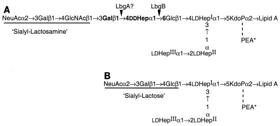
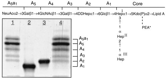
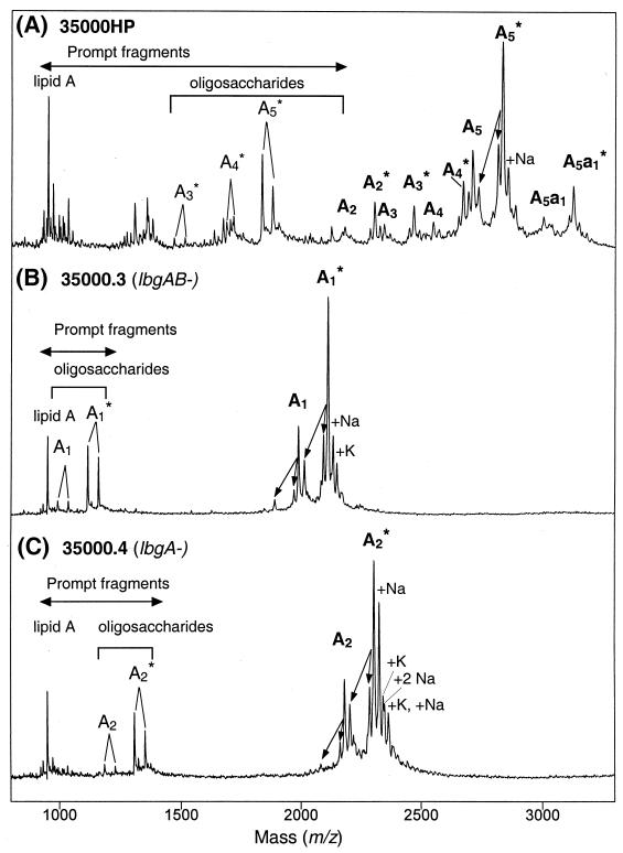
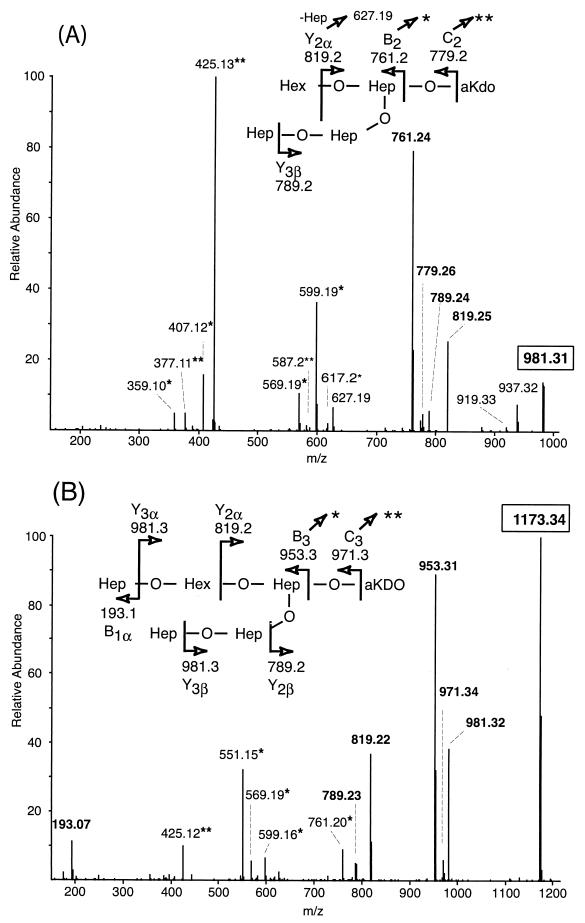
All Haemophilus ducreyi strains examined contain a lipooligosaccharide (LOS) consisting of a single but variable branch oligosaccharide that emanates off the first heptose (Hep-I) of a conserved Hep(3)-phosphorylated 3-deoxy-D-manno-octulosonic acid-lipid A core. In a previous report, identification of tandem genes, lbgA and lbgB, that are involved in LOS biosynthesis was described (Stevens et al., Infect. Immun. 65:651-660, 1997). In a separate study, the same gene cluster was identified and the lbgB (losB) gene was found to be required for transfer of the second sugar, D-glycero-D-manno-heptose (DD-Hep), of the major branch structure (Gibson et al., J. Bacteriol. 179:5062-5071, 1997). In this study, we identified the function of the neighboring upstream gene, lbgA, and found that it is necessary for addition of the third sugar in the dominant oligosaccharide branch, a galactose-linked beta1-->4, to the DD-Hep. LOS from an lbgA mutant and an lbgAB double mutant were isolated and were characterized by sodium dodecyl sulfate-polyacrylamide gel electrophoresis, carbohydrate analysis, mass spectrometry, and nuclear magnetic resonance spectroscopy. The results showed that the mutant strains synthesize truncated LOS glycoforms that terminate after addition of the first glucose (lbgAB) or the disaccharide DD-Hepalpha1-->6Glcbeta1 (lbgA) that is attached to the heptose core. Both mutants show a significant reduction in the ability to adhere to human keratinocytes. Although minor differences were observed after two-dimensional gel electrophoresis of total proteins from the wild-type and mutant strains, the expression levels of the vast majority of proteins were unchanged, suggesting that the differences in adherence and invasion are due to differences in LOS. These studies add to the mounting evidence for a role of full-length LOS structures in the pathophysiology of H. ducreyi infection.


Figures

FIG. 1.
FIG. 1.
Structures of the two major sialylated LOS glycoforms from H. ducreyi. (A) LOS with pentasaccharide branch terminting in N-acetyllactosamine that is partially substituted with alpha-2,3-linked sialic acid. (B) LOS with disaccharide lactose branch that is partially substituted with alpha-2,3-linked sialic acid.
FIG. 2.
FIG. 2.
Silver-stained SDS-polyacrylamide gel of LOS from the lbgAB (lane 2) and lbgA (lane 3) mutants of H. ducreyi strain 35000HP (lanes 1 and 4). The designations on the right (A5a1, A5, etc.) refer to the proposed LOS glycoforms to which the bands are believed to correspond. Only the bottom portion of the gel where the LOS migrated is shown. The phosphate of Kdo can be partially substituted with PEA (asterisk). The structure corresponding to the LOS glycoform designated A1 was determined previously (16).
FIG. 3.
FIG. 3.
Negative-ion MALDI-TOF spectra of O-deacylated LOS from the lbgAB (B) and lbgA (C) mutant strains, as well as parental strain 35000HP (A). Strain 35000HP is a human-passaged isolate of parent strain 35000 and produces an LOS profile similar to that of strain 35000 (6). There were prompt fragments corresponding to cleavage of the oligosaccharide from the O-deacylated lipid A, as well as fragments corresponding to loss of H2O and H3PO4 (indicated by arrows). Two additional fragments arose from each oligosaccharide via loss of CO2 (44 Da) from the terminal Kdo (17). Sodium and potassium adducts, primarily of PEA-containing LOS species, are also present. The singly deprotonated molecular ions (M-H) are designated (in boldface type) according to the nomenclature shown in Fig. 1. An asterisk indicates addition of PEA. Oligosaccharide prompt fragments arising from the O-deacylated LOS glycoforms are indicated by lightface type. The lipid A prompt fragments observed are diphosphoryl N-diacyl lipid A (O-deacylated lipid A).
FIG. 4.
FIG. 4.
Positive-ion MS/MS spectra of the dephosphorylated oligosaccharides from the LOS of lbgAB (A) and lbgA (B) H. ducreyi mutants. The singly charged parent ions are enclosed by boxes, and their structures are shown at the top of each panel. Anhydro-Kdo (aKDO) is formed by β-elimination of phosphate from phosphorylated Kdo during acetic acid hydrolysis. Peaks marked with one or two asterisks are internal ions that appear to originate from the highly abundant B3/B2 and C3/C2 ions that have lost the terminal anhydro-Kdo and subsequently undergo losses of Hex and/or Hep sugars from the nonreducing termini. For example, in spectrum A fragmentation of m/z 779.3 (C2) yields the internal ions at m/z 617.2 (-Hex), 587.2 (-Hep), and 425.1 (-Hex, Hep), and fragmentation of m/z 761.2 (B2) yields m/z 599.2 (-Hex), 569.2 (-Hep), 407.1 (-Hep, Hex), and 377.1 (-HepHep). In spectrum B fragmentation of m/z 953.3 (B3) yields the internal ions at m/z 761.2 (-Hep), 599.2 (-Hep, Hex), 569.2 (-HepHep), and 551.2 (-HepHep, H2O), and fragmentation of m/z 971.3 (C3) yields m/z 425.1 (-Hep, HepHex).

References

    1. Ahmed, H. J., A. Frisk, J. E. Mansson, E. K. Schweda, and T. Lagergard. 1997. Structurally defined epitopes of Haemophilus ducreyi lipooligosaccharides recognized by monoclonal antibodies. Infect. Immun. 65:3151-3158. - PMC - PubMed
    1. Alfa, M. J., and P. DeGagne. 1997. Attachment of Haemophilus ducreyi to human foreskin fibroblasts involves LOS and fibronectin. Microb. Pathog. 22:39-46. - PubMed
    1. Alfa, M. J., P. DeGagne, and P. A. Totten. 1996. Haemophilus ducreyi hemolysin acts as a contact cytotoxin and damages human foreskin fibroblasts in cell culture. Infect. Immun. 64:2349-2352. - PMC - PubMed
    1. Apicella, M. A., J. M. Griffiss, and H. Schneider. 1994. Isolation and characterization of lipopolysaccharides, lipooligosaccharides, and lipid A. Methods Enzymol. 235:242-252. - PubMed
    1. Apicella, M. A., R. E. Mandrell, M. Shero, M. E. Wilson, J. M. Griffiss, G. F. Brooks, C. Lammel, J. F. Breen, and P. A. Rice. 1990. Modification by sialic acid of Neisseria gonorrhoeae lipooligosaccharide epitope expression in human urethral exudates: an immunoelectron microscopic analysis. J. Infect. Dis. 162:506-512. - PubMed

Publication types

MeSH terms

LinkOut - more resources