. 2002 Jun;70(6):2982-8.
doi: 10.1128/IAI.70.6.2982-2988.2002.

Malaria-induced acquisition of antibodies to Plasmodium falciparum variant surface antigens

Affiliations

Malaria-induced acquisition of antibodies to Plasmodium falciparum variant surface antigens

Michael F Ofori et al. Infect Immun. 2002 Jun.

Abstract

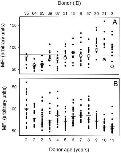
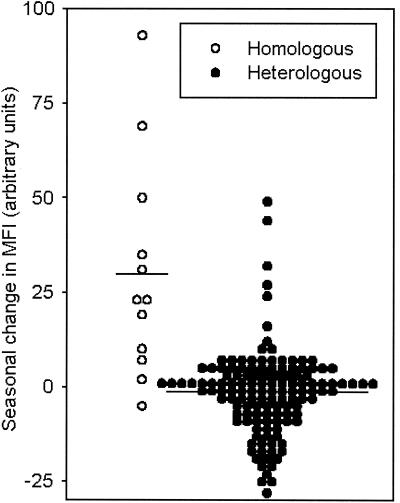
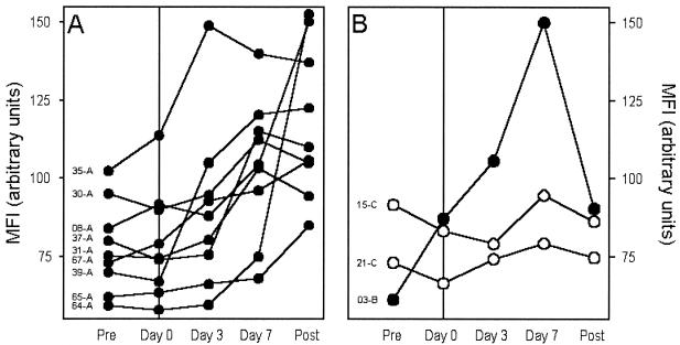
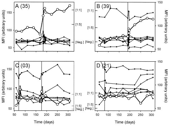
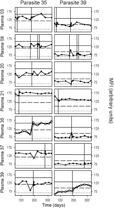
In areas of intense Plasmodium falciparum transmission, protective immunity is acquired during childhood in parallel with acquisition of agglutinating antibodies to parasite-encoded variant surface antigens (VSA) expressed on parasitized red blood cells. In a semi-immune child in such an area, clinical disease is caused mainly by parasites expressing VSA not recognized by preexisting VSA-specific antibodies in that child. Such malaria episodes are known to cause an increase in agglutinating antibodies specifically recognizing VSA expressed by the parasite isolate causing the illness, whereas antibody responses to other parasite isolates are relatively unaffected. However, the detailed kinetics of this VSA antibody acquisition are unknown and hence were the aim of this study. We show that P. falciparum malaria in Ghanaian children generally caused a rapid and sustained increase in variant-specific VSA antibody levels, while more transient and limited increases in levels of antibodies to VSA expressed by other parasite isolates were also seen. Plasma VSA antibody levels were positively correlated with the age of the healthy plasma donors but negatively correlated with the age of the parasite donors (the malaria patient). The data from this first detailed longitudinal study of acquisition of VSA antibodies support the hypothesis that naturally acquired protective immunity to P. falciparum malaria is mediated, at least in part, by VSA-specific antibodies.


Figures

FIG. 1.
FIG. 1.
Levels of VSA antibodies in plasma obtained at the beginning of the study (preseason), expressed as MFI values. (A) Increasing levels of VSA-specific plasma antibodies with increasing age of the plasma donor. For each plasma donor, the levels of plasma antibodies specifically recognizing VSA expressed by the isolate causing malaria in that individual (homologous isolate [○]) and by the 11 isolates causing malaria in the other children (heterologous isolates [•]) are shown. The overall mean VSA antibody reactivity is indicated by a horizontal line. (B) Decreasing VSA-specific plasma antibody recognition of parasites with increasing age of the parasite donor (malaria patient). For each parasite isolate, the levels of isolate-specific VSA antibodies in plasma from each of the 25 children who suffered malaria attacks during the study period, as well as means (long bars) and medians (short bars), are shown. The donor ID and age of the donors of the plasma samples (A) or parasite isolates (B) are indicated at the top and bottom of the figure, respectively.
FIG. 2.
FIG. 2.
Seasonal change in antibodies specifically recognizing VSA expressed by the parasite isolate causing clinical disease (homologous response [○]) and by 11 other parasites (heterologous responses [•]). For each child-parasite combination, the difference between postseason and preseason MFI is shown. In addition, the overall means are indicated by horizontal bars.
FIG. 3.
FIG. 3.
Temporal changes in plasma levels of antibodies recognizing VSA expressed by the causative (homologous) parasite isolate relative to the time of the P. falciparum malaria episode. Plasma VSA antibody levels (expressed as MFI values) at the beginning of the study (Pre), at diagnosis (Day 0), during convalescence (Day 3 and Day 7), and at the end of the study (Post) for individual patients are indicated. Donor identification codes are indicated in the left side of each panel. (A) Changes in individuals with rapid, marked, and sustained disease-induced increases in VSA antibody levels. (B) Changes in individuals with transient (•) or without conspicuous (○) changes in VSA antibody levels.
FIG. 4.
FIG. 4.
Detailed analysis of temporal changes in levels of VSA antibodies in plasma obtained at monthly intervals throughout the study, expressed as MFI values. Antibody responses to the homologous isolate (○) and heterologous isolates (•) are shown, and the timings of P. falciparum malaria episodes in the children are indicated by vertical lines. In all the panels, MFI values obtained using an undiluted ([1:1]) and 5-fold-diluted ([1:5]) hyperimmune plasma pool have been indicated by inward ticks to allow quantitative evaluation of the changes in antibody levels. The mean MFI + 2 standard deviations obtained from seven nonexposed donors is similarly indicated ([Neg.]). (A and B) Changes in two individuals (A, 35; B, 39) in whom the malaria episode caused rapid and sustained increases in VSA antibodies specific for the homologous isolate. (C) Changes in an individual (03) in whom the malaria episode caused limited and transient increases in VSA antibodies specific for the homologous isolate from the first of two malaria attacks after each of two clinical episodes with this individual (note marked disease-induced changes in levels of antibodies specific for VSA expressed by several heterologous isolates in this individual). (D) Changes in an individual (21) in whom the malaria episode caused no changes in VSA antibodies specific for the homologous isolate.
FIG. 5.
FIG. 5.
Detailed analysis of temporal changes in levels of VSA antibodies in plasma obtained at monthly intervals throughout the study, expressed as MFI values. Temporal changes in antibody responses to VSA expressed by two parasite isolates (35 [left panels] and 39 [right panels]) as seen in seven representative individuals (rows) are shown. The timing of P. falciparum malaria episodes in individual children is indicated by vertical lines. In all panels, the MFI obtained using a hyperimmune plasma pool undiluted (1:1) (horizontal solid line) and diluted 1:5 (dashed line), and the mean MFI + 2 standard deviations using plasma from seven nonexposed donors (dotted line) are shown.

Comment in

Similar articles

Cited by

References

    1. Afari, E. A., M. Appawu, S. Dunyo, A. Baffoe-Wilmot, and F. K. Nkrumah. 1995. Malaria infection, morbidity and transmission in two ecological zones in southern Ghana. Afr. J. Health Sci. 2:312-316. - PubMed
    1. Afari, E. A., K. A. Koram, S. K. Dunyo, and F. K. Nkrumah. 1992. The epidemiology of malaria with special emphasis on transmission, morbidity, mortality and disease control in Ghana. Internal report. Noguchi Memorial Institute for Medical Research, Legon, Ghana.
    1. Barnwell, J. W., A. S. Asch, R. L. Nachman, M. Yamaya, M. Aikawa, and P. Ingravallo. 1989. A human 88-kD membrane glycoprotein (CD36) functions in vitro as a receptor for a cytoadherence ligand on Plasmodium falciparum-infected erythrocytes. J. Clin. Investig. 84:765-772. - PMC - PubMed
    1. Barnwell, J. W., C. Ockenhouse, and D. M. Knowles II. 1985. Monoclonal antibody OKM5 inhibits the in vitro binding of Plasmodium falciparum infected erythrocytes to monocytes, endothelial, and C32 melanoma cells. J. Immunol. 135:3494-3497. - PubMed
    1. Baruch, D. I., B. L. Pasloske, H. B. Singh, et al. 1995. Cloning the P. falciparum gene encoding PfEMP1, a malarial variant antigen and adherence receptor on the surface of parasitized human erythrocytes. Cell 82:77-87. - PubMed

Publication types

MeSH terms

LinkOut - more resources