. 2002 Jun;70(6):2995-3003.
doi: 10.1128/IAI.70.6.2995-3003.2002.

Lipopolysaccharide down regulates both scavenger receptor B1 and ATP binding cassette transporter A1 in RAW cells

Affiliations

Lipopolysaccharide down regulates both scavenger receptor B1 and ATP binding cassette transporter A1 in RAW cells

Irina Baranova et al. Infect Immun. 2002 Jun.

Abstract

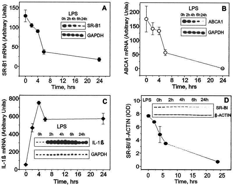
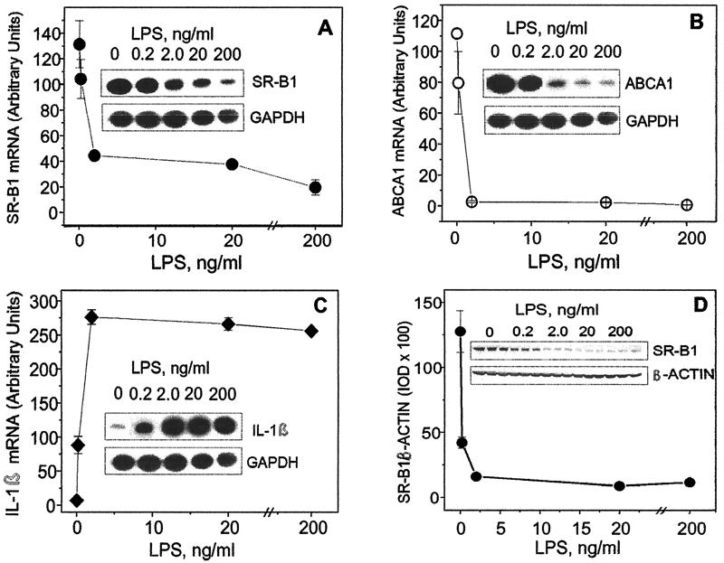
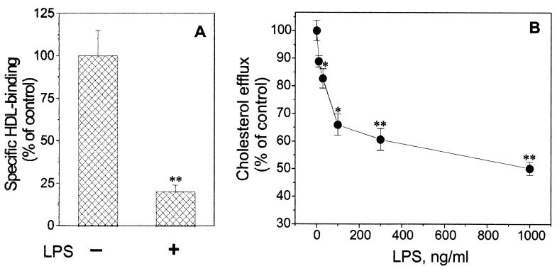
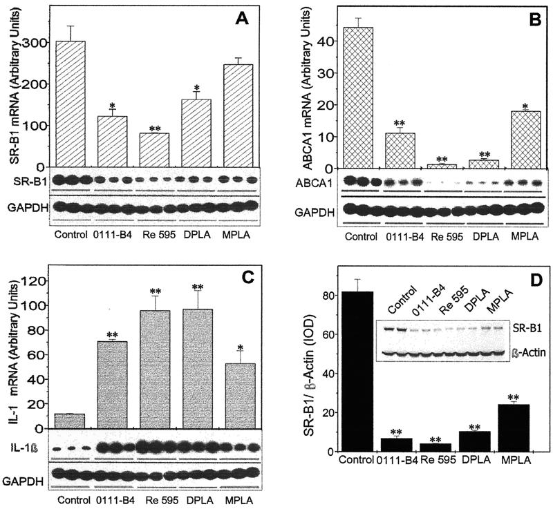
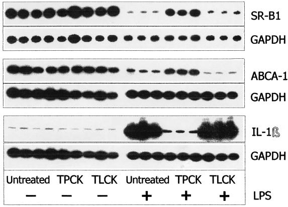
Lipopolysaccharide (LPS) has recently been shown to facilitate macrophage foam cell formation and has been suggested to be a proatherogenic factor. The mechanism of LPS induced cholesterol accumulation, however, is unclear. In this report, using the macrophage-like RAW 264.7 cell line, we provide experimental evidence that LPS's proatherogenic effects may at least in part reflect altered cholesterol metabolism. Data presented demonstrate that in a dose-dependent manner, LPS is able to down regulate the mRNA expression of the two primary high-density lipoprotein (HDL) receptors, scavenger receptor B1 (SR-B1) and ATP binding cassette A1 (ABCA1), with a 50% inhibitory concentration of less than 0.2 ng/ml, as well as to decrease SR-B1 protein expression by 80%. We also found that LPS treatment resulted in a significant decrease (to 20% of the control level) of the specific (125)I-HDL binding as well as in 50% inhibition of the HDL-mediated cholesterol efflux compared to untreated cells. In addition, we compared the potencies of various modified LPS preparations and demonstrated that the phosphorylated lipid A portion of LPS, which is highly conserved among gram-negative microorganisms, including Chlamydia, is primarily responsible for the effects of LPS on SR-B1 and ABCA1 expression. Inhibitors of NF-kappaB activation were observed to efficiently block the suppressive effect of LPS on SR-B1 and ABCA1, suggesting a mechanism involving NF-kappaB. These data indicate that the LPS effects on cholesterol metabolism may contribute to the proatherogenic properties of LPS.


Figures

FIG. 1.
FIG. 1.
Effect of LPS on the time course of SR-B1 (A), ABCA1 (B), and IL-1β (C) mRNA and SR-B1 protein (D) expression. (A, B, and C) Total mRNA was isolated from RAW cells after 0, 2, 4, 6, and 24 h of incubation with LPS (1 μg/ml) in serum-free medium. Levels of mRNA expression for the indicated genes were tested by RT-PCR analyses. Corresponding samples were analyzed for GAPDH mRNA as controls. Levels of mRNA were quantitated by scanning densitometry and corrected relative to the levels of housekeeping gene mRNA. Data are presented as the ratio of integral optical density of the indicated gene to that of the GADPH gene multiplied by 100. (D) Cultured cells were treated with LPS for the indicated periods of time, and SR-B1 protein expression was estimated by Western blot analysis. Simultaneously, β-actin levels in the corresponding samples were determined to confirm equal protein loading. Nitrocellulose membranes were scanned, and the integral optical density (IOD) values of the protein bands were estimated with the GelPro computer program. Data are expressed as the ratio of the SR-B1 band integral optical density to the corresponding β-actin band integral optical density. Error bars indicate standard deviations.
FIG. 2.
FIG. 2.
Dose-dependent effect of LPS on the expression of ABCA1 (A), SR-B1 (B), and IL-1β (C) genes and SR-B1 protein (D). Cultured cells were exposed to the increasing concentrations (0, 0.2, 2, 20, and 200 ng/ml) of LPS for 24 h. Levels of mRNA expression for the indicated genes were tested by RT-PCR analyses, and SR-B1 protein expression was estimated by Western blot analysis. For further steps, see the Fig. 1 legend. The results shown represent one of two experiments that yielded similar results. IOD, integrated optical density. Error bars indicate standard deviations.
FIG. 3.
FIG. 3.
Down regulation of specific HDL binding and HDL-mediated cholesterol efflux by LPS. (A) Effect of LPS on 125I-HDL specific binding in RAW cells. The cells were incubated with 1 μg of LPS per ml for 24 h. Following three PBS washes, the specific binding of 125I-HDL (5 μg/ml) was determined at 4°C as the difference between the total and nonspecific binding (in the absence or presence of a 50-fold excess of unlabeled HDL). (B) Dose-dependent response of HDL-mediated [3H]cholesterol efflux to LPS stimulation. RAW cells preloaded with cholesterol were labeled with 1 μCi of [1,2-3H]cholesterol (50 Ci/mmol) per ml. Before the cholesterol efflux determination, the cells were pretreated with the increasing concentrations (0 to 1,000 ng/ml) of LPS for 24 h. After HDL (100 μg/ml) as the cholesterol acceptor was added, the [3H]cholesterol efflux assay was performed after an additional 24 h. Cholesterol efflux was calculated as the amount of radioactivity present in the medium divided by the total radioactivity (medium plus cell) in each well. The data shown represent one of two independent experiments that yielded similar results. ∗, P < 0.05; ∗∗, P < 0.01 (compared to untreated control samples). Error bars indicate standard deviations.
FIG. 4.
FIG. 4.
Comparison of Re595, 0011:B4, DPLA, and MPLA potency to modulate LPS-sensitive gene expression and to suppress SR-B1 protein expression. RAW cells were exposed to different LPS preparations (10 ng/ml) for 24 h in serum-free medium. (A, B, and C) Total mRNA was isolated and treated as described in the Fig. 1 legend. The levels of mRNA expression for the indicated genes were tested by RT-PCR analyses. (D) The level of SR-B1 protein expression was determined by Western blot analysis. The results represent one of two separate experiments that yielded similar results. ∗, P < 0.05; ∗∗, P < 0.01 (compared to control untreated samples). IOD, integrated optical density. Error bars indicate standard deviations.
FIG. 5.
FIG. 5.
Effect of protease inhibitor TPCK and its structural analogue TLCK on LPS-induced changes in IL-1β, SR-B1, and ABCA1 mRNA expression. The cells were incubated with TPCK or TLCK (negative control) alone for 2 h prior to LPS addition and then for 22 h in the presence of LPS (10 ng/ml). After the incubation, total mRNA was isolated and the samples were analyzed by RT-PCR. The results shown represent one of two separate experiments that yielded similar results.

References

    1. Acton, S. L., A. Rigotti, K. T. Landschulz, S. Xu, H. H. Hobbs, and M. Krieger. 1996. Identification of scavenger receptor SR-BI as a high density lipoprotein receptor. Science 271:518-520. - PubMed
    1. Baeuerle, P. A., and V. R. Baichwal. 1997. NF-kappa B as a frequent target for immunosuppressive and anti-inflammatory molecules. Adv. Immunol. 65:111-137. - PubMed
    1. Bocharov, A. V., T. G. Vishnyakova, I. N. Baranova, A. P. Patterson, and T. L. Eggerman. 2001. Characterization of a 95 kDa high affinity human high density lipoprotein-binding protein. Biochemistry 140:4407-4416. - PubMed
    1. Bradford, M. M. 1976. A rapid and sensitive method for the quantitation of microgram quantities of protein utilizing the principle of protein-dye binding. Anal. Biochem. 72:248-254. - PubMed
    1. Buechler, C., M. Ritter, C. D. Quoc, A. Agildere, and G. Schmitz. 1999. Lipopolysaccharide inhibits the expression of the scavenger receptor Cla-1 in human monocytes and macrophages. Biochem. Biophys. Res. Commun. 262:251-254. - PubMed

MeSH terms