. 2002 Jun;70(6):3040-52.
doi: 10.1128/IAI.70.6.3040-3052.2002.

Differential regulation of the mitogen-activated protein kinases by pathogenic and nonpathogenic mycobacteria

Affiliations

Differential regulation of the mitogen-activated protein kinases by pathogenic and nonpathogenic mycobacteria

Shannon K Roach et al. Infect Immun. 2002 Jun.

Abstract

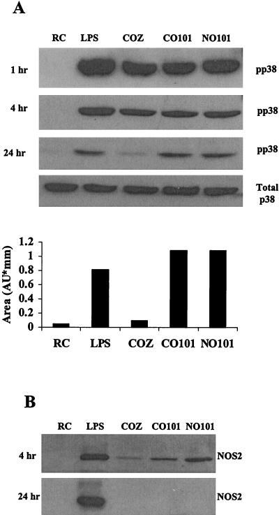
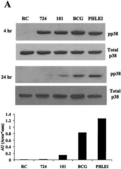
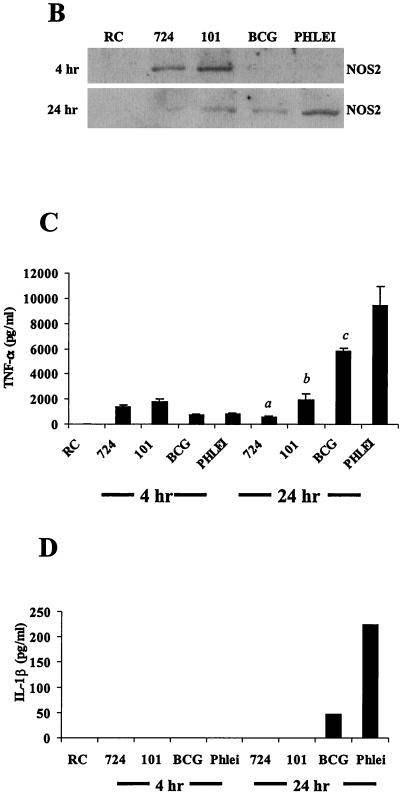
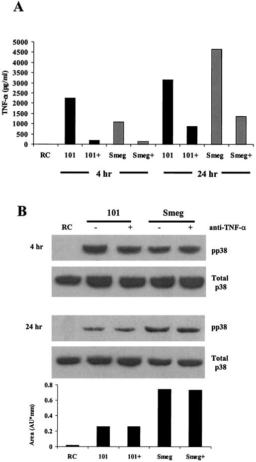
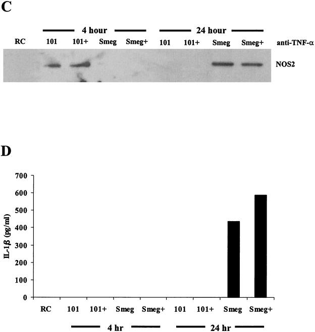
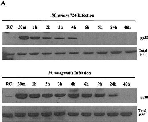
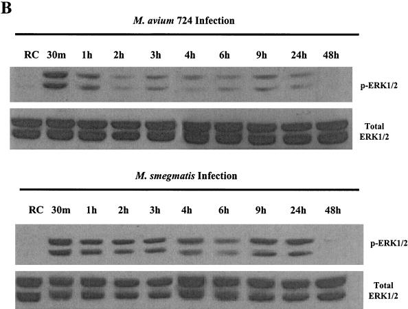
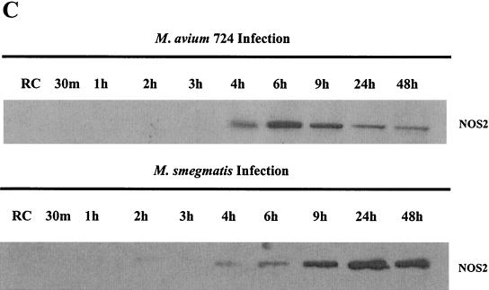
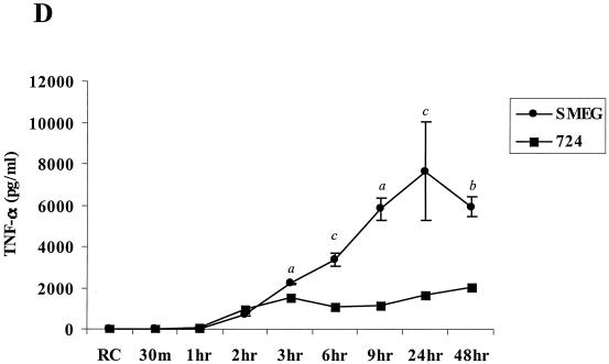
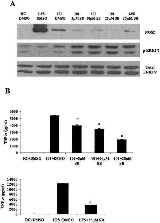
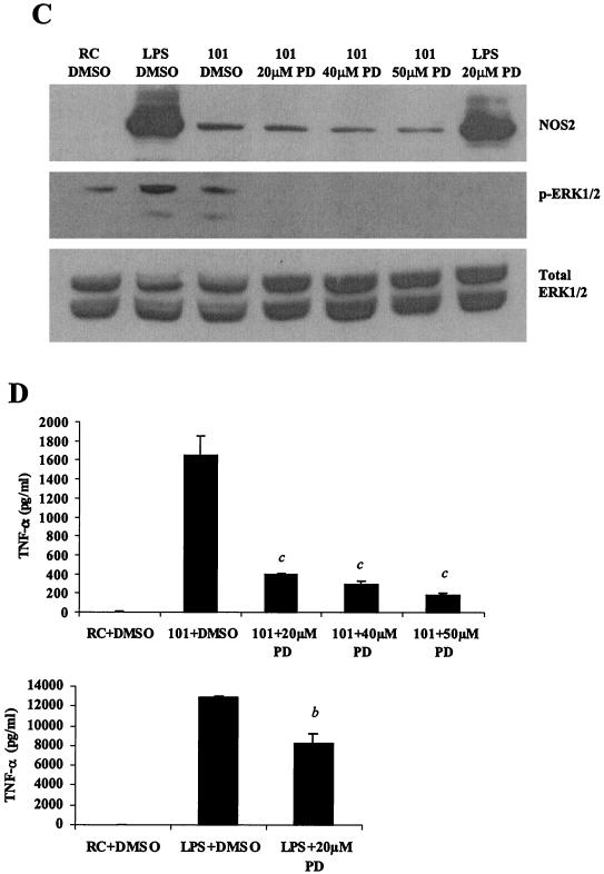
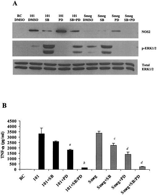
Mycobacteria are the etiologic agents of numerous diseases which account for significant morbidity and mortality in humans and other animal species. Many mycobacteria are intramacrophage pathogens and therefore the macrophage response to infection, which includes synthesis of cytokines such as tumor necrosis factor alpha (TNF-alpha) and production of nitric oxide, has important consequences for host immunity. However, very little is known about the macrophage cell signaling pathways initiated upon infection or how pathogenic mycobacteria may modulate the macrophage responses. Using primary murine bone marrow macrophages, we established that p38 and extracellular signal-regulated kinases 1 and 2 of the mitogen-activated protein kinase (MAPK) pathways are activated upon infection with different species of mycobacteria. However, we observed decreased MAPK activity over time in macrophages infected with pathogenic Mycobacterium avium strains relative to infections with nonpathogenic mycobacteria. Furthermore, macrophages infected with M. avium produced lower levels of TNF-alpha, interleukin 1beta, and inducible nitric oxide synthase 2 than macrophages infected with nonpathogenic species. Inhibitor studies indicate that the MAPKs are required for the Mycobacterium-mediated induction of these effector proteins. Our data indicate that MAPKs are activated in macrophages upon invasion by mycobacteria and that this activation is diminished in macrophages infected with pathogenic strains of M. avium, resulting in decreased production of important immune effector proteins. The decreased MAPK activation associated with M. avium infections suggests a novel point of immune intervention by this mycobacterial species.


Figures

FIG. 1.
FIG. 1.
Infection of murine bone marrow macrophages with M. avium 101 activates p38 MAPK. (A) Western blot analysis of p38 phosphorylation (pp38) was performed following treatment of bone marrow macrophages with LPS or infection with complement-opsonized zymosan (COZ), complement-opsonized M. avium 101 (CO101) or nonopsonized M. avium 101 (NO101) or left untreated as resting cells (RC). Equal protein, as determined by the Micro BCA assay, was loaded in each lane. All blots were stripped and reprobed with a p38 antibody to show equal amounts of this protein, but only the 24-h total p38 blot is shown here. The relative densities of the 24-h phosphorylated p38 bands were analyzed by densitometry. AU, arbitrary units. (B) NOS2 expression from cell lysates was detected by Western blotting. Results are representative of three separate experiments.
FIG. 2.
FIG. 2.
Macrophages infected with pathogenic mycobacteria show decreased MAPK activation and downstream production of inflammatory mediators compared to macrophages infected with nonpathogenic mycobacteria. (A) Western blot analysis of p38 phosphorylation (pp38) following infection with M. avium 724, M. avium 101, M. bovis BCG, and M. phlei. Noninfected macrophages (RC) were treated the same as infected macrophages for all incubations. Blots were stripped and reprobed with a p38 antibody. Densitometry was performed with the 24-h phospho-p38 blot. AU, arbitrary units. (B) NOS2 expression was detected by Western blotting. (C and D) TNF-α and IL-1β present in the culture supernatants at each time point were detected by ELISA. a, P < 0.001 compared to M. bovis BCG and to M. phlei; b, P < 0.01 compared to M. bovis BCG and P < 0.001 compared to M. phlei; c, P < 0.01 compared to M. phlei. Results are representative of three separate experiments.
FIG. 2.
FIG. 2.
Macrophages infected with pathogenic mycobacteria show decreased MAPK activation and downstream production of inflammatory mediators compared to macrophages infected with nonpathogenic mycobacteria. (A) Western blot analysis of p38 phosphorylation (pp38) following infection with M. avium 724, M. avium 101, M. bovis BCG, and M. phlei. Noninfected macrophages (RC) were treated the same as infected macrophages for all incubations. Blots were stripped and reprobed with a p38 antibody. Densitometry was performed with the 24-h phospho-p38 blot. AU, arbitrary units. (B) NOS2 expression was detected by Western blotting. (C and D) TNF-α and IL-1β present in the culture supernatants at each time point were detected by ELISA. a, P < 0.001 compared to M. bovis BCG and to M. phlei; b, P < 0.01 compared to M. bovis BCG and P < 0.001 compared to M. phlei; c, P < 0.01 compared to M. phlei. Results are representative of three separate experiments.
FIG. 3.
FIG. 3.
An autocrine effect of TNF-α is not the cause for the extended p38 phosphorylation seen after 24 h. (A) TNF-α in the culture supernatants was detected by ELISA for the 4- and 24-h time points. Each supernatant was incubated with Protein G to remove any antibodies and bound TNF-α prior to the ELISA. (B) Western blot analysis of p38 phosphorylation (pp38) following treatment with LPS or infection with M. avium 101 or M. smegmatis. Cells were treated with either anti-TNF-α neutralizing antibody (+) or antibody control (−) during the 24-h infection. Noninfected macrophages, i.e., resting cells (RC), were incubated with the IgG control antibody. Blots were stripped and reprobed for total p38. Densitometry was performed with the 24-h phospho-p38 blot. (C) NOS2 expression from the cell lysates was detected by Western blotting. (D) IL-1β in the culture supernatants was detected by ELISA. Results are representative of two separate experiments. AU, arbitrary units; Smeg, M. smegmatis.
FIG. 3.
FIG. 3.
An autocrine effect of TNF-α is not the cause for the extended p38 phosphorylation seen after 24 h. (A) TNF-α in the culture supernatants was detected by ELISA for the 4- and 24-h time points. Each supernatant was incubated with Protein G to remove any antibodies and bound TNF-α prior to the ELISA. (B) Western blot analysis of p38 phosphorylation (pp38) following treatment with LPS or infection with M. avium 101 or M. smegmatis. Cells were treated with either anti-TNF-α neutralizing antibody (+) or antibody control (−) during the 24-h infection. Noninfected macrophages, i.e., resting cells (RC), were incubated with the IgG control antibody. Blots were stripped and reprobed for total p38. Densitometry was performed with the 24-h phospho-p38 blot. (C) NOS2 expression from the cell lysates was detected by Western blotting. (D) IL-1β in the culture supernatants was detected by ELISA. Results are representative of two separate experiments. AU, arbitrary units; Smeg, M. smegmatis.
FIG. 4.
FIG. 4.
Kinetics of MAPK activation and inflammatory responses differ between M. avium 724 and M. smegmatis infections. Western blot analyses of phosphorylated p38 (A) and ERK1/2 (B) following infections with either M. avium 724 or M. smegmatis over 48 h are shown. Blots were stripped and reprobed for total p38 and ERK1/2. (C) Western blot of NOS2 production. (D) The amount of TNF-α in the supernatants was analyzed by ELISA. a, P < 0.001; b, P < 0.01; c, P < 0.05. The results are representative of two separate experiments.
FIG. 4.
FIG. 4.
Kinetics of MAPK activation and inflammatory responses differ between M. avium 724 and M. smegmatis infections. Western blot analyses of phosphorylated p38 (A) and ERK1/2 (B) following infections with either M. avium 724 or M. smegmatis over 48 h are shown. Blots were stripped and reprobed for total p38 and ERK1/2. (C) Western blot of NOS2 production. (D) The amount of TNF-α in the supernatants was analyzed by ELISA. a, P < 0.001; b, P < 0.01; c, P < 0.05. The results are representative of two separate experiments.
FIG. 5.
FIG. 5.
p38 and ERK1/2 MAPKs are essential for NOS2 and TNF-α production during M. avium infection. (A and C) Western blot analysis of NOS2 production and ERK1/2 phosphorylation in cell lysates. Cells were treated with varying concentrations of the p38 inhibitor SB203580 (A) or the MEK1 inhibitor PD98059 (C) (Calbiochem, San Diego, Calif.) at 37°C for 1 h before LPS treatment or M. avium 101 for a 4-h infection. DMSO vehicle control was added to resting cells (RC) and LPS treated- and M. avium 101-infected cells. Blots were probed with total ERK1/2 antibody to show equal protein loading. (B and D) TNF-α in the respective culture supernatants was analyzed by ELISA. a, P < 0.001compared to M. avium 101 plus DMSO; b, P < 0.001 compared to LPS plus DMSO; c, P < 0.01 compared to M. avium 101 plus DMSO. The results are representative of two separate experiments.
FIG. 5.
FIG. 5.
p38 and ERK1/2 MAPKs are essential for NOS2 and TNF-α production during M. avium infection. (A and C) Western blot analysis of NOS2 production and ERK1/2 phosphorylation in cell lysates. Cells were treated with varying concentrations of the p38 inhibitor SB203580 (A) or the MEK1 inhibitor PD98059 (C) (Calbiochem, San Diego, Calif.) at 37°C for 1 h before LPS treatment or M. avium 101 for a 4-h infection. DMSO vehicle control was added to resting cells (RC) and LPS treated- and M. avium 101-infected cells. Blots were probed with total ERK1/2 antibody to show equal protein loading. (B and D) TNF-α in the respective culture supernatants was analyzed by ELISA. a, P < 0.001compared to M. avium 101 plus DMSO; b, P < 0.001 compared to LPS plus DMSO; c, P < 0.01 compared to M. avium 101 plus DMSO. The results are representative of two separate experiments.
FIG. 6.
FIG. 6.
p38 and ERK1/2 MAPKs are required for NOS2 and TNF-α production by pathogenic and nonpathogenic mycobacteria. (A) Western blot analysis of NOS2 production and phosphorylated ERK1/2 in macrophages pretreated with 25 μM SB203580 and/or 50 μM PD98059. The treated and control macrophages were infected for 4 h with M. avium 101 or M. smegmatis. Blots were probed for total ERK1/2 to show equal protein loading. (B) TNF-α in the culture supernatant was analyzed by ELISA. a, P < 0.01 compared to M. avium 101 plus DMSO; b, P < 0.001 compared to M. avium 101 plus DMSO; c, P < 0.05 compared to M. smegmatis. plus DMSO; d, P < 0.001 compared to M. smegmatis. Results are representative of three separate experiments.

Similar articles

Cited by

References

    1. Adams, J. L., and C. J. Czuprynski. 1994. Mycobacterial cell wall components induce the production of TNF-alpha, IL-1, and IL-6 by bovine monocytes and the murine macrophage cell line RAW 264.7. Microb. Pathog. 16:401-411. - PubMed
    1. Ajizian, S. J., B. K. English, and E. A. Meals. 1999. Specific inhibitors of p38 and extracellular signal-regulated kinase mitogen-activated protein kinase pathways block inducible nitric oxide synthase and tumor necrosis factor accumulation in murine macrophages stimulated with lipopolysaccharide and interferon-gamma. J. Infect. Dis. 179:939-944. - PubMed
    1. Appelberg, R., A. Sarmento, and A. G. Castro. 1995. Tumour necrosis factor-alpha (TNF-alpha) in the host resistance to mycobacteria of distinct virulence. Clin. Exp. Immunol. 101:308-313. - PMC - PubMed
    1. Balcewicz-Sablinska, M. K., J. Keane, H. Kornfeld, and H. G. Remold. 1998. Pathogenic Mycobacterium tuberculosis evades apoptosis of host macrophages by release of TNF-R2, resulting in inactivation of TNF-alpha. J. Immunol. 161:2636-2641. - PubMed
    1. Bellmann, K., V. Burkart, J. Bruckhoff, H. Kolb, and J. Landry. 2000. p38-dependent enhancement of cytokine-induced nitric-oxide synthase gene expression by heat shock protein 70. J. Biol. Chem. 275:18172-18179. - PubMed

Publication types

MeSH terms

Substances

LinkOut - more resources