. 2002 Jun;70(6):3101-10.
doi: 10.1128/IAI.70.6.3101-3110.2002.

Purification, characterization, and immunogenicity of the refolded ectodomain of the Plasmodium falciparum apical membrane antigen 1 expressed in Escherichia coli

Affiliations

Purification, characterization, and immunogenicity of the refolded ectodomain of the Plasmodium falciparum apical membrane antigen 1 expressed in Escherichia coli

Sheetij Dutta et al. Infect Immun. 2002 Jun.

Abstract

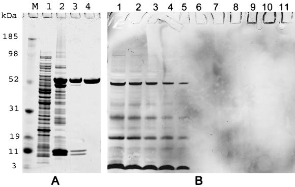
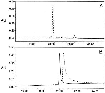
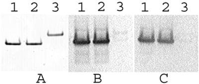
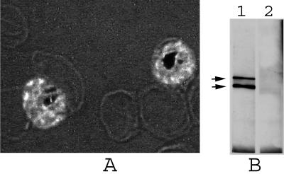
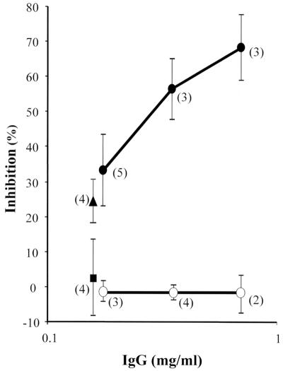
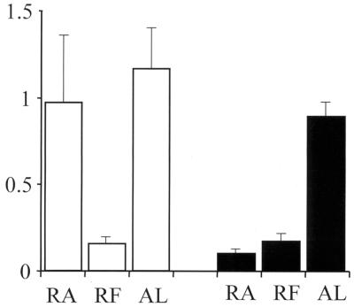
The apical membrane antigen 1 (AMA1) has emerged as a promising vaccine candidate against malaria. Advanced evaluation of its protective efficacy in humans requires the production of highly purified and correctly folded protein. We describe here a process for the expression, fermentation, refolding, and purification of the recombinant ectodomain of AMA1 (amino acids 83(Gly) to 531(Glu)) of Plasmodium falciparum (3D7) produced in Escherichia coli. A synthetic gene containing an E. coli codon bias was cloned into a modified pET32 plasmid, and the recombinant protein was produced by using a redox-modified E. coli strain, Origami (DE3). A purification process was developed that included Sarkosyl extraction followed by affinity purification on a Ni-nitrilotriacetic acid column. The recombinant AMA1 was refolded in the presence of reduced and oxidized glutathione and further purified by using two ion-exchange chromatographic steps. The final product, designated AMA1/E, was homogeneous, monomeric, and >99% pure and had low endotoxin content and low host cell contamination. Analysis of AMA1/E showed that it had the predicted primary sequence, and tertiary structure analysis confirmed its compact disulfide-bonded nature. Rabbit antibodies made to the protein recognized the native parasite AMA1 and inhibited the growth of the P. falciparum homologous 3D7 clone in an in vitro assay. Reduction-sensitive epitopes on AMA1/E were shown to be necessary for the production of inhibitory anti-AMA1 antibodies. AMA1/E was recognized by a conformation-dependent, growth-inhibitory monoclonal antibody, 4G2dc1. The process described here was successfully scaled up to produce AMA1/E protein under GMP conditions, and the product was found to induce highly inhibitory antibodies in rabbits.


Figures

FIG. 1.
FIG. 1.
(A) SDS-PAGE analysis of AMA1/E during purification. The results shown are from a lab-grade purification starting with 20 g of cell paste. Protein analysis was done on a 4 to 12% gradient gel under reduced conditions, and the gel was stained with Coomassie blue. The elutions of DEAE and SP Sepharose columns were concentrated on a 3.5-kDa cutoff Centricon concentrator before being loaded. Lane M, molecular mass markers; lane 1, E. coli lysate loaded on the Ni2+ column; lane 2, Ni2+ column elution; lane 3, DEAE Sepharose column elution (5 μg of protein); lane 4, SP Sepharose column elution (5 μg of protein; lab-grade AMA1/E product). (B) Western blot analysis for E. coli protein detection (Cygnus HCP detection kit). Lanes 1 to 5, Origami (DE3) bacterial lysate with 4,000, 2,000, 1,000, 500, or 250 ng of protein, respectively; lanes 6 to 11, AMA-1/E product with 1, 10, 100, 500, 1,000, or 2,000 ng of protein per well, respectively.
FIG. 2.
FIG. 2.
Analytical HPLC profile of the AMA1/E product. The detector output at 215 nm with absorbance units (AU) plotted against time (in minutes) is shown. (A) Gel permeation Shodex Protein KW-803 column elution profile with 10 μg of AMA1/E injected (broken line) and an equal volume of final formulation buffer with no protein injected (solid line); (B) reversed-phase C8 Aquapore RP-300 Å column (7 μm, 30 by 2.1 mm) elution profile with a 4-μg AMA1/E injection (solid line) or with 12 μg of AMA1/E reduced in the presence of 6 M GuHCl and 25 mM DTT and injected (broken line), for which a shift in retention time was observed under the same chromatographic conditions. See Materials and Methods for solvent and gradient information.
FIG. 3.
FIG. 3.
Relative SDS-PAGE mobility, alkylation analysis, and immune reactivity of AMA1/E. Lanes 1, AMA1/E protein (∼200 ng) in 4 M urea; lanes 2, AMA1/E in 4 M urea treated with iodoacetamide; lanes 3, AMA1/E in 4 M urea reduced with DTT followed by iodoacetamide treatment (see Materials and Methods for reaction details). (A) Proteins separated on a nonreducing 4 to 12% gradient gel and stained with Coomassie blue; (B) Western blot of the gel shown in panel A immunostained with monoclonal antibody 4G2dc1 and developed with HRP-POD substrate; (C) Western blot of the gel shown in panel A immunostained with an immune serum pool collected from western Kenya and developed with HRP-POD substrate.
FIG. 4.
FIG. 4.
Recognition of the parasite AMA1 with anti-AMA1/E antibodies produced in rabbit. (A) Anti-AMA1/E antibodies (R-1) used for IFA on P. falciparum (3D7) parasites fixed with methanol. Merozoites contained within the late-stage schizonts are shown with bright fluorescence. Magnification, ×1,000. (B) Western blot of P. falciparum 3D7 parasite late schizont proteins extracted with SDS-PAGE loading buffer, separated on a nonreducing gel, Western blotted and immunostained with anti-AMA1/E antibodies, and developed with a chemiluminescent substrate. The top and bottom arrows represent ∼76 and 62 kDa, respectively. Lane 1, postimmune rabbit sera (R-1); lane 2, preimmune serum control.
FIG. 5.
FIG. 5.
Purified anti-AMA1/E IgG inhibited growth by P. falciparum 3D7 parasite in a GIA, and the inhibition was reversed by the addition of the antigen. Shown are the results of a one-cycle GIA with 0.18, 0.35, or 0.7 mg of purified rabbit IgG ml−1 in suspension culture GIA in 48-well plates. •, anti-AMA1/E IgG; ○, anti-RA-AMA1/E IgG. Antigens (5.3 μg ml−1) were added to anti-AMA1/E IgG concentrations of 0.18 mg ml−1. ▴, RA-AMA1/E antigen added; ▪, AMA1/E antigen added (symbols are offset for clarity). The means ± standard deviations are shown, with the number of experiments for each data point represented within parentheses.
FIG. 6.
FIG. 6.
Competitive ELISA with individual sera. Serum samples at a 1:1,000 dilution were preincubated with either RA-AMA1/E (RA), AMA1/E (RF), or BSA protein (AL). ELISA was done to assay for anti-AMA1/E-specific antibodies. Shown here are the average OD405s at a 1:16,000 dilution for AMA1/E-immunized rabbits (R-1, R-2, and R-3) (clear bars) and RA-AMA1/E-immunized rabbits (R-7 and R-10) (black bars). The standard deviations are represented by lines on top of the bars.

Similar articles

Cited by

References

    1. Anders, R. F., P. E. Crewther, S. Edwards, M. Margetts, M. L. Matthew, B. Pollock, and D. Pye. 1998. Immunization with recombinant AMA-1 protects mice against infection with Plasmodium chabaudi. Vaccine 16:240-247. - PubMed
    1. Bessette, P. H., F. Aslund, J. Beckwith, and G. Georgiou. 1999. Efficient folding of proteins with multiple disulfide bonds in the Escherichia coli cytoplasm. Proc. Natl. Acad. Sci. USA 96:13703-13708. - PMC - PubMed
    1. Burgess, R. R. 1996. Purification of over produced E. coli RNA polymerase σ factors by solubilizing inclusion bodies and refolding from sarkosyl. Methods Enzymol. 273:145-149. - PubMed
    1. Cheng, Q., and A. Saul. 1994. Sequence analysis of the apical membrane antigen 1 (AMA-1) of Plasmodium vivax. Mol. Biochem. Parasitol. 65:183-187. - PubMed
    1. Collins, W. E., D. Pye, P. E. Crewther, K. L. Vandenberg, G. G. Galland, A. J. Sulzer, D. J. Kemp, S. J. Edwards, R. L. Coppel, J. S. Sullivan, et al. 1994. Protective immunity induced in squirrel monkeys with recombinant apical membrane antigen-1 of Plasmodium fragile. Am. J. Trop. Med. Hyg. 51:711-719. - PubMed

MeSH terms

LinkOut - more resources