. 2002 Jun;70(6):3130-42.
doi: 10.1128/IAI.70.6.3130-3142.2002.

Role of nitric oxide in host defense in murine salmonellosis as a function of its antibacterial and antiapoptotic activities

Affiliations

Role of nitric oxide in host defense in murine salmonellosis as a function of its antibacterial and antiapoptotic activities

Mohammad Samiul Alam et al. Infect Immun. 2002 Jun.

Abstract

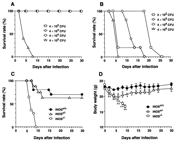
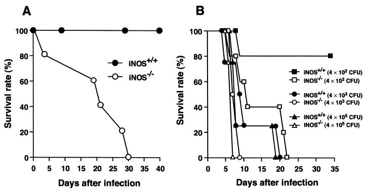
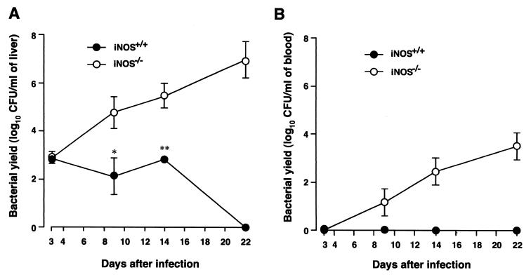
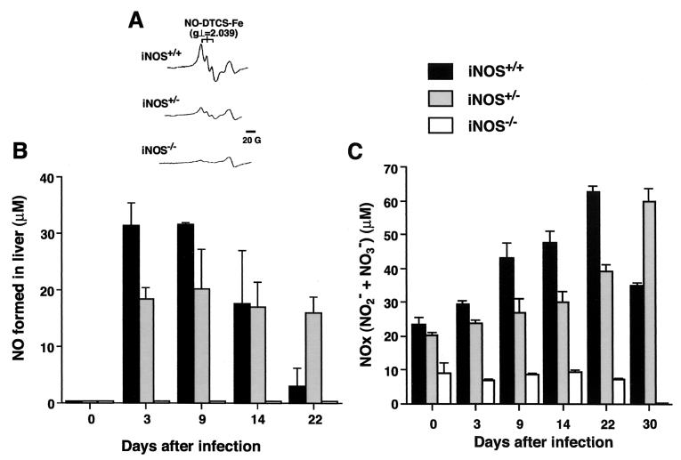
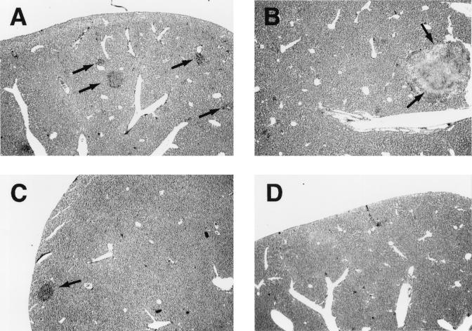
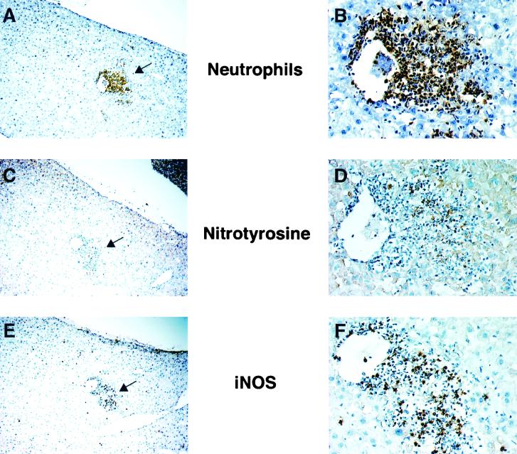
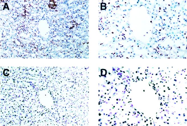
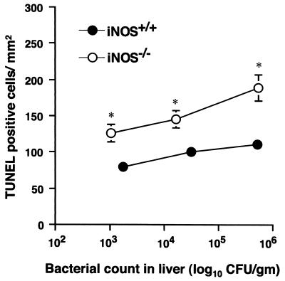
Host defense functions of nitric oxide (NO) are known for many bacterial infections. In this study, we investigated the antimicrobial effect of NO in murine salmonellosis by using inducible NO synthase (iNOS)-deficient mice infected with an avirulent or virulent Salmonella enterica serovar Typhimurium strain. All iNOS-deficient mice died of severe septicemia within 6 days after intraperitoneal injection with an avirulent strain (LT2) to which wild-type mice were highly resistant; 50% lethal doses (LD(50)s) of the LT2 strain for iNOS-deficient and wild-type mice were 30 CFU and 7 x 10(4) CFU, respectively. Lack of NO production in iNOS-deficient mice was verified directly by electron spin resonance spectroscopy. Bacterial yields in liver and blood were much higher in iNOS-deficient mice than in wild-type mice throughout the course of infection. Very small amounts of a virulent strain of serovar Typhimurium (a clinical isolate, strain Gifu 12142; LD(50), 50 CFU) given orally caused severe septicemia in iNOS-deficient animals; wild-type mice tolerated higher doses (LD(50), 6 x 10(2) CFU). Histopathology of livers from infected iNOS-deficient mice revealed extensive damage, such as diffuse hepatocellular apoptosis and increased neutrophil infiltration, but livers from infected wild-type mice showed a limited number of microabscesses, consisting of polymorphonuclear cells and macrophages and low levels of apoptotic change. The LT2 strain was much more susceptible to the bactericidal effect of peroxynitrite than the Gifu strain, suggesting that peroxynitrite resistance may contribute to Salmonella pathogenicity. These results indicate that NO has significant host defense functions in Salmonella infections not only because of its direct antimicrobial effect but also via cytoprotective actions for infected host cells, possibly through its antiapoptotic effect.


Figures

FIG. 1.
FIG. 1.
Percent survival of wild-type and iNOS-deficient mice infected with serovar Typhimurium. Both wild-type (A) and iNOS−/− (B) mice (7-week-old males) were infected with i.p. doses of serovar Typhimurium LT2 ranging from 4 × 10² to 4 × 10⁵ CFU/mouse. n = 6 for each dose. Survival rate was monitored until 30 days after infection. Survival curve (C) and changes in body weight (D) of wild-type (iNOS+/+; n = 10), heterozygous (iNOS+/−; n = 8), and iNOS-deficient homozygous (iNOS−/−; n = 14) littermate mice infected with 5 × 10⁴ CFU/mouse i.p. at different time points after infection. Body weight data are expressed as means ± SEM (∗, P < 0.05, and ∗∗, P < 0.01, versus iNOS+/+ mice).
FIG. 2.
FIG. 2.
Percent survival of wild-type and iNOS-deficient mice after oral challenge with serovar Typhimurium LT2 or Gifu 12142 strain. (A) Five-week-old male iNOS+/+ (n = 5) and iNOS−/− (n = 5) mice were orally infected with the LT2 strain at 109 CFU/mouse. (B) iNOS+/+ and iNOS−/− mice (5 weeks old, males; n = 4) were orally infected with various doses of the Gifu strain.
FIG. 3.
FIG. 3.
Bacterial growth in liver (A) and blood (B) of wild-type and iNOS-deficient mice infected with serovar Typhimurium LT2. Both iNOS−/− and iNOS+/+ mice were infected i.p. with 4 × 10² CFU of the LT2 strain per mouse. Bacterial counts in liver and blood samples were determined at different time points after infection. The number of bacteria was determined by the colony-forming assay. Data are means ± SEM (n = 3 or 4); ∗, P < 0.05, and ∗∗, P < 0.01, versus iNOS+/+ mice.
FIG. 4.
FIG. 4.
In vivo NO generation detected by ESR spectroscopy and production of NOx (NO2 + NO3) in the plasma during the course of infection. iNOS+/+, iNOS+/−, and iNOS−/− littermate mice were infected i.p. with 4 × 10² CFU of serovar Typhimurium LT2. (A) For each group of littermate mice, typical ESR spectra of the NO-DTCS-Fe complex produced in the liver on day 3 after infection are shown. For each group of mice, the amount of NO generated in the liver was determined directly by ESR spectroscopy (B), and the level in plasma of NOx (C) was measured by use of a high-performance liquid chromatography-based flow reactor with Griess reagent at various time points after infection. Data are expressed as means ± SEM (n = 3 to 6 per group at each time point).
FIG. 5.
FIG. 5.
Histopathology of livers of wild-type and iNOS-deficient mice infected with serovar Typhimurium strain LT2. Liver sections obtained at days 3 and 14 after infection (i.p.; 4 × 10² CFU/mouse) were stained with hematoxylin and eosin. Liver sections from iNOS−/− mice (A and B) and iNOS+/+ mice (C and D) were obtained on day 3 (A and C) and day 14 (B and D). The most typical result from at least six mice of each group is shown. Arrows indicate microabscess formations. Magnification, ×16.
FIG. 6.
FIG. 6.
Immunohistochemical analysis of livers of wild-type mice on day 3 after infection for neutrophil infiltration (A and B), nitrotyrosine formation (C and D), and iNOS expression (E and F). Three sequential sections of the liver (A→C→E; B→D→F) were immunostained using specific antibodies to neutrophils, nitrotyrosine, and iNOS. The area with the most intensive stain with each antibody, indicated with arrows in panels A, C, and E, is shown at higher magnification in panels B, D, and F. Mice were infected in the same manner as described for Fig. 3. Magnifications, ×36 (A, C, and E) and ×180 (B, D, and F).
FIG. 7.
FIG. 7.
Infiltration of neutrophils (A and B) and apoptotic change (C and D) in livers of iNOS-deficient mice during salmonellosis. iNOS−/− mice were infected with the serovar Typhimurium LT2 strain in the same manner as described for Fig. 3. Sequential sections of liver tissue obtained at 22 days after infection were examined for apoptosis by the TUNEL method and for neutrophil infiltration by immunohistochemical analysis as described for Fig. 6. The apoptotic cells are stained deep blue (C and D). Magnifications, ×80 (A and C) and ×160 (B and D).
FIG. 8.
FIG. 8.
Quantitative morphometric analyses for the size of the lesion (microabscess) (A) and the numbers of neutrophils infiltrated (B) and of apoptotic cells (C) in livers of wild-type and iNOS-deficient mice infected with serovar Typhimurium LT2 (i.p.; 4 × 10² CFU/mouse). Columns and error bars indicate means ± SEM (n = 6). ∗, P < 0.05, and ∗∗, P < 0.01, versus wild-type controls.
FIG. 9.
FIG. 9.
TUNEL analysis in liver tissues from both wild-type and iNOS−/− mice with similar levels of bacterial growth in the liver. After wild-type and iNOS−/−mice were infected i.p. with different numbers of CFU of serovar Typhimurium LT2, the infected mice were sacrificed and viable bacterial counts were done with the livers. The mice (n = 3) with a similar range of bacterial growth in liver tissues were then analyzed for TUNEL experiments. Columns and error bars indicate means ± SEM (n = 3). ∗, P < 0.05, and ∗∗, P < 0.01, versus wild-type controls.
FIG. 10.
FIG. 10.
Salmonella killing by H2O2, t-BuOOH, and peroxynitrite. H2O2 (1, 5, and 10 mM) (A) or t-BuOOH (25 and 50 mM) (B) was added to suspensions of LT2 or Gifu 12142 at stationary-phase growth in BHI broth. (C) Peroxynitrite at 10, 25, and 50 mM in 10 mM NaOH was infused into 1.9 ml of bacterial suspension (M9 medium) at a flow rate of 3.3 μl/min. The concentrations of peroxynitrite in the reaction mixture during peroxynitrite infusion were assumed to be maintained at a constant 9.7, 15.34, and 21.7 μM, respectively. Decomposed peroxynitrite in M9 medium was infused in the same manner. At different intervals during the treatment with these oxidants, aliquots were removed from the reaction mixture and the number of viable bacteria was determined by means of the colony-forming assay. Data are means ± SEM from three independent experiments. ∗, P < 0.05, and ∗∗, P < 0.01, versus the LT2 strain.

References

    1. Akaike, T. 2000. Mechanisms of biological S-nitrosation and its measurement. Free Radic. Res. 33:461-469. - PubMed
    1. Akaike, T. 2001. Role of free radicals in viral pathogenesis and mutation. Rev. Med. Virol. 11:87-101. - PMC - PubMed
    1. Akaike, T., Y. Noguchi, S. Ijiri, K. Setoguchi, M. Suga, Y. M. Zheng, B. Dietzschold, and H. Maeda. 1996. Pathogenesis of influenza virus-induced pneumonia: involvement of both nitric oxide and oxygen radicals. Proc. Natl. Acad. Sci. USA 93:2448-2453. - PMC - PubMed
    1. Beckman, J. S., T. W. Beckman, J. Chen, P. A. Marshall, and B. A. Freeman. 1990. Apparent hydroxyl radical production by peroxynitrite: implications for endothelial injury from nitric oxide and superoxide. Proc. Natl. Acad. Sci. USA 87:1620-1624. - PMC - PubMed
    1. Beckman, J. S., and W. H. Koppenol. 1996. Nitric oxide, superoxide, and peroxynitrite: the good, the bad, and ugly. Am. J. Physiol. 271:C1424-C1437. - PubMed

Publication types

MeSH terms